Abstract
De Winter pattern in anterior leads has been extensively described. However, there is only one case report of this pattern in inferior leads in English literature. Here, we describe a case of acute inferior wall myocardial infarction with thrombotic right coronary artery occlusion who presented with the classical De Winter sign in inferior leads.
Key words: De Winter left anterior descending, equivalent to ST-elevation myocardial infarction, inferior leads
INTRODUCTION
De Winter sign was reported for the first time by De Winter et al. in 2008.[1] A pattern of upsloping ST depression at J point with positive T wave was seen in 2% of proximal left anterior descending (LAD) occlusion with a positive predictive value of 95%–100%.[2,3] After that, many case reports were published in literature, but mostly limited to precordial leads. To the best of our knowledge, only one case has been reported so far showing electrocardiography (ECG) changes in inferior leads equivalent to De Winter LAD pattern.[4] Our report describes the first case from India depicting De Winter-type ECG changes in inferior leads.
CASE PRESENTATION
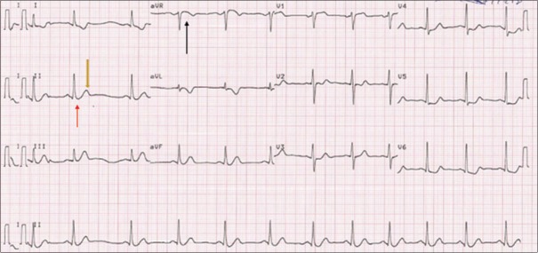
A 55-year-old male chronic smoker presented to the emergency department with complaints of retrosternal chest pain associated with perspiration of 3-h duration. He had two episodes of presyncope before reaching the hospital. At the time of presentation, his pulse rate was 48/min and systolic blood pressure was 90 mmHg. 12-lead ECG was obtained [Figure 1] that showed upsloping ST depression at J point in II, III, and aVF with positive T wave along with minimal ST elevation in aVR. Other leads showed horizontal or downsloping ST depression. On echocardiography, LV function was found to be normal with no regional wall motion abnormality at rest. Blood pressure responded adequately with intravenous fluids alone. Dual antiplatelets, unfractionated heparin, and nitrates were started. In view of global ST depression and ST elevation in aVR, significant coronary artery disease, possibly involving the left main or ostial LAD, was suspected, and the patient was advised for early revascularization therapy. However, the patient and his relatives were not willing for coronary angiography at that time, and hence conservative management was continued. Troponin I was significantly raised. The patient became pain free over the next 2–3 h of medical management.
Figure 1.
Upsloping ST depression at J point in the inferior lead (de Winter sign) (red arrow) with positive T wave (yellow arrow) along with minimal ST elevation in aVR (black arrow)
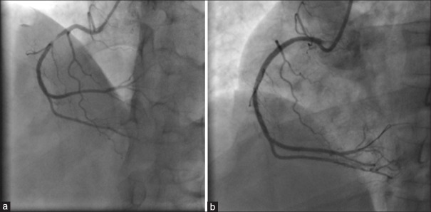
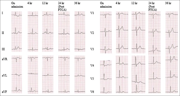
He had recurrence of angina the next morning and after counseling again, he gave consent for revascularization. Coronary angiography was done which showed normal left main and LAD artery with a plaque in the left circumflex artery. There was critical 99% thrombotic occlusion in the mid right coronary artery (RCA) with TIMI II flow in the distal RCA. Coronary angioplasty was done with sirolimus-eluting stent (3 mm × 20 mm) [Figure 2]. There was complete pain resolution after angioplasty with normalization of ST segment in all leads [Figure 3]. He was discharged on the 3rd postoperative day without any complication.
Figure 2.
(a) 99% thrombotic occlusion seen in the mid right coronary artery. (b) After placement of drug-eluting stent
Figure 3.
ST elevation was not noted in serial electrocardiography. Post-post angioplasty resolution of De Winter pattern in inferior leads
DISCUSSION
De Winter sign in ECG is one of the not so uncommon types of presentation for acute anterior wall myocardial infarction; however, it is often missed by physicians as well as cardiologists, leading to less aggressive initial management. The exact mechanism for this ECG pattern is not known. Such ECG changes might be associated with variation in the anatomy of Purkinje fibers, leading to endocardial conduction delay as described by De Winter et al. Till now, there is no clear recommendation regarding the reperfusion therapy for such ECG changes.[5,6] Cardiologists and physicians should be able to recognize that such unique pattern of ECG changes is not limited to anterior leads, but can occur in inferior leads as well, and such patients should be offered early reperfusion therapy as for acute ST-elevation myocardial infarction (STEMI).
CONCLUSION
De Winter sign in inferior leads is uncommon. It is equivalent to STEMI. Cardiologists and physicians should recognize such ECG changes and advice for early revascularization.
Declaration of patient consent
The authors certify that they have obtained all appropriate patient consent forms. In the form the patient(s) has/have given his/her/their consent for his/her/their images and other clinical information to be reported in the journal. The patients understand that their names and initials will not be published and due efforts will be made to conceal their identity, but anonymity cannot be guaranteed.
Financial support and sponsorship
Nil.
Conflicts of interest
There are no conflicts of interest.
Acknowledgment
We are sincerely thankful to the Cath laboratory staff, Department of Cardiology, Shree Krishna Hospital and Pramukhswami Medical College, Anand, Gujarat, India.
REFERENCES
- 1.De Winter RJ, Verouden NJ, Wellens HJ, Wilde AA. Interventional Cardiology Group of the Academic Medical Center. A new ECG sign of proximal LAD occlusion. N Engl J Med. 2008;359:2071–3. doi: 10.1056/NEJMc0804737. [DOI] [PubMed] [Google Scholar]
- 2.Goebel M, Bledsoe J, Orford JL, Mattu A, Brady WJ. A new ST-segment elevation myocardial infarction equivalent pattern? Prominent T wave and J-point depression in the precordial leads associated with ST-segment elevation in lead aVR. Am J Emerg Med. 2014;32:287.e5-8. doi: 10.1016/j.ajem.2013.09.037. [DOI] [PubMed] [Google Scholar]
- 3.Morris NP, Body R. The de winter ECG pattern: Morphology and accuracy for diagnosing acute coronary occlusion: Systematic review. Eur J Emerg Med. 2017;24:236–42. doi: 10.1097/MEJ.0000000000000463. [DOI] [PubMed] [Google Scholar]
- 4.Tsutsumi K, Tsukahara K. Is the diagnosis ST-segment elevation or non-ST-segment elevation myocardial infarction? Circulation. 2018;138:2715–7. doi: 10.1161/CIRCULATIONAHA.118.037818. [DOI] [PubMed] [Google Scholar]
- 5.Ibanez B, James S, Agewall S, Antunes MJ, Bucciarelli-Ducci C, Bueno H, et al. 2017 ESC guidelines for the management of acute myocardial infarction in patients presenting with ST-segment elevation: The task force for the management of acute myocardial infarction in patients presenting with ST-segment elevation of the European Society of Cardiology (ESC) Eur Heart J. 2018;39:119–77. doi: 10.1093/eurheartj/ehx393. [DOI] [PubMed] [Google Scholar]
- 6.O'Gara PT, Kushner FG, Ascheim DD, Casey DE, Jr, Chung MK, de Lemos JA, et al. 2013 ACCF/AHA guideline for the management of ST-elevation myocardial infarction: A report of the American College of Cardiology Foundation/American Heart Association Task force on Practice Guidelines. Circulation. 2013;127:e362–425. doi: 10.1161/CIR.0b013e3182742cf6. [DOI] [PubMed] [Google Scholar]


