. 2019 Apr 25;8:e41331. doi: 10.7554/eLife.41331

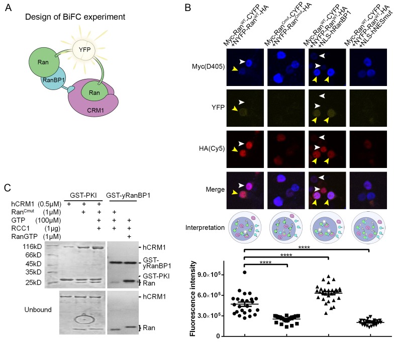
Figure 5. RanBP1 tetrameric complex exists in the cell nucleus.

(A) Design of BiFC experiment. When the RanBP1 tetramer is formed, a YFP signal could be detected. This assay may not discriminate which Ran binds to RanBP1 (or CRM1) in the tetramer. (B) Representative immunofluorescence (HA and Myc) and fluorescence (YFP) images on cells co-transfected with fusion plasmids (labelled on top). NLS-hRanBP1 or NLS-hNESmut (hNESmut: human RanBP1 with NES mutation) was used to increase nuclear level of RanBP1 or its mutant. Nucleus was not stained so that the two transfected proteins could be stained without contaminating the YFP channel. The boundary of nucleus could be estimated by Ran fusions, which is mainly localized in the nucleus (see Figure 5—figure supplement 3). Results interpretation (middle panel) explains why the yellow fluorescence is observed or not. Bottom panel shows the nuclear YFP intensities of at least 20 cells (only florescent cells transfected with two Ran fusions) in the corresponding samples under the same level of illumination. **** denotes p<0.0001. (C) RanCmut is able to bind to CRM1 and NES (left panel) but not able to bind yRanBP1 (right panel).

Figure 5.

Figure 5—figure supplement 1. Pull down of 293 T cells expressed Ran fusion proteins using GST-mRanBP1 or GST, in the presence or absence of Ran knock down.
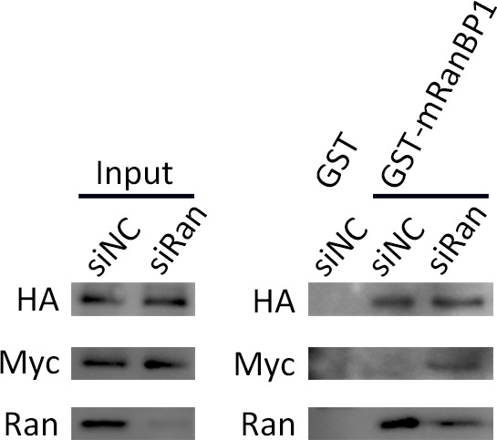
Figure 5—figure supplement 1.

Input lanes were used to ensure similar amount of Ran fusions present in each pull down sample. In the presence of Ran knock down, Ran fusions bound more to RanBP1, functionally rescues the reduction of bound RanWT.
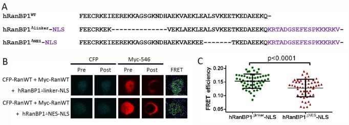
Figure 5—figure supplement 2. Acceptor photobleach-FRET and statistical analysis of FRET efficiencies.

Figure 5—figure supplement 2.

293 T cells were seeded in 24-well plates containing circular coverslips slides. After adhesion overnight, cells were transfected with Myc-Ran and CFP-Ran plasmids in addition to HA-tagged hRanBP1Δlinker-NLS or hRanBP1ΔNES-NLS (panel A, showing only the C terminus). Pull down experiments verified that RanBP1 tetramer formation is possible for hRanBP1Δlinker-NLS but not hRanBP1ΔNES-NLS. 24 hr after transformation, cells were fixed and stained with primary antibodies against Myc (ProteinTech, Mouse) and HA (CST, Rabbit) separately. The slides were then incubated with the secondary antibody anti-Mouse-Alexa Fluor 546 (ThermoFisher) and anti-Rabbit-Cy5 (ThermoFisher). FRET measurements were performed by acquiring pre- and post- acceptor bleach images of donor and acceptor using the Olympus FV-1000 acceptor photobleach FRET, followed by calculation of FRET efficiency for acquired images (B). Only cells transfected with two Ran plasmids and one RanBP1 plasmid were analyzed. Statistical significance (C) was calculated using two-tailed unpaired Student’s t test. Each group contains more than 40 cells.
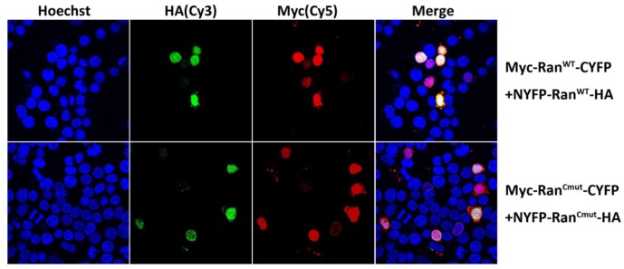
Figure 5—figure supplement 3. Immunofluorescence (HA and Myc) on cells co-transfected with two fusion plasmids.

Figure 5—figure supplement 3.

All the four Ran fusion constructs are mainly localized in the cell nucleus, similar as RanWT.