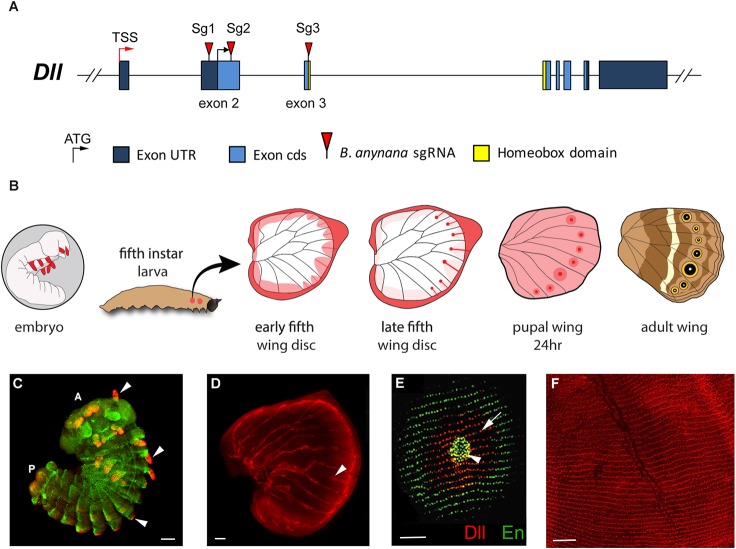
Fig. 1.
Expression of Distal-less in embryos, and larval and pupal wings. (A) Dll gene structure indicating the exons targeted by guide RNAs in this work (red triangles). TSS, transcription start site. (B) Summary diagram of relevant expression patterns of Dll in embryo limbs and different stages of fifth instar and pupal wings. Dll is represented as a gradient of pink to red illustrating weaker to stronger expression, respectively, with highest expression in the wing margin and also fingers terminating in an eyespot center. This temporal expression pattern of Dll in the larval and pupal wings has been replicated in numerous studies (Brakefield et al., 1996; Monteiro et al., 2013; Oliver et al., 2013, 2014; Reed and Serfas, 2004). (C-F) Fluorescent immunostainings of Dll (red) and Engrailed (En, green). In pupal wings, Dll is expressed in all scale-building cells (at low levels) and at higher levels in the scale cells that will become the black disc of an eyespot. (C) Dll is expressed in antennae, thoracic legs, and abdominal prolegs of embryos (arrowheads), A, anterior; P, posterior. En is also expressed in embryos. Photo credit: Xiaoling Tong (Southwest University, Chongqing, China). (D) Dll is expressed in eyespot centers (arrowhead) and along the wing margin in late larval wings. (E) Dll is expressed in eyespot centers (arrowhead) and in black scale cells of pupal wings (arrow). En is expressed in the eyespot center and area of the gold ring. (F) Dll expression in rows of scales across the entire surface of a 24-h pupal wing. Scale bars: 100 µm in C,D; 50 µm in E,F.

