



Abstract
Background and objectives
MicroRNA‐9 is frequently dysregulated in many human carcinoma types, including gastric cancer (GC). Previous studies demonstrated that the expression of TNFAIP8 in GC is correlated with tumour occurrence, development, invasion, metastasis and prognosis. However, till now, the relationship between MicroRNA‐9 and TNFAIP8 in GC has not been reported.
Materials and methods
Levels of miR‐9 and TNFAIP8 expression in GC tissues and in human GC cell lines were studied using qualitative real‐time PCR (qRT‐PCR) and Western blotting. Cell viability was detected using the CCK‐8 and clone formation assays. A dual‐luciferase reporter system was used to confirm the target gene of miR‐9.
Results
We found that the expression level of MicroRNA‐9 in GC tissues and cell lines was significantly lower than that in adjacent non‐cancerous tissues and human immortalized gastric epithelial cell (GES) line, respectively. In addition, overexpression of MicroRNA‐9 markedly inhibited GC cell proliferation in vitro and tumour growth in vivo. Further experiments revealed that TNFAIP8 was a direct and functional target of MicroRNA‐9 in GC and overexpression of MicroRNA‐9 obviously down‐regulated the expression of TNFAIP8, which was involved in the gastric carcinogenesis and cancer progression.
Conclusion
Our results suggested that MicroRNA‐9‐TNFAIP8 might represent a promising diagnostic biomarker for GC patients and could be a potential therapeutic target in the prevention and treatment of GC.
Keywords: miR‐9, TNFAIP8, gastric cancer, proliferation, biomarker
1. Introduction
Gastric cancer (GC) is the fifth most common malignant cancer and ranks third worldwide as a cause of cancer‐related deaths in both sexes as WHO GLOBOCAN reported. 1 It is proposed that the incidence of the GC has significant regional differences, particularly in Asian regions.2 Particular efforts are currently underway to improve prevention and treatment for the GC patients, including advanced surgical techniques, anti‐Her‐2 molecular‐targeted therapies, radiation and chemotherapeutic treatments; nevertheless, the 5‐year survival rates with localized GC have increased minimally in the past few years. Therefore, we urgently need an extensive understanding of underlying mechanism by which it can consummate the diagnosis, therapy and prognosis prediction in GC.
TNFAIP8 is primordially identified from an expression profile by means of comparing a primary human head and neck squamous cell carcinoma cell line with its metastatic cell line.3 More convincingly, number of data support that the overexpressed TNFAIP8 is associated with adverse clinical outcomes in several cancer types, including GC,4, 5, 6, 7 osteosarcoma8 and oesophageal squamous cell carcinoma.9 Recently, experimental evidences have shown that TNFAIP8 acts as an oncogene in human cancers and is related with enhanced DNA synthesis, cell survival and inhibition of apoptosis through inhibiting caspase 3 and caspase 8.10, 11, 12, 13, 14 Moreover, the expression level of TNFAIP8 can be up‐regulated by NF‐κB and TNF‐α in diversified cell lines.10 However, the role of TNFAIP8 and its upstream regulation in GC remains to be clarified.
MicroRNAs (miRNAs), a kind of small non‐coding RNAs with 18‐22 nucleotides in length, interact directly with the 3′‐untranslated region (3′‐UTR) of specific target mRNAs; subsequently, the interactions between miRNAs and 3′‐UTR regulate their expression at the post‐transcriptional levels.15, 16, 17 In GC, unlike the miRNAs with anti‐tumour activity, such as miR‐129‐5p,18 miR‐361‐5p19 and miR‐506,20 which are significantly down‐regulated, oncogenic miRNAs such as miR‐21,21 miR‐296‐5p22 and miR‐36223 are abnormally up‐regulated. However, little is known about the role of miRNA‐9 (miR‐9) described to date in the pathogenesis of GC.
With in vitro study, we have shown that TNFAIP8 was up‐regulated and able to promote GC cells proliferation; on the contrary, downregulation of TNFAIP8 inhibited the cell viability and growth. More particularly, we also found that miR‐9 was suppressive in GC tissues and cell lines. Restoration of miR‐9 expression could reduce cell viability and growth in vitro and suppress tumorigenicity in vivo. In addition, our results demonstrated that miR‐9 could down regulate the expression of TNFAIP8 by directly targeting the latter 3′‐UTR. Taken together, our findings collectively show that miR‐9 acts as a tumour suppressor via regulation of TNFAIP8 and provide new therapeutic targets for future treatment of GC.
2. Materials and methods
2.1. Tissue samples and cell culture
Gastric cancer tissue samples from 16 GC patients were collected and archived at the Affiliated Hospital of Xuzhou Medical University between December 2015 and January 2016. Sixteen non‐cancerous gastric tissues (controls) were also collected and archived. Detailed clinical information was listed in Table 1. TNM stage was classified according to the seventh edition of the Union for International Cancer Control TNM Classification of Malignant Tumors. None of the patients included in our study received chemotherapy or radiotherapy before surgical resection. A total of 16 fresh GC tissues and paired non‐cancerous gastric mucosal tissues were immediately snap‐frozen in liquid nitrogen and stored at −80°C until their use in Western blotting and quantitative RT‐PCR (qPCR) assays. All patients provided written informed consent to participate in the study. GC (MKN‐45, MGC‐803, SGC‐7901, MKN28, AGS) and GES cell lines were purchased from the Shanghai Institute of Biochemistry and Cell Biology, Chinese Academy of Sciences (Shanghai, China). Both cells were cultured in RPMI Medium 1640 (RPMI1640; Gibco, New York, USA) supplemented with 10% foetal bovine serum (Zhejiang, Zhejiang Tianhang Biotechnology Co.,Ltd. China), 100 U/mL penicillin and 100 μg/mL streptomycin (Beyotime, Jiangsu, China) in a humidified with 5% CO2 atmosphere at 37°C.
Table 1.
The basic information of patients
| Variables | Case |
|---|---|
| Total cases | 16 |
| Gender | |
| Male | 11 |
| Female | 5 |
| Age (years) | |
| ≥60 | 9 |
| <60 | 7 |
| Pathological grading | |
| Well differentiated | 0 |
| Moderately differentiated | 8 |
| Poorly differentiated | 8 |
| Tumour size (maximum diameter) | |
| ≤5.0 cm | 7 |
| >5.0 cm | 9 |
| Vascular invasion | |
| Yes | 10 |
| No | 6 |
| Lymphatic node metastasis | |
| No | 2 |
| Yes | 14 |
| TNM stage | |
| I | 2 |
| II | 3 |
| III | 9 |
| IV | 2 |
| Tumour stage | |
| T1 | 0 |
| T2 | 2 |
| T3 | 4 |
| T4 | 10 |
2.2. Cell transfection
MiRNA‐9 mimics, inhibitor, si‐TNFAIP8 and their respective negative controls were purchased from Genepharma (Shanghai, China). Si‐TNFAIP8 was designed: sense strand: 5′‐ACCUUAAUAGACGACACAATT‐3′, antisense strand: 5′‐UUGUGUCGUCUAUUAAGGUGG‐3′. Their final concentration was 20 μmol/L. Cells were grown to 50%‐70% confluence and transfected using Lipofectamine 2000 (Invitrogen, Waltham, MA, USA) following the manufacturer's protocol. After 48 hours of transfection, the efficiency of knocking down or overexpression was assayed by Western blotting and qPCR.
2.3. Cell infection and stable cell line generation
MiR‐9 gene was subcloned into the lentiviral expression vector, pGLVH1 (Genepharma). An empty pGLVH1 vector was used as the negative control. MKN45 and MGC803 cells (1.0 × 10⁵) were infected with a lentivirus encoding miR‐9 mimics or an empty lentivirus (control) for 96 hours and then selected with puromycin (Santa Cruz Biotechnology, Santa Cruz, CA, USA) for 3 weeks. The efficiency of infection was assayed by Western blotting and qPCR.
2.4. Quantitative real‐time RT‐PCR
Total RNA was extracted from GC tissues and cell lines using TRIzol reagent (ambion; Invitrogen), and reverse transcription was performed using a reverse transcription kit (Cat. no. RR037A; Takara, Tokyo, Japan), following the manufacturers' instructions. qRT‐PCR reactions were performed on an ABI 7500 real‐time PCR system (Applied Biosystems, Waltham, MA, USA) using 2× SYBR Premix Ex Taq (Cat. no. RR420A, Takara). The primers used in this study are listed as follows: miR‐9‐RT: 5′‐GTCGTATCCAGTGCAGGGTCCGAGGTATTCGCACTGGATACGACCACAAGA‐3′; miR‐9‐forward: 5′‐GCTGGAGAACCCGTAGATCCGAT‐3′; miR‐9‐reverse: 5′‐GTGCAGGGTCCGAGGT‐3′; U6‐forward: 5′‐GCTTCGGCAGCACATATACTAAAAT‐3′; U6‐reverse: 5′‐CGCTTCACGAATTTGCGTGTCAT‐3′; TNFAIP8‐forward: 5′‐TTCCATCAGGTGGATTATAC‐3′; TNFAIP8‐reverse: 5′‐AGGTGGCGCTGAATGATTTG‐3′; GADPH‐forward: 5′‐AGAAGGCTGGGGCTCATTTG‐3′, GADPH‐reverse: 5′‐AGGGGCCATCCACAGTCTTC‐3′. The levels of miR‐9 and TNFAIP8 were normalized by U6 and GADPH levels, respectively. Each sample was run in triplicate. The relative expression was calculated using the relative quantification equation (RQ) = 2−ΔΔCt.
2.5. Western blotting
Total protein was extracted from GC tissues and cells by lysing in ice‐cold lysis buffer. One hundred microgram proteins were electrophoresed on an SDS‐PAGE gel (12.5%), transferred onto nitrocellulose membranes, and probed with a primary antibody targeted against TNFAIP8 (1:200, ab64988; Abcam, Cambridge, MA, USA). After incubating overnight at 4°C with the primary antibody, membranes were washed with TBS/0.05% Tween‐20 (TBST) and incubated with a secondary antibody (1:10 000, VICMED) for 2 hours at room temperature. After washing three times with (TBST) for 15 minutes, the membranes were scanned with Odyssey (Tanon, Shanghai, China).
2.6. In vitro cell proliferation assays
Clone formation assays and CCK‐8 were used to determine the biological effect of miR‐9 and TNFAIP8 on GC cell proliferation, following previously described methods.20, 24
2.7. Xenograft studies in athymic nude mice
Four‐week‐old BALB/c nude mice were purchased from the Institute of Laboratory Animal sciences, Cams & Pumc (Beijing, China). BALB/c nude mice were randomly divided into two groups (n=5/group). The suspending MKN45 stable cells (0.2 mL) were subcutaneously injected into either side of the posterior flank of the nude mouse. Thirty days after injection, the mice were killed and tumour weights were measured. All experiments were performed under the guidelines for animal experiments.
2.8. Luciferase reporter plasmids and assays
Predicted miRNA‐binding regions for miR‐9 in the 3′‐UTR of TNFAIP8 were subcloned into the GP‐miRGLO Luciferase miRNA expression vector (Genepharma). Mutants of the binding sites were used as the negative control. MKN45 cells were seeded in 24‐well plates at a density of 5 × 10⁴ cells per well and transiently transfected with mutant or wild‐type luciferase reporter plasmids (miR‐9 and the negative control) at a final concentration of 50 nmol/L. Following 24 hours of incubation, luciferase activity was measured using a dual‐luciferase reporter system (Cat. no. E1910; Promega, Fitchburg, WI, USA).
2.9. Statistical analysis
Data were analysed using the statistical software, spss 16.0 (IBM, Armonk, NY, USA). Values were expressed as the mean ± SD of at least three independent experiments. The difference between groups was determined by an unpaired two‐tailed Student's t‐test. P value <.05 was considered as statistically significant.
3. Results
3.1. TNFAIP8 expression is up‐regulated in GC tissues and cell lines
Overexpression of TNFAIP8 has been reported in numerous human cancers,8, 9, 12, 14 including GC.5 Quantitative RT‐PCR results demonstrated that the mRNA expression levels of TNFAIP8 in cell lines and GC tissues (n=16) were significantly higher than those in GES and adjacent non‐cancerous tissues (ANT), respectively (Figure 1A,C). The protein levels of TNFAIP8 in samples prepared from five GC cell lines and GES were analysed by Western blotting. Compared with GES, all cancer cell lines expressed higher levels of TNFAIP8 protein (Figure 1B). Moreover, as determined by Western blotting, TNFAIP8 protein levels were higher in GC tissues than in ANT (n=16, Figure 1D). Results extracted from Western blotting demonstrated that higher TNFAIP8 protein expression was evidently associated with GC. These results suggest that TNFAIP8 may function as an oncogene in GC.
Figure 1.
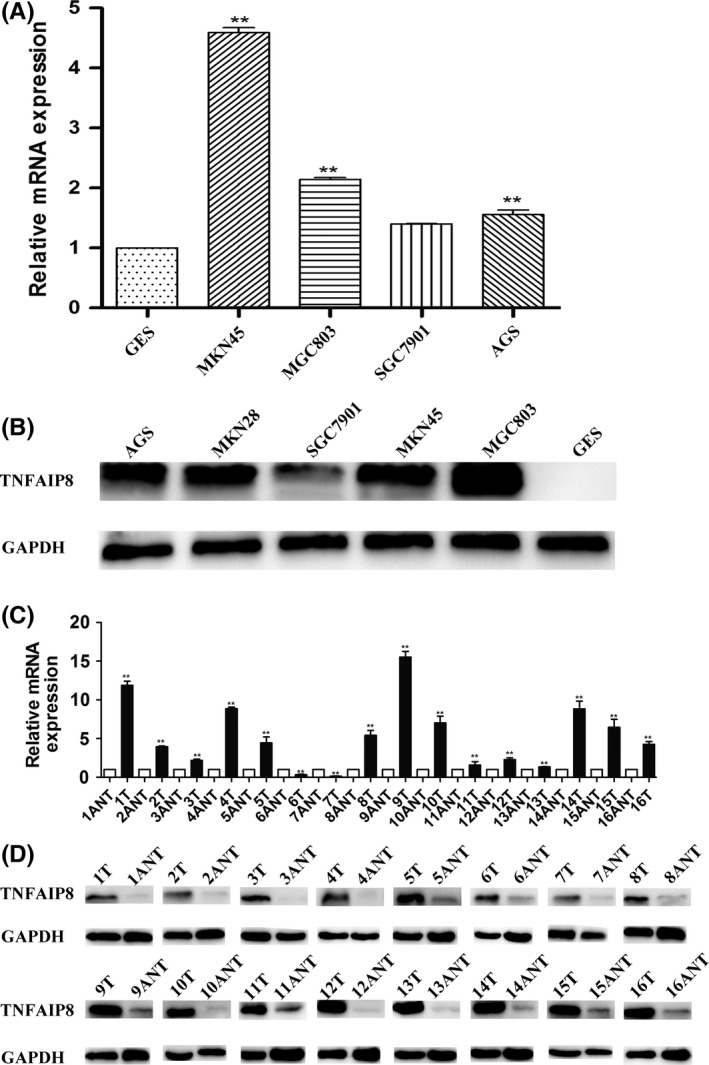
Expression of TNFAIP8 is up‐regulated in gastric cancer tissues and cell lines. A, Quantitative PCR results of TNFAIP8 mRNA levels in GC cell lines. B, Western blotting results of TNFAIP8 protein levels in different GC cell lines. TNFAIP8 protein levels are higher in the five GC cell lines than in the human immortalized gastric epithelial cell line (GES). C, Quantitative PCR results of TNFAIP8 mRNA levels in GC tissues (T) and in adjacent non‐cancerous gastric tissues (ANT). TNFAIP8 mRNA levels are higher in the GC tissues (T) than in adjacent non‐cancerous gastric tissues (ANT) (n=16). D, Western blotting results of TNFAIP8 protein levels in GC tissues and adjacent non‐cancerous gastric tissues. TNFAIP8 protein levels are higher in GC tissues (T) than in adjacent non‐cancerous gastric tissues (ANT) from the same patient (n=16). Each bar represents the mean ± SD of three independent experiments. **P<.01
3.2. TNFAIP8 promotes GC cell proliferation
To investigate the role of TNFAIP8 in GC cell proliferation, we conducted loss‐of‐function and gain‐of‐function experiments (Figure 2A,D). Cell counting Kit‐8 (CCK8) assays showed that MKN45 and MGC803 cell proliferations were significantly suppressed after knocking down TNFAIP8 (Figure 2B), whereas cell proliferation was enhanced after TNFAIP8 overexpression (Figure 2E). Furthermore, clone formation assays also showed that down‐regulating TNFAIP8 inhibited GC cell proliferation (Figure 2C) and overexpressing TNFAIP8 promoted cell proliferation (Figure 2F), which is consistent with our CCK8 assay results. Taken together, these results show that TNFAIP8 promotes GC cell proliferation.
Figure 2.
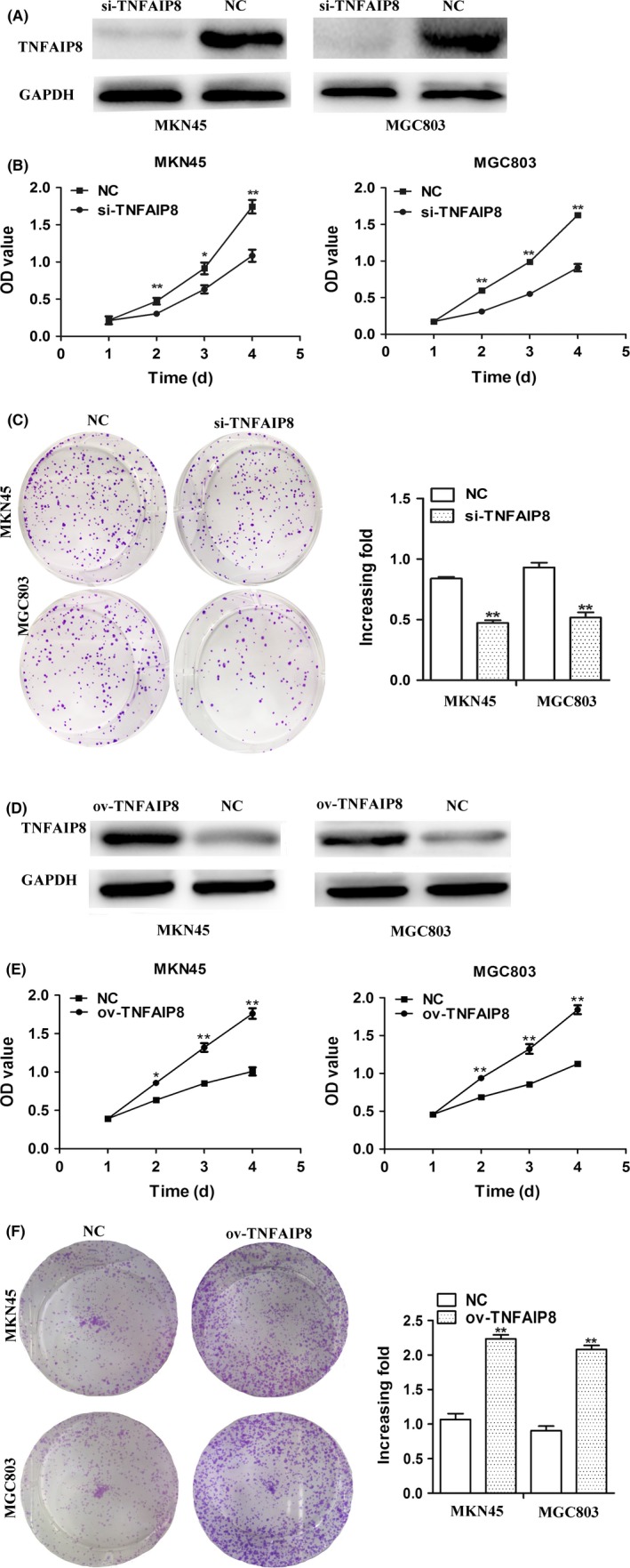
TNFAIP8 promotes GC cell proliferation. A, Western blotting results of TNFAIP8 protein levels after knocking down TNFAIP8 in different GC cell lines. Knocking‐down TNFAIP8 decreased the protein level of TNFAIP8 in MKN45 and MGC803 cells. B, Cell proliferation was examined by CCK8 assay after knocking down TNFAIP8. Knocking‐down TNFAIP8 inhibited cell proliferation of MKN45 and MGC803 cells compared to cells in the control mock‐treated group. C, Knocking‐down TNFAIP8 suppressed clone formation in MKN45 and MGC803 cells. D, Overexpressing TNFAIP8 increased the protein level of TNFAIP8 in MKN45 and MGC803 cells. E, Overexpressing TNFAIP8 promoted cell proliferation of MKN45 and MGC803 cells compared to cells in the control mock‐treated group. F, Overexpressing TNFAIP8 promoted clone formation in MKN45 and MGC803 cells. Each bar represents the mean ± SD of three independent experiments. *P<.05, **P<.01
3.3. TNFAIP8 is a direct target of miR‐9
The mechanisms underlying regulation of TNFAIP8 expression by miR‐9 have not been well characterized. We identified putative miRNAs that might regulate TNFAIP8 via bioinformatics methods. Four miRNAs, including miR‐205, miR‐182, miR‐96 and miR‐9, were predicted by two independent miRNA databases: TargetScan (http://www.targetscan.org/) and miRBase (http://www.mirbase.org/) (Figure 3A), among which miR‐9 has the highest aggregate PCT score. Western blotting results showed that overexpressing miR‐9 significantly reduced TNFAIP8 protein expression in MKN45 and MGC‐803 cells. Conversely, knocking‐down miR‐9 remarkably increased TNFAIP8 protein expression (Figure 3C). By qPCR, our results showed that overexpressing miR‐9 significantly down‐regulated TNFAIP8 mRNA levels in MKN45 and MGC‐803 cells, whereas knocking‐down miR‐9 led to the opposite results (Figure 3D).
Figure 3.
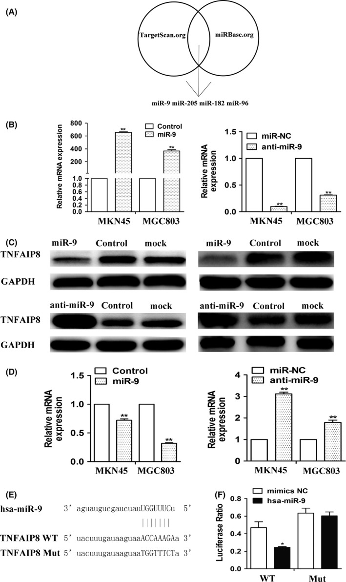
TNFAIP8 is a direct target of miR‐9. A, miRNAs were computationally predicted using two independent miRNA databases. B, qPCR results of miR‐9 mRNA levels after transfecting GC cells with miR‐9 mimics or anti‐miR‐9. Overexpression of miR‐9 markedly increased the mRNA level of miR‐9. Conversely, knocking‐down miR‐9 significantly decreased the mRNA level of miR‐9. C, Western blotting results showed that the overexpression of miR‐9 significantly reduced TNFAIP8 protein levels in MKN45 and MGC803 cells, whereas knocking‐down miR‐9 has the opposite results. D, qPCR results of TNFAIP8 mRNA levels after transfecting GC cells with miR‐9 mimics or anti‐miR‐9. E, Bioinformatics predicted wild type or mutant TNFAIP8 3′‐UTR miRNA binding sites were inserted into luciferase reporter plasmids. F, In luciferase activity assays, miR‐9 suppressed luciferase activity of the wild type but not mutant TNFAIP8 3′‐UTR constructs in MKN45. Each bar represents the mean ± SD of three independent experiments. *P<.05, **P<.01
To determine whether miR‐9 repressed TNFAIP8 expression by targeting its 3′‐UTR, wherein the predicted binding sites were disrupted, were cloned into luciferase reporter vectors and tested using luciferase activity assays (Figure 3E). Luciferase expression from the wild type but not from mutant 3′‐UTR constructs was significantly suppressed by miR‐9 (Figure 3F). These data support that the TNFAIP8 3′‐UTR is a direct target of miR‐9 in GC cells.
3.4. MiR‐9 regulates GC cell proliferation
Several studies have proved that miR‐9 is decreased in human cancers, suggesting that it functions as tumour suppressor.24, 25, 26, 27, 28 However, till now, the expression level of miR‐9 in GC has not been reported. This is supported by our observation that overexpressing miR‐9 suppressed the proliferation of GC cells and knocking‐down miR‐9 promoted the proliferation of GC cells in vitro (Figure 4A). Clone formation assays also showed that overexpressing miR‐9 inhibited GC cell proliferation; conversely, knocking‐down miR‐9 significantly promoted GC cell proliferation (Figure 4B), which is consistent with our CCK8 assay results. Moreover, to explore the role of miR‐9‐TNFAIP8 pathway in GC tumorigenicity, we conducted the restoration of TNFAIP8 in miR‐9‐treated cells. Importantly, co‐transfecting miR‐9 with overexpressing TNFAIP8 rescued miR‐9‐mediated inhibition of TNFAIP8 expression levels and cell proliferation in MKN45 and MGC803 cells (Figure 4C,D). Furthermore, in order to determine the role of miR‐9 in the growth of GC xenografts in nude mice, we performed a contrast experiment with the lentivirus vectors mediated overexpression of miR‐9 in MKN45 and MGC803 cells (Figure 4E‐G). Compared to a negative control lentivirus, lentivirus‐mediated overexpression of miR‐9 significantly inhibited growth of MKN45 tumour xenografts (Figure 4H). Taken together, these results suggest that miR‐9‐TNFAIP8 plays a vital role in the development and progression of GC.
Figure 4.
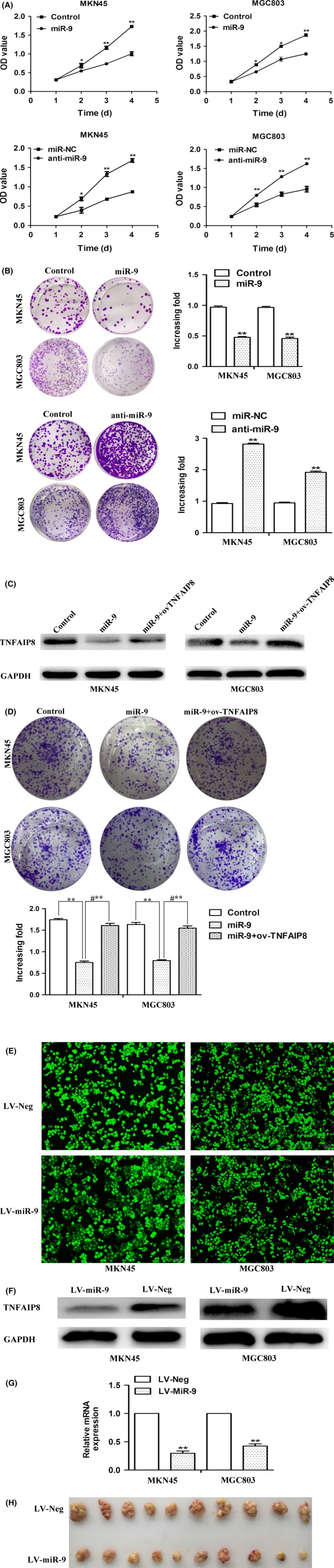
miR‐9 inhibits cell proliferation. A, Overexpression of miR‐9 inhibits MKN45 and MGC‐803 cell proliferation in CCK8 assays. However, knocking down of miR‐9 promotes MKN45 and MGC‐803 cell proliferation. B, Overexpressing miR‐9 suppressed clone formation in MKN45 and MGC803 cells. However, knocking‐down miR‐9 promotes clone formation in MKN45 and MGC803 cells. C, Overexpressing TNFAIP8 rescued miR‐9‐mediated inhibition of TNFAIP8 expression levels in MKN45 and MGC803. D, Restoration of TNFAIP8 rescued miR‐9‐mediated inhibition of cell proliferation. E, MKN45 and MGC803 cells were infected with a lentivirus encoding miR‐9 mimics or an empty lentivirus (control) for 96 h. F, After 96 h of infection, the TNFAIP8 protein levels were examined by Western blotting in MKN45 and MGC803. G, After 96 h of infection, the TNFAIP8 mRNA levels were examined by qPCR in MKN45 and MGC803. H, Representative images of tumours from mice in each group. Each bar represents the mean ± SD of three independent experiments. *P<.05, **P<.01. **P<.01 vs control, #**P<.01 vs miR‐9+ov‐TNFAIP8
3.5. Clinical relevance between miR‐9 and TNFAIP8 in fresh GC tissues
In addition to TNFAIP8 proteins, we detected the expression of miR‐9 in 16 pairs of fresh GC tissues (Figure 5A). We observed that the expression of miR‐9 was inversely correlated with protein levels of TNFAIP8 (Figure 5B, R=−.687, P=.0033). Taken together, these results suggest that miR‐9‐TNFAIP8 plays a vital role in the development and progression of GC.
Figure 5.
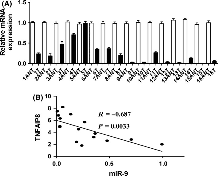
Clinical relevance of miR‐9‐TNFAIP8 in fresh GC tissues. A, The relative levels of miR‐9 expression in 16 pairs of fresh GC tissues were determined by qPCR. B, Statistical analysis suggested an inverse correlation between miR‐9 expression and TNFAIP8 protein expression. Each bar represents the mean ± SD of three independent experiments
4. Discussion
Recent researches have shown that dysregulation of miRNAs is a common event that affects cell survival, proliferation and apoptosis, invasion, immigration in tumour development and progression. Our results provide that the expression of miR‐9, a member of the miRNA family, is significantly down‐regulated in GC tissues. We investigated the biological functions of miR‐9 in GC, and our in vitro findings demonstrate that overexpression of miR‐9 could remarkably suppress GC cell proliferation, while inhibition of miR‐9 might significantly promote cell proliferation. TNFAIP8 was identified to be a target of miR‐9 using two independent databases. Our findings showed that a significant inverse correlation was identified between TNFAIP8 and miR‐9. As indicated on luciferase reporter assay, miR‐9 repressed the construct with GP‐miRGLO‐TNFAIP8‐3′‐UTR. Overexpression of miR‐9 inhibited the expression of TNFAIP8 at both mRNA and protein levels. It suppressed TNFAIP8‐3′‐UTR luciferase report activity, and this effect was abolished by mutation of the miR‐9 seed binding site. Our results suggest that miR‐9 might function as a negative regulator or tumour suppressor for cell growth in GC, which is partly mediated by repressing TNFAIP8 expression.
Meanwhile, it has been demonstrated that miR‐9 was also down‐regulated in numerous cancers, including oral and oropharyngeal squamous cell carcinomas,25 chronic lymphocytic leukaemia,26 as well as hepatocellular carcinoma,27 cervical adenocarcinoma28 and multiple myeloma.29 Zhang et al.27 demonstrated that miR‐9 restoration retarded cell proliferation in HCC. MiR‐9 may function as a tumour suppressor through inhibition of cell proliferation in HNSCC,25 and miR‐9 could repress tumorigenesis through inhibiting the activity of IL‐6/Jak/STAT3 pathway in Hela cell.28 And what we have done about miR‐9 as described above are in line with this. Furthermore, an increasing number of evidence indicates that TNFAIP8 can function as an oncogene in different tumour types, such as prostate cancer,11 endometrial cancer,30 epithelial ovarian cancer,31, 32 oesophageal squamous cell carcinoma,9, 13 cervical cancer,14 pancreatic cancer33 and multiple myeloma,34 thus may be used as a prognostic molecular biomarker for these cancers. Eisele et al. demonstrated that overexpression of TNFAIP8 induced the resistance to anti‐cancer drug in acute myeloid leukaemia patients.35 Moreover, a nude mice of lung metastasis model of breast cancer cells revealed that TNFAIP8 suppression decreased lung metastasis by inhibiting MMP1 and MMP2 in tumour cells and VEGFR2 in endothelial cells.36 The increased expression levels of TNFAIP8 in ESCC cells were correlated with cancer progression and poor prognosis.13 TNFAIP8 expression is positively associated with what have been shown, including EGFR levels, tumour staging and lymph metastasis in pancreatic cancer.33 However, the mechanism underlying the regulation of TNFAIP8 have not been well characterized in GC. In our study, we found that the expression of TNFAIP8 in GC tissues and cells was significantly higher than that in adjacent ANT and GES, respectively. Therefore, our results suggested that in GC, TNFAIP8 functioned as an oncogene. The further investigations, thus, are warranted to spell out what is exactly happening to the mechanism of carcinogenesis for TNFAIP8 in GC.
Although the research associated with TNFAIP8 in different cancer types have been reported previously, less is well known about the accurate mechanisms underlying the link between TNFAIP8 and miRNAs in GC. Numerous studies have shown that miRNAs can regulate various physiological processes.37, 38, 39 Recently, some members of the TNFAIP family of proteins were shown to be regulated by miRNAs. For instance, miR‐99a targets TNFAIP8 to inhibit cell proliferation in osteosarcoma cells,8 and miRNA‐21 regulates T‐cell apoptosis by directly targeting the Tipe2.40 Here, using bioinformatics and luciferase reporter assays, we identified that the miRNA, miR‐9, directly target the 3′‐UTR of TNFAIP8 transcripts. Furthermore, we verified that miR‐9 suppressed GC cell proliferation by directly targeting TNFAIP8. The miR‐9‐TNFAIP8 we have described here is likely to have important clinical implications, as we also observed highly significant correlations among miR‐9 and TNFAIP8 in GC tissue samples.
In summary, our study demonstrates that TNFAIP8 is up‐regulated in patients with GC. We also demonstrated that perturbations to miR‐9‐TNFAIP8 contributed to gastric oncogenesis. Our characterization of this signalling pathway contributes to a better understanding of the development and progression of GC and may provide novel therapeutic targets for the future treatment of GC.
Acknowledgements
This work was supported by the National Natural Science Foundation of China (no. 81572349) and the Science and Technology Department of Jiangsu Province (BK20141149).
Conflicts of interest
We have no conflicts of interest.
Gao H‐Y, Huo F‐C, Wang H‐Y, Pei D‐S. MicroRNA‐9 inhibits the gastric cancer cell proliferation by targeting TNFAIP8. Cell Prolif. 2017;50:e12331. 10.1111/cpr.12331
Contributor Information
Hai‐Yan Wang, Email: xzhaiyan68@163.com.
Dong‐Sheng Pei, Email: dspei@xzhmu.edu.cn.
References
- 1. Baniak N, Senger JL, Ahmed S, Kanthan SC, Kanthan R. Gastric biomarkers: a global review. World J Surg Oncol. 2016;14:212. [DOI] [PMC free article] [PubMed] [Google Scholar]
- 2. Shen L, Shan YS, Hu HM, et al. Management of gastric cancer in Asia: resource‐stratified guidelines. Lancet Oncol. 2013;14:e535–e547. [DOI] [PubMed] [Google Scholar]
- 3. Patel S, Wang FH, Whiteside TL, Kasid U. Identification of seven differentially displayed transcripts in human primary and matched metastatic head and neck squamous cell carcinoma cell lines: implications in metastasis and/or radiation response. Oral Oncol. 1997;33:197–203. [DOI] [PubMed] [Google Scholar]
- 4. Yang M, Zhao Q, Wang X, et al. TNFAIP8 overexpression is associated with lymph node metastasis and poor prognosis in intestinal‐type gastric adenocarcinoma. Histopathology. 2014;65:517–526. [DOI] [PubMed] [Google Scholar]
- 5. Li Y, Jing C, Chen Y, et al. Expression of tumor necrosis factor alpha‐induced protein 8 is upregulated in human gastric cancer and regulates cell proliferation, invasion and migration. Mol Med Rep. 2015;12:2636–2642. [DOI] [PMC free article] [PubMed] [Google Scholar]
- 6. Chen L, Yang X, Yang X, et al. Association between the expression levels of tumor necrosis factor‐alpha‐induced protein 8 and the prognosis of patients with gastric adenocarcinoma. Exp Ther Med. 2016;12:238–244. [DOI] [PMC free article] [PubMed] [Google Scholar]
- 7. Hu R, Qiu X, Hong S, et al. Clinical significance of TIPE expression in gastric carcinoma. Onco Targets Ther. 2016;9:4473–4481. [DOI] [PMC free article] [PubMed] [Google Scholar]
- 8. Xing B, Ren C. Tumor‐suppressive miR‐99a inhibits cell proliferation via targeting of TNFAIP8 in osteosarcoma cells. Am J Transl Res. 2016;8:1082–1090. [PMC free article] [PubMed] [Google Scholar]
- 9. Sun Z, Liu X, Song JH, et al. TNFAIP8 overexpression: a potential predictor of lymphatic metastatic recurrence in pN0 esophageal squamous cell carcinoma after Ivor Lewis esophagectomy. Tumour Biol. 2016;37:10923–10934. [DOI] [PubMed] [Google Scholar]
- 10. Lou Y, Liu S. The tipe (tnfaip8) family in inflammation, immunity, and cancer. Molecular Immunology. 2011;49:4–7. [DOI] [PubMed] [Google Scholar]
- 11. Romanuik TL, Ueda T, Le N, et al. Novel biomarkers for prostate cancer including noncoding transcripts. Am J Pathol. 2009;175:2264–2276. [DOI] [PMC free article] [PubMed] [Google Scholar]
- 12. Zhang C, Kallakury BV, Ross JS, et al. The significance of TNFAIP8 in prostate cancer response to radiation and docetaxel and disease recurrence. Int J Cancer. 2013;133:31–42. [DOI] [PMC free article] [PubMed] [Google Scholar]
- 13. Hadisaputri YE, Miyazaki T, Suzuki S, et al. TNFAIP8 overexpression: clinical relevance to esophageal squamous cell carcinoma. Ann Surg Oncol. 2012;19(Suppl 3):S589–S596. [DOI] [PubMed] [Google Scholar]
- 14. Shi TY, Cheng X, Yu KD, et al. Functional variants in TNFAIP8 associated with cervical cancer susceptibility and clinical outcomes. Carcinogenesis. 2013;34:770–778. [DOI] [PubMed] [Google Scholar]
- 15. Bartel DP. MicroRNAs: genomics, biogenesis, mechanism, and function. Cell. 2004;116:281–297. [DOI] [PubMed] [Google Scholar]
- 16. Su Z, Yang Z, Xu Y, Chen Y, Yu Q. MicroRNAs in apoptosis, autophagy and necroptosis. Oncotarget. 2015;6:8474–8490. [DOI] [PMC free article] [PubMed] [Google Scholar]
- 17. Dong L, Deng J, Sun ZM, et al. Interference with the beta‐catenin gene in gastric cancer induces changes to the miRNA expression profile. Tumour Biol. 2015;36:6973–6983. [DOI] [PubMed] [Google Scholar]
- 18. Wu Q, Yang Z, Xia L, et al. Methylation of miR‐129‐5p CpG island modulates multi‐drug resistance in gastric cancer by targeting ABC transporters. Oncotarget. 2014;5:11552–11563. [DOI] [PMC free article] [PubMed] [Google Scholar]
- 19. Ma F, Song H, Guo B, et al. MiR‐361‐5p inhibits colorectal and gastric cancer growth and metastasis by targeting staphylococcal nuclease domain containing‐1. Oncotarget. 2015;6:17404–17416. [DOI] [PMC free article] [PubMed] [Google Scholar]
- 20. Deng J, Lei W, Xiang X, et al. MicroRNA‐506 inhibits gastric cancer proliferation and invasion by directly targeting Yap1. Tumour Biol. 2015;36:6823–6831. [DOI] [PubMed] [Google Scholar]
- 21. Zhang Z, Li Z, Gao C, et al. miR‐21 plays a pivotal role in gastric cancer pathogenesis and progression. Lab Invest. 2008;88:1358–1366. [DOI] [PubMed] [Google Scholar]
- 22. Li T, Lu YY, Zhao XD, et al. MicroRNA‐296‐5p increases proliferation in gastric cancer through repression of Caudal‐related homeobox 1. Oncogene. 2014;33:783–793. [DOI] [PubMed] [Google Scholar]
- 23. Xia JT, Chen LZ, Jian WH, et al. MicroRNA‐362 induces cell proliferation and apoptosis resistance in gastric cancer by activation of NF‐kappaB signaling. J Transl Med. 2014;12:33. [DOI] [PMC free article] [PubMed] [Google Scholar]
- 24. Deng J, Lei W, Fu JC, Zhang L, Li JH, Xiong JP. Targeting miR‐21 enhances the sensitivity of human colon cancer HT‐29 cells to chemoradiotherapy in vitro. Biochem Biophys Res Commun. 2014;443:789–795. [DOI] [PubMed] [Google Scholar]
- 25. Minor J, Wang X, Zhang F, et al. Methylation of microRNA‐9 is a specific and sensitive biomarker for oral and oropharyngeal squamous cell carcinomas. Oral Oncol. 2012;48:73–78. [DOI] [PMC free article] [PubMed] [Google Scholar]
- 26. Wang LQ, Kwong YL, Kho CS, et al. Epigenetic inactivation of miR‐9 family microRNAs in chronic lymphocytic leukemia–implications on constitutive activation of NFkappaB pathway. Mol Cancer. 2013;12:173. [DOI] [PMC free article] [PubMed] [Google Scholar]
- 27. Zhang J, Cheng J, Zeng Z, et al. Comprehensive profiling of novel microRNA‐9 targets and a tumor suppressor role of microRNA‐9 via targeting IGF2BP1 in hepatocellular carcinoma. Oncotarget. 2015;6:42040–42052. [DOI] [PMC free article] [PubMed] [Google Scholar]
- 28. Zhang J, Jia J, Zhao L, et al. Down‐regulation of microRNA‐9 leads to activation of IL‐6/Jak/STAT3 pathway through directly targeting IL‐6 in HeLa cell. Mol Carcinog. 2016;55:732–742. [DOI] [PMC free article] [PubMed] [Google Scholar]
- 29. Zhang Q, Wang LQ, Wong KY, Li ZY, Chim CS. Infrequent DNA methylation of miR‐9‐1 and miR‐9‐3 in multiple myeloma. J Clin Pathol. 2015;68:557–561. [DOI] [PubMed] [Google Scholar]
- 30. Liu T, Gao H, Yang M, Zhao T, Liu Y, Lou G. Correlation of TNFAIP8 overexpression with the proliferation, metastasis, and disease‐free survival in endometrial cancer. Tumour Biol. 2014;35:5805–5814. [DOI] [PubMed] [Google Scholar]
- 31. Liu T, Xia B, Lu Y, Xu Y, Lou G. TNFAIP8 overexpression is associated with platinum resistance in epithelial ovarian cancers with optimal cytoreduction. Hum Pathol. 2014;45:1251–1257. [DOI] [PubMed] [Google Scholar]
- 32. Liu T, Gao H, Chen X, et al. TNFAIP8 as a predictor of metastasis and a novel prognostic biomarker in patients with epithelial ovarian cancer. Br J Cancer. 2013;109:1685–1692. [DOI] [PMC free article] [PubMed] [Google Scholar]
- 33. Liu K, Qin CK, Wang ZY, Liu SX, Cui XP, Zhang DY. Expression of tumor necrosis factor‐alpha‐induced protein 8 in pancreas tissues and its correlation with epithelial growth factor receptor levels. Asian Pac J Cancer Prev. 2012;13:847–850. [DOI] [PubMed] [Google Scholar]
- 34. Wang MC, Liu SX, Liu PB. Gene expression profile of multiple myeloma cell line treated by realgar. J Exp Clin Cancer Res. 2006;25:243–249. [PubMed] [Google Scholar]
- 35. Eisele L, Klein‐Hitpass L, Chatzimanolis N, et al. Differential expression of drug‐resistance‐related genes between sensitive and resistant blasts in acute myeloid leukemia. Acta Haematol. 2007;117:8–15. [DOI] [PubMed] [Google Scholar]
- 36. Zhang C, Chakravarty D, Sakabe I, et al. Role of SCC‐S2 in experimental metastasis and modulation of VEGFR‐2, MMP‐1, and MMP‐9 expression. Mol Ther. 2006;13:947–955. [DOI] [PubMed] [Google Scholar]
- 37. Tong F, Cao P, Yin Y, Xia S, Lai R, Liu S. MicroRNAs in gastric cancer: from benchtop to bedside. Dig Dis Sci. 2014;59:24–30. [DOI] [PubMed] [Google Scholar]
- 38. Bao W, Fu HJ, Xie QS, et al. (2011) HER2 interacts with CD44 to up‐regulate CXCR4 via epigenetic silencing of microRNA‐139 in gastric cancer cells. Gastroenterology. 141:2076–2087.e6. [DOI] [PubMed] [Google Scholar]
- 39. Bedi U, Mishra VK, Wasilewski D, Scheel C, Johnsen SA. Epigenetic plasticity: a central regulator of epithelial‐to‐mesenchymal transition in cancer. Oncotarget. 2014;5:2016–2029. [DOI] [PMC free article] [PubMed] [Google Scholar]
- 40. Ruan Q, Wang P, Wang T, et al. MicroRNA‐21 regulates T‐cell apoptosis by directly targeting the tumor suppressor gene Tipe2. Cell Death Dis. 2014;5:e1095. [DOI] [PMC free article] [PubMed] [Google Scholar]