INTRODUCTION
Single-sided deafness (SSD) refers to normal hearing in one ear and a severe-profound sensorineural hearing loss in the contralateral ear. The prevalence of unilateral hearing loss (UHL) is estimated to affect between 3 to 8.3 percent of the general population, with the incidence increasing as children progress to adolescence (1–2). Individuals with SSD experience difficulties that include sound localization and speech perception in background noise (3–7). In addition, children with UHL are at greater risk for developmental delays in speech and language, educational delays, and behavioral problems, highlighting the need for effective and timely intervention (1).
Treatment options for UHL include hearing aids, contralateral routing of signal (CROS) hearing aids, bone-anchored hearing devices, and frequency modulated (FM) systems (2). In the case of SSD, success with these devices is limited since adequate amplification allowing for speech perception improvements is not often achievable. Moreover, the ability for true sound localization is not possible with currently available devices. While FDA approval for cochlear implantion (CI) as a treatment for SSD has not yet been established in the United States, several studies show promising results in adults and children with SSD and other non-traditional implant candidates (i.e. asymmetric hearing loss) (8–14).
In bilaterally deaf children, CI has proven successful when implantation occurs within a sensitive period for central auditory development, wherein the auditory pathways are maximally plastic (16). The benefits of early implantation are sustained by electrophysiological (15–19) and behavioral data (23–24), which demonstrate optimal cortical development and speech and language outcomes in early- compared to late-implanted children.
In the case of bilateral congenital deafness, there is growing evidence that poor outcomes in cochlear implanted patients may be explained by cross-modal reorganization, whereby a sensory modality (i.e., vision and somatosensation) may recruit another sensory system (i.e., audition) as compensation for deficits in the deprived modality (23–32). Such re-organization of cortical auditory areas by other sensory systems may be maladaptive in nature, inhibiting an individual’s ability to make use of incoming auditory information via the implant (31). Cross-modal plasticity in the case of SSD has never been documented. Further, it is unknown whether cross-modal changes, if present, are reversible following CI.
The purpose of this study was to examine developmental cortical neuroplasticity in a child with SSD before and after CI. The findings presented in this article track auditory, visual, and somatosensory neuroplasticity and behavioral performance over time with the child’s continued use of the implant. Since a CI for children with SSD is not currently approved in many countries, data from case studies are valuable in assessing the efficacy of CI in this population.
MATERIALS AND METHODS
Subjects
The subject was a female child who had a progressive idiopathic hearing loss in her right ear beginning at age 5 years. She underwent trials with a CROS hearing aid and FM system. However, when her hearing loss progressed to severe she was no longer benefiting from their use. Due to complications with insurance coverage, she was denied the FDA approved bone-anchored hearing device. Unaided audiometric thresholds indicated a severe-profound hearing loss in the right ear (masked air conduction thresholds at 250, 500, 1000, 2000, 4000, 8000 Hz were 85, 90, 90, 90, 80, 90 dB HL) and normal hearing in the left ear (masked air conduction thresholds at 250, 500, 1000, 2000, 4000, 8000 Hz were 0 dB HL). Her unaided speech awareness threshold was 80 dB HL in the right ear and speech recognition threshold was 5 dB in the left ear. At the age of 9.86 years, the child received a CI in her right ear.
EEG Recording
Evoked potentials are a non-invasive technique to record electrical activity in response to sensory stimulation. Cortical auditory evoked potentials (CAEPs), cortical visual evoked potentials (CVEPs), and cortical somatosensory evoked potentials (CSSEPs) were recorded from the patient’s scalp using a 128-channel electrode net (Electrical Geodesic, Inc.). Informed consent was obtained as per the University of Colorado institutional review board.
For the CAEP recordings in the pre-CI condition, a speech syllable, /ba/ was presented via insert earphones to the patient’s normal hearing (left) ear at 65 dB HL (20) and the SSD (right) ear at 100 dB HL and post-CI, the /ba/ was presented to the normal hearing (left) ear at 65 dB HL via a speaker located at 45 degrees to the left ear, with the right ear implant turned off. Post-CI, the speech sound was presented to the SSD (right) ear at a level of 65 dB HL with her implant turned on. The left normal hearing ear was plugged with a deep insertion disposable earplug over which an earmuff was placed. Masking noise was not used due to potential for the noise to distort the CAEP recording. While we acknowledge the possibility of cross-over, our results suggest that the morphology of the CAEP response from the two ears was clearly distinct. The subject was asked to ignore the stimulus while watching a movie with subtitles, to ensure she remained awake and alert. Each /ba/ stimulus was 90 milliseconds (ms) in duration and was presented at an inter-stimulus interval of 610ms; 1200 sweeps were collected for each ear at each session.
For the CVEP recordings, the patient was shown a high contrast sinusoidal concentric grating morphing into a radially modulated grating or circle-star pattern (36) providing the percept of apparent motion and shape change (36). A total of 300 stimulus sweeps were recorded. The child was instructed to direct her gaze to the center of the star/circle at a black dot and to not shift gaze during testing.
For the CSSEP recordings, a 250 Hz tone was presented via a bone oscillator adhered to the right index finger. Throughout the recording, steady state noise was presented through a speaker to mask audibility of the bone oscillator (51). The subject confirmed that the stimulus was felt and not heard. A total of 1200 sweeps were collected. The subject was asked to ignore the stimulus while watching a movie, with the sound off and subtitles on, to ensure the subject remained awake and alert.
EEG Analysis
Post-processing of the EEG data included baseline correction, eye-blink and artifact rejection, bad channel rejection and interpolation of bad channels, and averaging. Data were pruned according to amplitudes and latencies for all obligatory CAEP peaks (i.e. P1, N1, and P2), CVEP peaks (i.e. P1, N1, and P2), and CSSEP peaks (P50, N70, P100, N140). For CAEP recordings the cochlear implant artifact was minimized using previously described procedures from our laboratory (32).
Current Density Reconstructions
CAEP, CVEP, and CSSEP data were baseline corrected, artifact rejected, and down-sampled to 250 Hz. An independent component analysis (ICA) was performed on the concatenated EEG sweeps, allowing for observation of spatially fixed and temporally independent components underlying the evoked potential. Pruned waveforms were used for source modeling. Current density reconstruction (CDR) was conducted via the sLORETA algorithm using the boundary element method for each component. The activations are represented by a graded color scale superimposed on an average MRI.
Behavioral Testing
At 33 months post-CI the patient was tested with a battery of speech perception tests. In a single-loudspeaker, audiometric booth environment, speech understanding in quiet using the AzBio sentence material was tested using direct, cable input to the cochlear implant. The patient was also tested in an 8-loudspeaker ‘surround sound’ environment (37). In this environment, directionally appropriate restaurant noise was presented from each loudspeaker. In one condition, sentences in noise were presented from the loudspeaker on the side of the patient’s CI. In this condition the CI signal processor was turned off and performance was a function of the normal-hearing ear. In another condition, the cochlear implant was activated and the signal was directed again to the loudspeaker on the side of the cochlear implant. At issue was the gain in performance when the patient had access to information from her CI in addition to her normal hearing ear. In yet another test, sound source localization was assessed with methodology described by Dorman and colleagues (38).
RESULTS
EEG Results
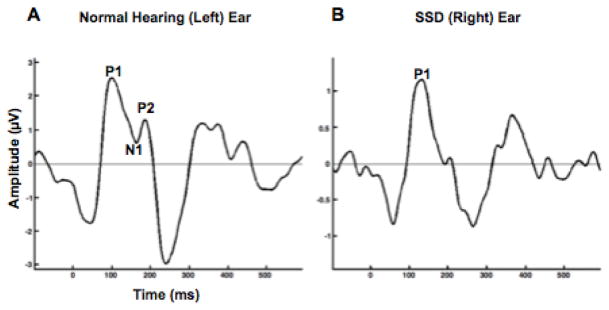
Figure 1 shows CAEP responses at age 9.86 years, shortly before the child received a CI in her right ear. The patient’s normal hearing (left) ear shows clear P1, N1, and P2 responses with normal morphology and peak latencies falling within normal limits for the child’s chronological age (left panel), suggestive of age-appropriate development of the central auditory pathways in response to stimulation of the normal hearing ear. However, the child’s SSD (right) ear shows a delayed CAEP response with abnormal morphology dominated by a single (P1) component (right panel); N1 and P2 components are absent from the response, suggestive of immature development of the pathways in response to stimulation of the SSD ear.
Figure 1.
(Left panel): CAEP response recorded from the midline Cz electrode when the speech stimulus was presented to the patient’s left (normal hearing) ear. (Right panel): CAEP response recorded from the midline Cz electrode when the speech stimulus was presented to the patient’s right (SSD) ear.
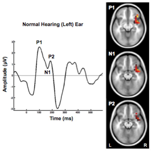
Figure 2 depicts current density reconstructions (CDR) for the P1, N1, and P2 components, pre-CI, when stimulating the patient’s normal hearing ear. As seen in Figure 2, the P1, N1, and P2 components activated auditory areas in the contralateral (right) superior temporal gyrus, medial temporal gyrus, inferior frontal gyrus, frontal gyrus, and insula. It is noteworthy that we saw primarily contralateral temporal cortex activation, suggesting that the activation of the cortex ipsilateral to the normal hearing (left) ear was significantly weaker and therefore not apparent in the CDR, consistent with reports in animal studies (33–35). This finding is also consistent with Gordon et al (2013) showing similar results in children with long periods of unilateral CI use prior to bilateral implantation, where there was a reduction in normal dominance of contralateral input in the auditory cortex (40). In addition to auditory temporal cortical areas of activation, inferior frontal gyrus was also activated when sound was presented to the subject’s normal hearing ear. Activation of frontal areas is consistent with evidence of increased cognitive load and listening effort during auditory processing in hearing impaired listeners (36, 41).
Figure 2.
Current density reconstructions (CDR) for the P1 (top panel), N1 (middle panel), and P2 (bottom panel) components of the CAEP elicited when stimulating the patient’s normal hearing (left) ear.
Figure 3 depicts the developmental trajectory of the subject’s SSD (right) ear, pre-CI and at 3, 8, and 14 months post-CI. As seen in Figure 3, P1 latency in the right ear decreases within a 3-month span post-CI. By 8 months post-CI, there is a clear morphological change in the CAEP response, marked by the emergence of the N1 and P2 response, reflecting an age-appropriate waveform morphology 14 months after implantation.
Figure 3.
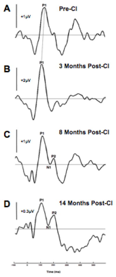
Developmental trajectory of patient’s CAEP response in her right ear (SSD-CI) ear pre-implantation (3A) and at 3, 8, and 14 months post-cochlear implantation (3B-3D).
Figure 4 shows the changes in current density reconstructions for the P1 CAEP pre-CI (left panel) and 14 months post-CI (right panel) when the auditory stimulus was presented to the patient’s SSD (right) ear. Although pre-CI we saw temporal and frontal activation of primarily the right hemisphere, i.e., ipsilateral to SSD ear, at 14 months post-CI we see robust contralateral activation of only auditory temporal areas in superior and medial temporal gyrus. The decrease in frontal activation post-CI is suggestive of a decrease in listening effort and the robust contralateral activation suggests more typical development of auditory pathways post implantation.
Figure 4.
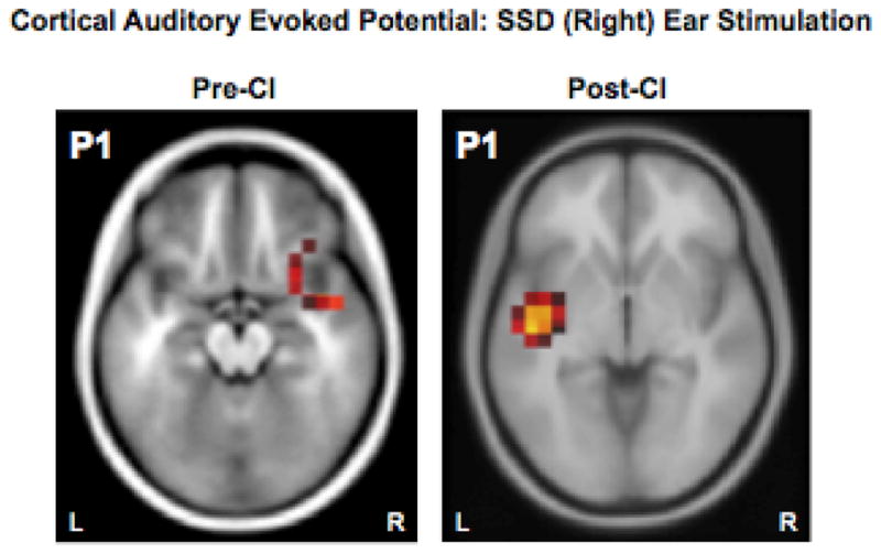
Current density reconstructions (CDR) of the P1 CAEP for the patient’s right (deaf) ear pre-cochlear implantation (left panel) and at 27 months post-cochlear implantation (Right panel).
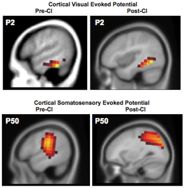
Figure 5 shows cortical activation (CDR) for visual stimuli pre-CI (upper left panel) and 27 months post-CI (upper right panel). Pre-CI, the subject showed activation of left frontal gyrus, and auditory areas in inferior temporal gyrus, and medial temporal gyrus consistent with cross-modal recruitment of temporal areas for visual processing. However, post-CI, the subject showed activation of activation of higher order visual areas including fusiform gyrus, consistent with more normal processing of these stimuli (50–51) as well as some residual activation in auditory areas of left superior and middle temporal gyrus. This response pattern suggests that while some auditory processing regions are still being recruited for visual processing in this patient post-CI, the dominant activation occurs in visual processing regions, suggesting a significant (but not complete) reversal of the visual cross-modal plasticity relative to the pre-CI condition. This result is consistent with recent findings from our laboratory showing that compensatory cross-modal recruitment of auditory processing areas by vision persists in children after cochlear implantation (50–51).
Figure 5.
Current density reconstructions (CDR) for the P2 visual evoked potential (CVEP) response to an apparent visual motion stimulus pre-implantation (upper left panel) at 27 months post-implantation (upper right panel). Current density reconstructions (CDR) for the P50 cortical somatosensory evoked potentials (CSSEP) in response to right finger stimulation recorded pre-implantation (lower left panel) and 27 months post-implantation (lower right panel).
Figure 5 also shows cortical activation (CDR) for somatosensory stimuli pre-CI (lower left panel) and elicited 27 months post-CI (lower right panel). Pre-CI, the subject shows activation of somatosensory regions (pre- and post-central gyrus, inferior parietal lobule, superior parietal lobule) in addition to activation of temporal regions (middle temporal gyrus) and the insula. This suggests recruitment of auditory regions for somatosensory processing and is evidence of cross-modal plasticity. Post-CI, the subject shows left somatosensory activation of pre- and post-central gyrus, inferior and superior parietal lobule, which is consistent with normal processing of these stimuli (51–54). No auditory areas are being recruited for somatosensory processing post CI, suggesting a complete reversal of cross-modal recruitment of auditory areas for somatosensory processing after cochlear implantation (51).
Behavioral Perception Results
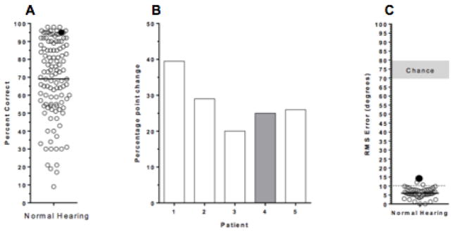
As seen in Figure 6 (left panel), speech perception testing at 33 months post-CI revealed a sentence score of 95% correct in the CI-only condition. This score is at the 90th percentile of scores on this test for adults fit with a cochlear implant (42). (Middle Panel): Testing in the surround sound environment revealed a 25-percentage point improvement in performance when the CI was used in addition to the normal hearing ear This improvement score is within the range of scores achieved by adult SSD subjects who have received a CI tested in the same environment. (Right Panel): Sound localization testing showed a root mean score error of 14 degrees. This score is just outside the range of normal-hearing adults [95th percentile for young normal hearing adults is approximately 10 degrees of error (39) but the patient’s score as good, or better than, for example, subjects fit with bilateral cochlear implants (43)]. Overall, the subject’s excellent behavioral perception scores are consistent with the child’s subjective perception of improved benefit with the CI.
Figure 6.
(Left panel): Performance of patient (solid black circle) on task of speech understanding in quiet (AzBio sentences) compared to 131 adult patients (open circles). Solid bar = mean score. (Middle panel): Percentage point improvement in speech understanding in a complex listening environment with noise masking when listening with CI and normal hearing ear compared to the only the normal hearing ear. Patient’s performance is indicated by grey bar. Open bars indicate performance of four adult SSD CI patients. (Right panel): Performance of patient (solid black circle) and normal hearing adults (open circles) in terms of RMS error on task of sound source localization to a wide-band noise stimulus. Dotted line = 95th percentile of normal performance.
DISCUSSION
In this study we provide a first description of developmental neuroplasticity following CI in a pediatric case of SSD. A clear developmental difference in the CAEP waveforms between the two ears was evidenced pre-CI. While the patient’s normal hearing ear showed an age-appropriate CAEP response with normal P1 morphology and developmentally appropriate emergence of higher-order (N1 and P2) components, stimulation of the SSD ear showed an immature response marked by a single P1 component occurring at a delayed latency (Figure 1). Such a present, but delayed, CAEP response in the SSD (right) ear is conceivably due to the child’s early experience with sound in the deaf ear before the hearing loss progressed and/or potential preservation of plasticity due to stimulation of crossed pathways arising from the normal hearing ear (33–35, 44–45).
Pre-CI, stimulation in each ear activated the right hemisphere, i.e., contralateral to the normal ear and ipsilateral to the SSD ear (Figure 2; Figure 4, left panel). In normal hearing subjects, monaural auditory stimulation results in bilateral activation with greater activation in the contralateral cortex to the ear stimulated, displaying asymmetrical activation of the central auditory system and dominance of contralateral pathways at the level of the cortex (44–45). Studies in animal models of congenital and early-onset SSD show a massive re-organization in aural preference for the normal hearing ear (46). That is, though bilateral activation is still present, the cortex contralateral to the normal hearing ear shows a strengthened response when either the normal hearing (left) ear or SSD (right) ear is stimulated (46). In our study, the fact that we did not see bilateral activation can be explained by the fact that in the context of a dominant underlying neural source, simultaneously active, less robust sources may not be visible using the sLORETA algorithm (47). Pre-CI, we saw representational dominance of contralateral cortex to the normal hearing ear, in that, stimulation of either the normal hearing ear or SSD ear resulted in activation of right auditory cortex. These results converge with the animal and human studies, showing clear changes in cortical auditory activation in preference of the normal hearing ear (40, 46, 57). Importantly, we saw a reversal of this dominance for the normal hearing ear once auditory stimulation was restored with the CI, with the patient demonstrating primarily contralateral activation when stimulating the CI ear (Figure 4, right panel). This demonstrates a restoration of cortical auditory activation patterns more typical of what would be expected in normal hearing.
A similar change was seen at 8 months post implantation in the morphology of the CAEP response, where previously absent N1 and P2 components became identifiable and age-appropriate (Figure 3). The dramatic changes in morphology and cortical activation indicate a significant potential benefit of CI in SSD. This result is in line with our previous studies that have shown that it takes approximately 6–8 months of implant use for important developmental landmarks to become apparent (46).
Overall the results of this study show that a very high degree of neuroplasticity was apparent in the auditory cortex even after implantation at age 9.86 years, well after the brief sensitive period previously described for deaf children who receive cochlear implants (20–22). It is possible that the progressive nature of the hearing loss may have restricted deterioration of the central auditory pathways, preserving plasticity well into the school-age years. Future studies are needed to determine the time limits for a sensitive period for CI in congenitally deaf children with SSD.
The CDR results showing activation of cortical sources further sheds light on how unilateral auditory deprivation affects cortical resource allocation. Pre-CI, auditory stimulation of the patient’s normal hearing ear resulted in temporal and frontal activation only in the contralateral cortex to this ear. As discussed above, this is consistent with patterns observed in normal hearing individuals, in which stimulation of one ear results in greater activation of the contralateral hemisphere (44–45). However, the presence of frontal cortical activation pre-CI in response to auditory stimulation appears to reflect compensatory plasticity indicative of increased cognitive load. The presence of frontal activation is consistent with previous fMRI and EEG studies in adults with hearing loss (39, 48–49) in which pre-frontal and frontal activation was correlated with increased listening effort measured through tasks of executive function and working memory. Our results suggest that untreated SSD results in increased listening effort, which may not be easily captured using traditional audiological testing. CDR results post-CI do not show frontal cortex activation by sound (Figure 4, right panel) suggesting a decrease in listening effort with the CI.
Our results suggest that CI in SSD may also reverse cross-modal re-organization from other modalities. In this case study, we saw a reversal of cross-modal recruitment of temporal areas for somatosensory processing by 27 months post-CI (Figure 5, lower left and right panels). However, visual stimulation at 27 months post-CI continued to show some residual recruitment of auditory temporal areas for visual processing (FIGURE 5, upper left and right panels) at 27 months post-CI. That is, by 27 months post-CI, visual stimulation resulted in activation of typical cortical visual processing regions, with only a small amount of residual cross-modal activation of temporal cortical areas traditionally associated with auditory processing including left superior and middle temporal gyrus. This is in line with recent results from our laboratory which show that, as a group, CI children who have worn their implant for several years continue to show evidence of cross-modal recruitment of auditory temporal areas by visual stimulation (50–51). Overall, this result is consistent with previous findings that cochlear implanted children as a whole are more attentive to visual stimulation compared with normal hearing peers (56). Our results also suggest that while compensatory plasticity from both visual and somatosensory modalities is present in auditory deprivation, reversal of visual cross-modal re-organization may take longer (compared to somatosensory re-organization) due to the functional role of visual processing in every day human communication and may even be considered beneficial as the auditory cortex adapts to a cochlear implant (55).
Finally, as predicted by the high degree of experience-driven neuroplasticity (described above) at 33 months post-CI, rigorous behavioral testing revealed that our patient was an excellent user of her cochlear implant. As can be seen from the results in Figure 6, she showed clear improvement from the addition of the cochlear implant and depicted adult levels of performance in speech perception and sound localization tasks.
In summary, in this case study of SSD, we describe evidence of deficits in cortical auditory development apparent in the delayed latency of the P1, absence of the N1/P2 components of the CAEP and cortical auditory activation showing a clear aural preference for pathways contralateral to the normal hearing ear. After cochlear implantation at a relatively late age (9.86 years) in childhood, we observed significant benefit as indicated by development of typical CAEP morphology, cortical activation contralateral to the implanted ear, decreased listening effort, reversal of somatosensory cross-modal plasticity and only residual visual cross-modal plasticity. Results from a single case should be interpreted with caution, and there is a clear need for larger studies using more subjects with rigorous statistical analyses to describe changes in cortical re-organization after cochlear implantation in SSD. Nevertheless, in our case report, results suggest that cochlear implantation in pediatric SSD cases can provide significant benefit by taking advantage of the high degree of neuroplasticity in childhood.
Acknowledgments
This research was supported by the National Institutes of Health [R01DC006257].
References
- 1.Sladen DP, Rothpletz A, Bess FH. Paediatric Audiological Medicine. 3. West Sussex: John Wiley & Sons; 2009. Children with unilateral sensorineural hearing loss; pp. 288–308. [Google Scholar]
- 2.Tharpe AM, Sladen DP. Causation of permanent unilateral and mild bilateral hearing loss in children. Trends Amplif. 2008;12:17–25. doi: 10.1177/1084713807313085. [DOI] [PMC free article] [PubMed] [Google Scholar]
- 3.Ching TY, van Wanrooy E, Hill M, et al. Performance in children with hearing aids or cochlear implants: Bilateral stimulation and binaural hearing. Int J Audiol. 2006;45:S108–12. doi: 10.1080/14992020600783087. [DOI] [PubMed] [Google Scholar]
- 4.Eapen RJ, Buss E, Adunka MC, et al. Hearing-in-noise benefits after simultaneous cochlear implantation continue to improve 4 years after implantation. Otol Neurotol. 2009;30:153–9. doi: 10.1097/mao.0b013e3181925025. [DOI] [PMC free article] [PubMed] [Google Scholar]
- 5.Bess FH, Tharpe AM. Unilateral hearing impairment in children. Pediatrics. 1984;74:206–16. [PubMed] [Google Scholar]
- 6.Ruscetta MN, Arjmand EM, Pratt SR. Speech recognition abilities in noise for children with severe-to-profound unilateral hearing impairment. Int J Pediat Otorhinolaryngol. 2005;69:771–9. doi: 10.1016/j.ijporl.2005.01.010. [DOI] [PubMed] [Google Scholar]
- 7.Dancer J, Burl NN, Waters S. Am Ann Deaf. Vol. 140. John Wiley & Sons; 1995. Effects of unilateral hearing loss on teacher responses to the SIFTER: Screening instrument for targeting educational risk; pp. 291–4. [DOI] [PubMed] [Google Scholar]
- 8.Firszt JB, Holden LK, Reeder RM, et al. Auditory abilities after cochlear implantation in adults with unilateral deafness: A pilot study. Otol Neurotol. 2012;33:1339–46. doi: 10.1097/MAO.0b013e318268d52d. [DOI] [PMC free article] [PubMed] [Google Scholar]
- 9.Arndt S, Aschendorff A, Laszig R, et al. Comparison of pseudobinaural hearing ot real binaural rehabilitation after cochlear implantation in patients with unilateral deafness and tinnitus. Otol Neurotol. 2011;32:39–47. doi: 10.1097/MAO.0b013e3181fcf271. [DOI] [PubMed] [Google Scholar]
- 10.Arndt S, Laszig R, Aschendorff A, et al. The University of Freiburg asymmetric hearing loss study. Audio Neurotol. 2011;16:S4–6. [Google Scholar]
- 11.Vermeire K, Van de Heyning P. Binaural hearing after cochlear implantation in subjects with unilateral sensorineural deafness and tinnitus. Audio Neurotol. 2009;14:163–71. doi: 10.1159/000171478. [DOI] [PubMed] [Google Scholar]
- 12.Hassepass F, Aschendorff A, Wesarg T, et al. Unilateral deafness in children: Audiologic and subjective assessment of hearing ability after cochlear implantation. Otol Neurotol. 2013;34:53–60. doi: 10.1097/MAO.0b013e31827850f0. [DOI] [PubMed] [Google Scholar]
- 13.Cadieux J, Firszt JB, Reeder RM. Cochlear implantation in non-traditional candidates: Preliminary results in adolescents with asymmetric hearing loss. Otol Neurotol. 2013;34:408–15. doi: 10.1097/MAO.0b013e31827850b8. [DOI] [PMC free article] [PubMed] [Google Scholar]
- 14.Firszt JB, Holden LK, Reeder RM, et al. Cochlear implantation in adults with asymmetric hearing loss. Ear Hear. 2012;33:521–33. doi: 10.1097/AUD.0b013e31824b9dfc. [DOI] [PMC free article] [PubMed] [Google Scholar]
- 15.Kral A, Hartmann R, Tillein J, et al. Congenital auditory deprivation reduces synaptic activity within the auditory cortex in a layer-specific manner. Cereb Cortex. 10:714–26. doi: 10.1093/cercor/10.7.714. 200. [DOI] [PubMed] [Google Scholar]
- 16.Kral A, Sharma A. Developmental neuroplasticity after cochlear implantation. Trends Neurosci. 2012;35:111–22. doi: 10.1016/j.tins.2011.09.004. [DOI] [PMC free article] [PubMed] [Google Scholar]
- 17.Ponton CW, Eggermont JJ, Kwong B, et al. Maturation of human central auditory activity: Evidence from multi-channel evoked potentials. Clin Neurophysiol. 2000;11:220–36. doi: 10.1016/s1388-2457(99)00236-9. [DOI] [PubMed] [Google Scholar]
- 18.Eggermont JJ, Ponton CW. They neurophysiology of auditory perception: From single units to evoked potentials. Audio Neurotol. 2002;7:71–99. doi: 10.1159/000057656. [DOI] [PubMed] [Google Scholar]
- 19.Eggermont JJ, Ponton CW. Auditory-evoked potential studies of cortical maturation in normal hearing and hearing impaired children: Correlations with changes in structure and speech perception. Acta Otolaryngol. 2003;123:249–52. doi: 10.1080/0036554021000028098. [DOI] [PubMed] [Google Scholar]
- 20.Sharma A, Dorman MF, Spahr AJ. A sensitive period for the development of the central auditory system in children with cochlear implants: Implications for age of implantation. Ear Hear. 2002;23:532–9. doi: 10.1097/00003446-200212000-00004. [DOI] [PubMed] [Google Scholar]
- 21.Sharma A, Dorman MF, Kral A. The influence of a sensitive period on central auditory development in children with unilateral and bilateral cochlear implants. Hear Res. 2005;203:134–43. doi: 10.1016/j.heares.2004.12.010. [DOI] [PubMed] [Google Scholar]
- 22.Sharma A, Dorman MF. Central auditory development in children with cochlear implants: Clinical implications. Adv Otorhinolaryngol. 2006;64:66–8. doi: 10.1159/000094646. [DOI] [PubMed] [Google Scholar]
- 23.Doucet ME, Bergeron F, Lassonde M, et al. Cross-modal reorganization and speech perception cochlear implant users. Brain. 2006;129:3376–83. doi: 10.1093/brain/awl264. [DOI] [PubMed] [Google Scholar]
- 24.Geers AE. Factors influencing spoken language outcomes in children following cochlear implantation. Adv Otorhinolaryngol. 2006;64:50–65. doi: 10.1159/000094644. [DOI] [PubMed] [Google Scholar]
- 25.Kral A, Schroder JH, Klinke R, et al. Absence of cross-modal reorganization in the primary auditory cortex of congenitally deaf cats. Exp Brain Res. 2003;153:805–13. doi: 10.1007/s00221-003-1609-z. [DOI] [PubMed] [Google Scholar]
- 26.Giraud AL, Price CJ, Graham JM, et al. Functional plasticity of language-related brain areas after cochlear implantation. Brain. 2001;124:1307–16. doi: 10.1093/brain/124.7.1307. [DOI] [PubMed] [Google Scholar]
- 27.Giraud AL, Price CJ, Graham JM, et al. Cross-modal plasticity underpins language recover after cochlear implantation. Neuron. 2001;30:657–63. doi: 10.1016/s0896-6273(01)00318-x. [DOI] [PubMed] [Google Scholar]
- 28.Kral AJ, Eggermont JJ. What’s to lose and what’s to learn: Development under auditory deprivation, cochlear implants and limits of cortical plasticity. Brain Res Rev. 2007;56:259–69. doi: 10.1016/j.brainresrev.2007.07.021. [DOI] [PubMed] [Google Scholar]
- 29.Nishimura H, Hashikawa K, Doi K, et al. Sign language ‘heard’ in the auditory cortex. Nature. 1999;397:116. doi: 10.1038/16376. [DOI] [PubMed] [Google Scholar]
- 30.Neville H, Bavelier D. Human brain plasticity: Evidence from sensory deprivation and altered language experience. Prog Brain Res. 2002;138:177–88. doi: 10.1016/S0079-6123(02)38078-6. [DOI] [PubMed] [Google Scholar]
- 31.Lee DS, Lee JS, Oh SH, et al. Cross-modal plasticity and cochlear implants. Nature. 2001;409:149–150. doi: 10.1038/35051653. [DOI] [PubMed] [Google Scholar]
- 32.Gilley PM, Sharma A, Dorman MF. Cortical re-organization in children with cochlear implants. Brain Res. 2008;1239:56–65. doi: 10.1016/j.brainres.2008.08.026. [DOI] [PMC free article] [PubMed] [Google Scholar]
- 33.Kral A, Hubka P, Heid S, Tillein J. Single-sided deafness leads to unilateral aural preference within an early sensitive period. Brain. 2013;136:180–93. doi: 10.1093/brain/aws305. [DOI] [PubMed] [Google Scholar]
- 34.Kral A, Heid S, Hubka P, Tillein J. Unilateral hearing during development: Hemispheric specificity in plastic reorganizations. Front Syst Neurosci. 2013;7:93. doi: 10.3389/fnsys.2013.00093. [DOI] [PMC free article] [PubMed] [Google Scholar]
- 35.Kral A, Hubka P, Tillein J. Strengthening of hearing ear representation reduces binaural sensitivity in early single-sided deafness. Otol Neurotol. 2015;20(S1):7–12. doi: 10.1159/000380742. [DOI] [PubMed] [Google Scholar]
- 36.Campbell J, Sharma A. Cross-modal re-organization in adults with early stage hearing loss. PLoS One. 2014;9 doi: 10.1371/journal.pone.0090594. [DOI] [PMC free article] [PubMed] [Google Scholar]
- 37.Gifford R, Dorman M, Skarzynski H, Lorens A, et al. Cochlear implantation with hearing preservation yields significant benefit for speech recognition in complex listening environments. Ear Hear. 2013;34(4):413–425. doi: 10.1097/AUD.0b013e31827e8163. [DOI] [PMC free article] [PubMed] [Google Scholar]
- 38.Dorman M, Loiselle L, Yost W, Stohl J, Spahr A, Brown C, Cook S. Interaural level differences and sound source localization for bilateral cochlear implant patients. Ear Hear. 2014;35(6):633–640. doi: 10.1097/AUD.0000000000000057. [DOI] [PMC free article] [PubMed] [Google Scholar]
- 39.Campbell J, Sharma A. Compensatory changes in cortical resource allocation in adults with hearing loss. Front Syst Neurosci. 2013;7:71. doi: 10.3389/fnsys.2013.00071. [DOI] [PMC free article] [PubMed] [Google Scholar]
- 40.Gordon K, Wong DD, Papsin BC. Bilateral input protects the cortex from unilaterally-driven reorganization in children who are deaf. Brain. 2013;136:1609–25. doi: 10.1093/brain/awt052. [DOI] [PubMed] [Google Scholar]
- 41.Dupont P, Sary G, Peuskens H, et al. Cerebral regions processing first- and higher-order motion in an opposed-direction discrimination task. Eur J Neurosci. 2003;17:1509–17. doi: 10.1046/j.1460-9568.2003.02571.x. [DOI] [PubMed] [Google Scholar]
- 42.Wilson R, Dorman M, Gifford R, McAlpine D. Cochlear implant design considerations. In: Young N, Kirk KI, editors. Cochlear Implants in Children: Learning and the Brain. Springer; In press. [Google Scholar]
- 43.Yost W, Loiselle L, Dorman M, Brown C, Burns J. Sound Source Localization of Filtered Noises by Listeners with Normal Hearing: A Statistical Analysis. J Acoust Soc Am. 2013;133(5):2876–82. doi: 10.1121/1.4799803. [DOI] [PMC free article] [PubMed] [Google Scholar]
- 44.Ponton CW, Vasama JP, Tremblay K, et al. Plasticity in the adult human central auditory system: Evidence from late-onset profound unilateral deafness. Hear Res. 2001;154:32–44. doi: 10.1016/s0378-5955(01)00214-3. [DOI] [PubMed] [Google Scholar]
- 45.Khosla D, Ponton CW, Eggermont JJ, et al. Differential ear effects of profound unilateral deafness on the adult human central auditory system. J Assoc Res Otolaryngol. 2003;4:235–49. doi: 10.1007/s10162-002-3014-x. [DOI] [PMC free article] [PubMed] [Google Scholar]
- 46.Sharma A, Dorman M, Spahr A. Rapid development of cortical auditory evoked potentials after early cochlear implantation. Neuroreport. 2002;13:1–4. doi: 10.1097/00001756-200207190-00030. [DOI] [PubMed] [Google Scholar]
- 47.Wagner M, Fuchs M, Kastner J. Evaluation of sLORETA in the presence of noise and multiple sources. Brain Topogr. 2004;16:277–80. doi: 10.1023/b:brat.0000032865.58382.62. [DOI] [PubMed] [Google Scholar]
- 48.Peelle JE, Jonstrude IS, Davis MH. Hierarchical processing for speech in human auditory cortex and beyond. Front Hum Neurosci. 2010;4:1–3. doi: 10.3389/fnhum.2010.00051. [DOI] [PMC free article] [PubMed] [Google Scholar]
- 49.Peelle JE, Troianai V, Grossman M, et al. Hearing loss in older adults affects neural systems supporting speech comprehension. J Neurosci. 2011;32:12638–42. doi: 10.1523/JNEUROSCI.2559-11.2011. [DOI] [PMC free article] [PubMed] [Google Scholar]
- 50.Campbell J. Unpublished doctoral dissertation. University of Colorado; Boulder: 2011. Visual cross-modal re-organization in children with cochlear implants. [DOI] [PMC free article] [PubMed] [Google Scholar]
- 51.Sharma A, Campbell J, Cardon G. Developmental and cross-modal plasticity in deafness: Evidence from the P1 and N1 event related potentials in cochlear implanted children. Int J Psychophysiol. 2015;95:135–44. doi: 10.1016/j.ijpsycho.2014.04.007. [DOI] [PMC free article] [PubMed] [Google Scholar]
- 52.Cardon G. Unpublished doctoral dissertation. University of Colorado; Boulder: 2015. Somatosensory cross-modal re-organization in children with cochlear implants. [Google Scholar]
- 53.Cardon G, Sharma A. Multimodal cortical mechanisms in sound processing. Poster: Annual meeting of the Cognitive Neuroscience Society; Boston, MA. 2014. [Google Scholar]
- 54.Cardon G, Sharma A. Somatosensory-to-auditory cross-modal plasticity in deafness. Poster: Annual meeting of the Society for Neuroscience; San Diego, CA. 2013. [Google Scholar]
- 55.Strelnikov K, Rouger J, Demonet JF, et al. Visual activity predicts auditory recovery from deafness after adult cochlear implantation. Brain. 2013;136:3682–3695. doi: 10.1093/brain/awt274. [DOI] [PubMed] [Google Scholar]
- 56.Schorr EA, Fox NA, Wassenhove V, et al. Auditory-visual fusion in speech perception in children with cochlear implants. Proc Natl Acad Sci USA. 2005;102:18748–50. doi: 10.1073/pnas.0508862102. [DOI] [PMC free article] [PubMed] [Google Scholar]
- 57.Gordon KA, Jiwani S, Papsin BC. What is the optimal timing for bilateral cochlear implantation in children? Cochlear Implants Int. 2011;12:S8–14. doi: 10.1179/146701011X13074645127199. [DOI] [PubMed] [Google Scholar]


