

Abstract
Introduction:
Intravascular migration of a double J stent into the inferior vena cava is an uncommon complication. The management of such complication is less reported in the literature. This study aimed to reveal the diagnosis and treatment process of migration of a double J stent into the inferior vena cava.
Patient concerns:
A 53-year-old male patients was transferred to our hospital because of migration of a double J stent into the inferior vena cava after left-side pyelolithotomy.
Diagnosis:
In accordance with manifestations on computed tomography urography, the patient was diagnosed with migration of a double J stent into the inferior vena cava.
Interventions:
Percutaneous nephroscope under C-arm guidance was performed to remove the migrated stent. After the operation, the patient was treated with continued anticoagulants and antibiotics.
Outcomes:
The migrated stent was removed successfully without any complications, and a new double J stent was placed and its location was confirmed under C-arm. The patient was discharged in good condition and the follow-up was uneventful.
Conclusion:
Intravascular migration of a double J stent into the inferior vena cava is an uncommon complication. Radiologic imaging after placement of ureteral stent is critical for prevention of this complication. Percutaneous nephroscope under C-arm guidance is a safe and effective approach to remove the migrated DJS in the IVC.
Keywords: complication, intravascular migration, ureteral stent, urolithiasis
1. Introduction
Double J stent (DJS), first reported by Zimskind et al in 1967,[1] that is used for maintaining urine flow from the kidney to the bladder and restore the continuity of the urinary tract in various urological conditions.[2] The common complications of DJS placement are dysuria, frequency, hematuria, bacteriuria, fragmentation, and stone formation.[3] Most of these complications are self-limited and easy to be managed. However, intravascular migration of DJS is an uncommon and severe complication. The management of this complication is less reported in the literature. In this case report, we present the successful endoscopic treatment of migration of a DJS into the inferior vena cava (IVC).
2. Case presentation
A 53-year-old male patient with a history of long-standing left renal calculi underwent left-side pyelolithotomy with postoperative placement of a DJS at a county hospital on April 2016. However, the stent placement was not performed under fluoroscopy guidance and postoperative imaging examinations were not performed to confirm the DJS position. The patient was advised for stent removal after 5 months. Surprisingly, kidney, ureters, and bladder (KUB) x-ray film showed that the DJS was not in the right location and appeared to enter into the IVC (Fig. 1).
Figure 1.
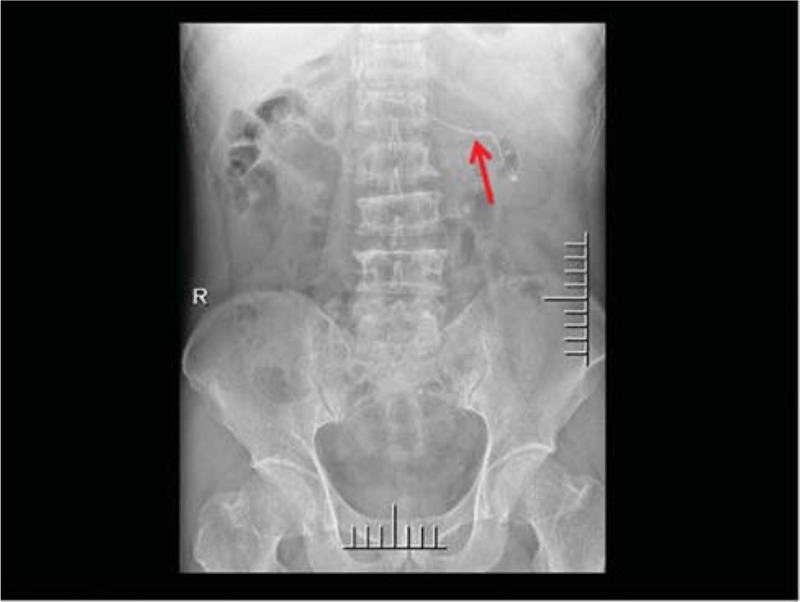
KUB x-ray film revealed an abnormal pathway of the double J stent that probably entered into the inferior vena cava. The red arrow shows the position of the double J stent. KUB = kidney ureters and bladder.
For further managements, the patient was transferred to our hospital. During hospitalization, moderate flank pain persisted. Blood analysis showed normal leukocytes counts and normal creatinine levels. Urine analysis showed increased leukocytes counts and increased erythrocyte counts. Further urine culture was positive with a growth of Enterococcus faecalis. Color Doppler flow imaging (CDFI) showed the formation of small mural thrombus in the IVC. Therefore, the patient was assigned to treatments with anticoagulants and antibiotics. The next-day computed tomography urography (CTU) was performed to assure the position of the stent. In combination of left-side hydronephrosis, migration of the DJS into the IVC was seen at the level of the left renal vein. The proximal coil was in the IVC and the distal coil was in the left renal pelvis (Fig. 2). Then, we performed percutaneous nephroscope under C-arm guidance by using 18G puncture needles and 20 Fr working sheaths. The distal coil of the DJS was visualized and removed successfully under nephroscope without any complications. A new DJS was placed and its location was confirmed by radiologic imaging (Fig. 3). After the operation, the patient was transferred to intensive care unit (ICU) and treated with continued anticoagulants and antibiotics. Fifteen days after the operation, CDFI showed there was no thrombus in the IVC. The patient was discharged 16 days after the operation with symptom free and the follow-up was uneventful.
Figure 2.

CTU scan confirmed that the double J stent was in the inferior vena cava (red arrow). CTU = computed tomography urography.
Figure 3.

KUB x-ray film showed a new double J stent was placed into the left ureter (red arrow). KUB = kidney ureters and bladder.
3. Discussion
The placement of a DJS is a common urological procedure. However, this procedure will cause complications that include dysuria, frequency, hematuria, bacteriuria, fragmentation, stone formation, and migration. Intravascular migration of the stent is one of the serious complications. There are few published cases about intravascular migration of DJS. Intravascular migration of DJS was first reported by Michalopoulos et al. in 2002[4]. The reported position of the DJS intravascular migration is the external iliac vein,[5] IVC,[6,7] right ventricle,[8] right atrium,[5] and pulmonary arterial.[9]
In our case, the DJS migrated into the IVC because of the following reasons. First, the ureter is adjacent to the IVC. Second, the placement of DJS has no standard operation procedure. For this reason, the occurrence of stent migration is mostly determined by the experience of the surgeon. Third, uretheral wall is fragile because of long-standing hydronephrosis, chronic inflammation, and urolithiasis, the surgeon had difficulty placing the guidewire and feeling resistance. Besides, the stent placement did not performed under imaging monitoring.
Several strategies have been mentioned to prevent this complication. First, the surgeon should be always aware of severe complications and pay attention to the length of DJS during the procedure of placement. If resistance is felt, it is important not to force the DJS. Second, the coil of DJS in the renal pelvis and bladder should be at least 180° and length of >2 cm. Third, the surgeon should pay attention to warning signs and symptoms of the patient, such as significant hematuria and severe abdominal pain. In addition, perioperative x-ray imaging should be performed to confirm the position of the DJS. It is very important to perform monitoring and follow-up after a DJS placement, in order to early diagnosis of complications. A migrated DJS must be removed early to avoid the development of more tough complications. For example, the migrated DJS may completely enter the IVC or enter the heart through IVC.[5,8] Secondary urosepsis, pulmonary embolism, and valvular disease are most severe outcomes of DJS migration. If the patient had urinary tract infection or thrombosis, anti-infective therapy is needed to prevent urosepsis and anti-coagulant therapy is needed to prevent thrombotic complications.
Four approaches have been reported for removal of a migrated DJS, including endoscopic operation,[6] laparoscopic operation,[10] endovascular operation,[9,11] and open surgery.[8] The treatment options are determined by the position of the stent, the condition of patient, the hemorrhagic condition, the formation of thrombus, and expertise of the surgeon. In this case, we used endoscopic approaches to remove the migrated DJS because of the following reasons. First, the distal coil of the DJS was in the renal pelvis. For this reason, we could visualize and remove the DJS by using nephroscope. Second, the surgeons are experienced in the endoscopic operation and had experienced several similar cases. Third, the vital signs of the patient were stable and the thrombus was small.
In conclusion, intravascular migration of a DJS into the IVC is an uncommon complication. Radiologic imaging after placement of ureteral stent is critical for prevention of this complication. Percutaneous nephroscope under C-arm guidance is a safe and effective approach to remove the migrated DJS in the IVC.
Author contributions
Formal analysis: Prashant Mishra and Deqiang Chen.
Funding acquisition: Yong Chen.
Investigation: Xiaohui Ni.
Writing – original draft: Changyi Jiang.
Writing – review & editing: Shi Fu, Jian Chen and Changxing Ke.
Footnotes
Abbreviations: CDFI = color Doppler flow imaging, CTU = computed tomography urography, DJS = double J stent, ICU = intensive care unit, IVC = inferior vena cava, KUB = kidney, ureters, and bladder.
JC and FS have contributed equally to this study.
This study was supported by the National Natural Science Foundation of China (No. 81660422), the Project of Yunnan Provincial Health Department (No. 2016NS260) and the 2018 Doctor Newcomer Award of Yunnan Province.
Ethical approval was obtained from Second Affiliated Hospital of Kunming Medical University. Informed written consent was obtained from the patient for publication of this case report and accompanying images.
The authors declare no conflict of interest.
References
- [1].Zimskind PD, Fetter TR, Wilkerson JL. Clinical use of long-term indwelling silicone rubber ureteral splints inserted cystoscopically. J Urol 1967;97:840–4. [DOI] [PubMed] [Google Scholar]
- [2].Brotherhood H, Lange D, Chew BH. Advances in ureteral stents. Transl Androl Urol 2014;3:314–9. [DOI] [PMC free article] [PubMed] [Google Scholar]
- [3].Richter S, Ringel A, Shalev M, et al. The indwelling ureteric stent: a ’friendly’ procedure with unfriendly high morbidity. BJU Int 2000;85:408–11. [DOI] [PubMed] [Google Scholar]
- [4].Michalopoulos AS, Tzoufi MJ, Theodorakis G, et al. Acute postoperative pulmonary thromboembolism as a result of intravascular migration of a pigtail ureteral stent. Anesth Analg 2002;95:1185–8. table of contents. [DOI] [PubMed] [Google Scholar]
- [5].Sabnis RB, Ganpule AP, Ganpule SA. Migration of double J stent into the inferior vena cava and the right atrium. Indian J Urol 2013;29:353–4. [DOI] [PMC free article] [PubMed] [Google Scholar]
- [6].Tilborghs S, Vaganee D, De Wachter S, et al. Intravascular double J stent migration: a case report, review, and management algorithm. Urol Ann 2019;11:93–7. [DOI] [PMC free article] [PubMed] [Google Scholar]
- [7].Marques V, Parada B, Rolo F, et al. Intracaval misplacement of a double-J ureteral stent. BMJ Case Rep 2018;2018: pii: bcr-2017-221713. [DOI] [PMC free article] [PubMed] [Google Scholar]
- [8].Hastaoglu IO, Tokoz H, Kavlak E, et al. Double J ureteral stent displaced through the right ventricle. Interact Cardiovasc Thorac Surg 2014;18:853–4. [DOI] [PubMed] [Google Scholar]
- [9].Arab D, Ardestani Zadeh A, Eskandarian R, et al. An extremely rare complication of ureteral pigtail stent placement: a case report. Nephrourol Mon 2016;8:e36527. [DOI] [PMC free article] [PubMed] [Google Scholar]
- [10].Mao XW, Xu G, Xiao JQ, et al. Ureteral double J stent displaced into vena cava and management with laparoscopy: a case report and review of the literature. World J Clin Cases 2018;6:1160–3. [DOI] [PMC free article] [PubMed] [Google Scholar]
- [11].Falahatkar S, Hemmati H, Gholamjani Moghaddam K. Intracaval migration: an uncommon complication of ureteral Double-J stent placement. J Endourol 2012;26:119–21. [DOI] [PubMed] [Google Scholar]