Abstract
Diabetes mellitus (DM) is a public problem closely associated with numerous oral complications, such as coated tongue, xerostomia, salivary dysfunction, etc. Tongue diagnosis plays an important role in clinical prognosis and treatment of diabetes in the traditional Chinese medicine (TCM). This study investigated discriminating tongue features to distinguish between type 2 DM and non-DM individuals through non-invasive TCM tongue diagnosis.
The tongue features for 199 patients with type 2 DM, and 372 non-DM individuals, serving as control, are extracted by the automatic tongue diagnosis system (ATDS). A total of 9 tongue features, namely, tongue shape, tongue color, fur thickness, fur color, saliva, tongue fissure, ecchymosis, teeth mark, and red dot. The demography, laboratory, physical examination, and tongue manifestation data between 2 groups were compared.
Patients with type 2 DM possessed significantly larger covering area of yellow fur (58.5% vs 22.5%, P < .001), thick fur (50.8% vs 29.2%, P < .001), and bluish tongue (P < .001) than those of the control group. Also, a significantly higher portion (72.7% vs 55.2%, P < .05) of patients with long-term diabetics having yellow fur color than the short-term counterparts was observed.
The high prevalence of thick fur, yellow fur color, and bluish tongue in patient with type 2 DM revealed that TCM tongue diagnosis can serve as a preliminary screening procedure in the early detection of type 2 DM in light of its simple and non-invasive nature, followed by other more accurate testing process. To the best of our knowledge, this is the first attempt in applying non-invasive TCM tongue diagnosis to the discrimination of type 2 DM patients and non-DM individuals.
Keywords: automatic tongue diagnosis system, tongue features, traditional Chinese medicine, type 2 diabetes mellitus
1. Introduction
With the rapid increase in population growth, aging, obesity, and inactivity of lifestyle, diabetes mellitus (DM) has become the major burden of adult public health.[1–3] According to an epidemiological study of the global prevalence of diabetes, there were 171 million people with diabetes worldwide in 2000 and was expected to keep increasing to 366 million by 2030.[3] Prolonged duration of diabetes and high blood sugar level would have risk leading to micro-vascular or macro-vascular complications.[4,5] Besides, most diabetic patients were found to have oral manifestations, such as periodontal disease, tooth loss, xerostomia, caries, burning mouth disorder, taste and salivary gland dysfunction, delayed wound healing, lichen planus, geographic tongue, and candidiasis.[6] Buccal alterations can also be easily observed in patients with DM.
Tongue diagnosis, serving as a vital non-invasive tool to provide useful clinical information, plays a pivotal role in TCM.[7–17] By observing tongue features, TCM practitioners can probe qi-blood, yin-yang disorders which are important in treatment selection and prognosis.[18,19] Clinically, practitioners observe tongue characteristics, such as tongue color and shape, fur color and thickness, and the amount of saliva, to help deduce the primary pattern of a patient. However, tongue diagnosis is often biased by subjective judgment, which originates from personal experience, knowledge, diagnostic skills, thinking patterns, and color perception/interpretation. The inconsistency of subjective diagnosis can be improved by using the development of validated instruments. An automatic tongue diagnosis system (ATDS), with a high degree of diagnostic consistency, was developed by our team.[20,21]
Previous studies have been conducted on exploring the association between tongue characteristics and specific diseases, including breast cancer,[8,10] metabolic syndrome,[12] and functional dyspepsia,[22] in the hope of deriving discriminating tongue features to distinguish individuals with/without the specific disease through a non-invasive means in the early stage. However, to the best of our knowledge, no study has yet been performed on the comprehensive scrutiny of tongue features in patients with type 2 DM. This is the first attempt in applying TCM tongue diagnosis to the discrimination of type 2 DM patients and non-DM individuals.
2. Methods
2.1. Ethics approval and consent to participate
An observational cross-sectional and clinical cohort study was conducted. All candidates received a standardized interview process. Participants were introduced to the purpose, procedures, potential risks and benefits of the study first, and signed the informed consent form. This study was approved by the Institutional Review Board of Changhua Christian Hospital, Taiwan (institutional review board reference number: 111,106 and 140,704).
2.2. Participants
The 199 subjects in the type 2 DM group were collected through the outpatient clinics of both Endocrinology and Traditional Chinese Medicine Departments of Changhua Christian Hospital in Taiwan, from January 2012 to December 2013. All individuals recruited were informed of the study purpose, procedures, potential risks and benefits, and then the consent form were signed. Patients with type 1 DM, cancer, end-stage renal disease, pregnancy, infection, and cerebrovascular disease were excluded from type 2 DM group. The participants, with written consent, older than 20 years old, and no specific medical history, namely, acute infection, unstable vital sign, and tongue or oral cancer, were enrolled in the control group from September 2014 to September 2016. A total of 372 participants in the control group, sex- and age-matched with type 2 DM group, were recruited in our study. The tongue images of both groups, that is, experimental and control groups, were collected by the validated Automatic Tongue Diagnosis System (ATDS) with the corresponding tongue features automatically extracted.
2.3. Data collection
Information including demography, body mass index (BMI, kg/m2), and fasting blood-glucose level (AC sugar, mg/dL), cholesterol and triglyceride (TG, mg/dL) were gathered for each subject. The duration of type 2 DM history and hemoglobin A1c (HbA1c, %) were also collected for the patients with type 2 DM. Tongue images were collected for each subject to further derive the relevant tongue features of every participant. All personal details and photographs of subjects recruited were encrypted to ensure confidentiality.
2.4. ATDS and tongue features
Observation diagnosis is often biased by subjective judgment, which originates from personal experience, knowledge, diagnostic skills, thinking patterns, and color perception/interpretation. A practitioner may reach inconsistent diagnoses on the same tongue examined at different time. In addition, different practitioners may pass disparate judgments on an identical tongue. The ATDS was developed by our team to capture tongue images and automatically extract features consistently to assist the diagnosis of TCM practitioners (Fig. 1). The value of ATDS hinges on its ability to calibrate brightness and color to compensate for variations in intensity and color temperatures from light sources and imaging hardware. The tongue region is segmented automatically to derive relevant tongue features. There are 9 primary features for TCM clinical tongue diagnosis, namely, tongue shape, tongue color, fur thickness, fur color, saliva, tongue fissure, ecchymosis, teeth mark, and red dot. Features extracted are further sub-divided according to the areas located, that is, spleen–stomach, liver–gall-left, liver–gall-right, kidney, and heart–lung areas.
Figure 1.
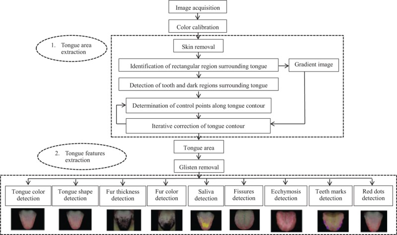
The processing flow of ATDS analysis. ATDS = automatic tongue diagnosis system.
These features are extracted by ATDS to further generate detailed information regarding length, area, moisture, and number of relevant tongue features. The experience of TCM practitioners and multiple professional opinions are integrated to forge the final classification based on the aforementioned characteristics of tongue features derived.
A listing of the tongue features extracted is summarized below:
-
1.
Tongue shape: includes small, median, and enlarged
-
2.
Tongue color: includes pale, pink, red, and bluish
-
3.
Fur thickness: degree of thickness (thin or thick)
-
4.
Fur color: includes fur color (white or yellow)
-
5.
Saliva: the proportion of saliva area to total tongue area (dry or normal)
2.5. Data analysis
The tongue features of subjects participated were extracted by ATDS, and then compared between the type 2 DM and control groups to identify features with significant difference (P < .05). Moreover, the type 2 DM group was further stratified into 2 subgroups of sickness duration, that is, <15 years and ≥15 years, and another 2 subgroups of AC sugar, HbA1C <6.5% and HbA1C ≥6.5%, based on the length of time interval since his/her first diagnosis, and the amount of blood glucose level, respectively. The data were expressed as mean ± standard deviation (SD), percentage, or number where appropriate. The tongue features in different groups were analyzed and compared using independent t test for continuous variables and chi-square test (or Fisher exact) for categorical variables. Furthermore, one-way analysis of variance (ANOVA) was applied to compare difference between group means. Statistical analysis was performed using SPSS statistical software for Windows version 19 (SPSS for Windows, Version 19; SPSS Inc., Chicago, IL). A P-value <.05 was considered statistically significant.
3. Results
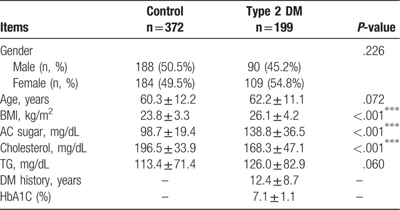
The clinical characteristics of the control group and the patients with type 2 DM are listed in Table 1. A total of 372 participants of the control group and 199 patients with type 2 DM were enrolled in the study. The clinical characteristics of the patients with type 2 DM were as follows: mean age (62.2 ± 11.1 years); mean diabetes duration (12.4 ± 8.7 years), and HbA1c level (7.1 ± 1.1%). The patients with type 2 DM had significantly higher degrees of variations in BMI and AC sugar levels but significantly lower changes in total cholesterol than those in the control group. The mean BMI (26.1 ± 4.2 vs 23.8 ± 3.3, P < .001) and AC sugar (138.8 ± 36.5 vs 98.7 ± 19.4 mg/dL, P < .001) in type 2 DM group were significantly higher than the control counterpart. The cholesterol level in control group was significantly higher than the type 2 DM one (196.5 ± 33.9 vs 168.3 ± 47.1 mg/dL, P < .001).
Table 1.
Demography of study participants.
Differences of tongue features between the type 2 DM and control groups are shown in Table 2. There was a higher proportion of bluish tongue (P < .001) in the type 2 DM group. In terms of fur thickness, the proportion of thick fur in type 2 DM group was significantly higher than the control counterpart (50.8% vs 29.2%, P < .001). In type 2 DM group, 58.5% patients possessed yellow fur, as shown in Fig. 2, a figure significantly higher than that from control group (58.5% vs 22.5%, P < 0.001). The proportion of dry mouth in the type 2 DM group was higher than that in control (8.5% vs 4.5%), yet with no significant difference (P = .079). There were fewer red dots in the type 2 DM group than those of control one (84.8 ± 74.4 vs 24.8 ± 30.2, P < .001). No significant difference in tongue shape between the type 2 DM and the control groups was observed.
Table 2.
Comparison of tongue features between patients with type 2 diabetes mellitus (DM) and control subjects.
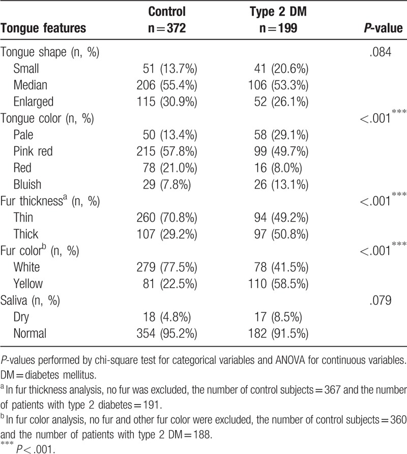
Figure 2.
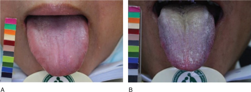
The tongue features of control group and type 2 DM group. Control group: (A) pink tongue, thin, and white fur. Type 2 DM group: (B) bluish tongue, thick, and yellow fur. DM = diabetes mellitus.
The tongue features for the 2 diabetic duration subgroups, that is, <15 years and ≥15 years, in the type 2 DM group were tabulated and compared in Table 3. A significant difference in fur color was identified among these 2 subgroups. The proportions of yellow fur color and dry saliva in the prolonged DM duration ≥15 years subgroup were higher than the other counterpart (72.7% vs 55.2%, P = .046) and (10.4% vs 6.4%, P = .515), respectively, yet with no significant difference. The remaining tongue features did not vary significantly between these 2 subgroups.
Table 3.
Comparison of tongue features between the type 2 diabetes mellitus (DM) with history <15 years and ≥15 years groups in patients with type 2 DM (n = 157).
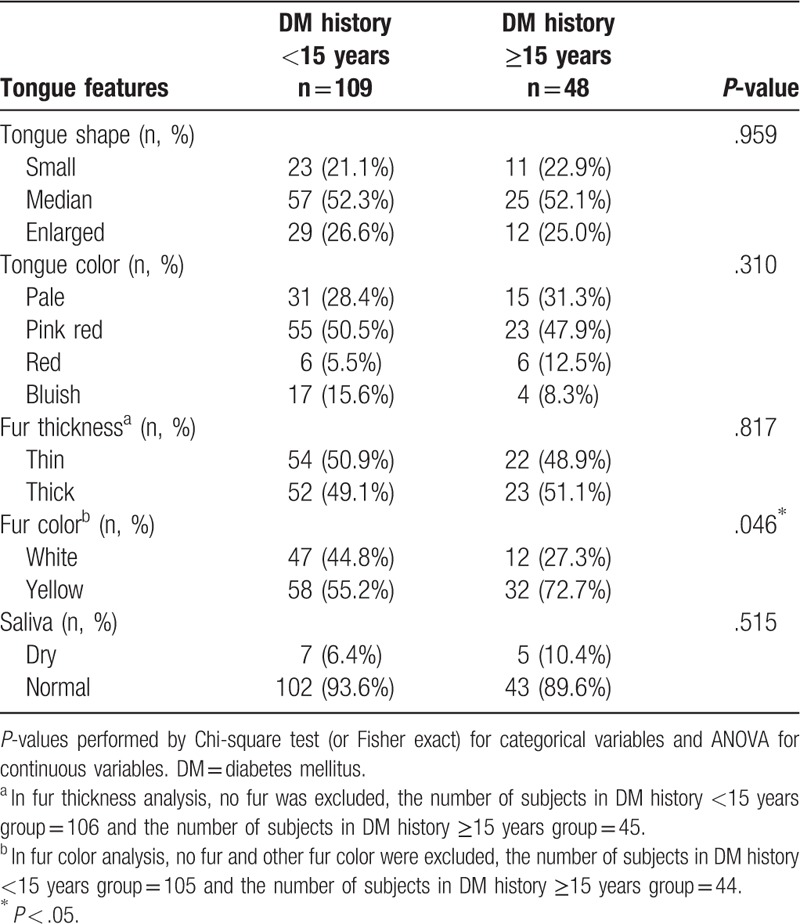
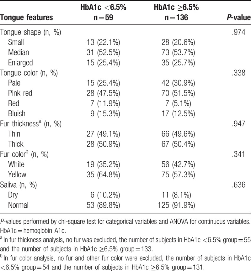
As for the 2 subgroups of AC sugar, HbA1C <6.5% and HbA1C ≥6.5%, in the type 2 DM group, the tongue features were tabulated and compared in Table 4. No tongue feature was identified to significantly link to the amount of blood glucose level between these 2 subgroups.
Table 4.
Comparison of tongue features between the glycated hemoglobin (HbA1c) <6.5% and HbA1c ≥6.5% groups in patients with type 2 diabetes mellitus (DM) (n = 195).
4. Discussion
To the best of our knowledge, this is the first study to apply ATDS to thoroughly investigate the tongue features associated with type 2 DM. The tongue appearance is a crucial indicator in the TCM assessment.[19] Tongue inspection refers to visual evaluation of the shape, color, and fur color, fur thickness, and other tongue characteristics.[23] In the past few years, there were many reports investigating the relationship between tongue features and specific diseases. Studies have shown that tongue diagnosis played an important role in clinical prognosis and treatment of diabetes.[9,24] In this study, ATDS was used to identify the tongue features due to the high degree of diagnostic consistency. In light of the aforementioned observation, the differences in tongue features between type 2 DM and control groups were performed. Moreover, the tongue features in different diabetic duration and blood glucose level were analyzed and compared in this study.
Most diabetic patients are found to have oral manifestations, e.g., periodontal disease, xerostomia, burning mouth, salivary gland dysfunction, geographic tongue, candidiasis, etc.[6] Buccal alterations could also be easily observed in patients with DM, especially coated tongue. Tongue fur represents the retention of exfoliated mucosa cells, debris, and proliferation of microorganisms, especially on the tongue's surface.[25] In terms of fur thickness, the proportion of thick fur in type 2 DM group was significantly higher than control counterpart (50.8% vs 29.2%, P < .001). Our previous study revealed that 47.1% of patients with type 2 DM had thick fur.[11] According to TCM theory, tongue fur is associated with the Yang organs, especially the digestive system. The thick fur is usually accompanied with patterns of phlegm-dampness and blood stasis.[18,19] The proportion of patients with type 2 DM possessing yellow fur was significantly higher than those in control group (58.5% vs 22.5%, P < .001). Another study revealed that yellow tongue coating was associated with higher prevalence of DM and tended to be linked with pre-diabetes.[26] Based on these aforementioned studies, the appearance of coated tongue should be alerted among patients with diabetes.
The presence of xerostomia and hyposalivation is often encountered among DM patients. These studies showed higher prevalence of xerostomia in DM patients in relation to non-DM population, 12.5% to 53.5% versus 0% to 30%. The determination of xerostomia is based solely on the responses to a pre-formatted questionnaire, such as “Does your mouth feel dry frequently?”[27–29] The other study showed that dry mouth is generally associated with decreased saliva production and is present in 10% to 30% patients with DM.[25] In TCM, diabetes-related symptoms are referred to as “Xiaoke,” which corresponds to increased thirst (or polydipsia).[30] However, even though the proportion of dry saliva in type 2 DM group was higher than the control counterpart (8.5% vs 4.5%), yet no significant difference can be reached (P = .079). ATDS classified the amount of saliva into dry, normal, and wet categories based on the proportion of tongue area covered by saliva. Different stratification regarding the amount of saliva might be employed shall the need arise in the future.
We speculate that the tongue features of type 2 DM patients with poor control or prolonged duration would show more significant changes, such as red tongue color, thick fur, yellow fur, and dry saliva. Therefore, the type 2 DM group was further stratified into 2 subgroups of sickness duration, that is, <15 years and ≥15 years, and another 2 subgroups of AC sugar, HbA1C <6.5% and HbA1C ≥6.5%, based on the length of time interval since his/her first diagnosis, and the amount of blood glucose level, respectively. The result demonstrated that significantly higher portion of patients with prolonged diabetic duration possessed yellow fur color than the counterparts with shorter sick period. Yellow fur color usually results from poor oral hygiene as food and bacteria can accumulate on the filiform papillae on the surface. According to TCM theory, yellow fur color mirrors an interior heat pattern and the deeper yellow color indicates an even more severe heat condition.[31,32]
Understanding and interpreting these tongue manifestations of type 2 DM are important for both theoretical and clinical applications. This study applied ATDS, a modern instrument with high degree of consistency, to derive tongue features automatically to reduce human vision bias. However, there were several limitations in our study. First, some participants might have comorbidity, such as hypertension, hyperlipidemia, etc., and the diabetic complications were not mentioned. These comorbidities and vascular complications may affect the tongue appearance and the features extracted, although there is not enough evidence to support the above statement. Secondly, the sublingual collateral vessels have not yet been analyzed. Abnormal collateral vessels may be associated with vascular complications. The association between diabetic vascular complications and tongue features will be explored in our future research.
5. Conclusions
This study showed that significantly high prevalence of thick fur, yellow fur color, and bluish tongue in patients with type 2 DM compared with those in the control group. In the future, observing the tongues of patients with different diseases as comparators to show that the findings we have made are either unique to type 2 DM or statistically more prevalent in type 2 DM.
Acknowledgments
The authors would like to thank the Ministry of Health and Welfare (MOHW103-CMAP-M-114–000416, MOHW104-CMAP-M-114–123405, MOHW105-CMAP-M-114–133402, and MOHW108-TDU-B-212–133004), China Medical University Hospital (CRS-106–002, and DMR-108–005), and Ministry of Science and Technology of Taiwan (MOST 107–2320-B-039–068) for the grant support, and Miss Li-Jyun Deng, and all of the colleagues who helped contribute to this study. The authors would like to thank Miss Li-Jyun Deng, and all of the colleagues who helped contribute to this study. The authors are grateful to the anonymous reviewers for their valuable comments.
Author contributions
Po-Chi Hsu and Lun-Chien Lo conceived of the study. The study was mainly conducted by Po-Chi Hsu and Yi-Ping Chen. Yu-Chuen Huang and Yi-Ping Chen performed the statistical analysis. Po-Chi Hsu, Yi-Ping Chen, John Y. Chiang, and Lun-Chien Lo led the writing of the manuscript. Han-Kuei Wu, Hen-Hong Chang and Tsung-Chieh Lee participated in the design and coordination of the study. All authors commented on the analytic plan and interpretation, and contribution to the editing and final approval of the manuscript.
Conceptualization: Po-Chi Hsu, Lun-Chien Lo.
Formal analysis: Yu-Chuen Huang, Yi-Ping Chen.
Methodology: Po-Chi Hsu, Han-Kuei Wu, Yu-Chuen Huang, Lun-Chien Lo, John Y. Chiang.
Supervision: Han-Kuei Wu, Yu-Chuen Huang, Hen-Hong Chang, Tsung-Chieh Lee, Lun-Chien Lo.
Writing – original draft: Po-Chi Hsu, Yi-Ping Chen.
Writing – review & editing: John Y. Chiang, Lun-Chien Lo.
Footnotes
Abbreviations: AC sugar = fasting blood-glucose level, ATDS = automatic tongue diagnosis system, BMI = body mass index, DM = diabetes mellitus, HbA1c = hemoglobin A1c, SD = standard deviation, TCM = traditional Chinese medicine, TG = triglyceride.
The authors declare that they have no competing interests.
References
- [1].Yang W, Lu J, Weng J, et al. Prevalence of diabetes among men and women in China. N Engl J Med 2010;362:1090–101. [DOI] [PubMed] [Google Scholar]
- [2].Engelgau MM, Geiss LS, Saaddine JB, et al. The evolving diabetes burden in the United States. Ann Inter Med 2004;140:945–50. [DOI] [PubMed] [Google Scholar]
- [3].Wild S, Roglic G, Green A, et al. Global prevalence of diabetes: estimates for the year 2000 and projections for 2030. Diabetes Care 2004;27:1047–53. [DOI] [PubMed] [Google Scholar]
- [4].Cade WT. Diabetes-related microvascular and macrovascular diseases in the physical therapy setting. Phys Ther 2008;88:1322–35. [DOI] [PMC free article] [PubMed] [Google Scholar]
- [5].Kitada M, Zhang Z, Mima A, et al. Molecular mechanisms of diabetic vascular complications. J Diabetes Investig 2010;1:77–89. [DOI] [PMC free article] [PubMed] [Google Scholar]
- [6].Nazir MA, AlGhamdi L, AlKadi M, et al. The burden of diabetes, its oral complications and their prevention and management. Open Access Maced J Med Sci 2018;6:1545–53. [DOI] [PMC free article] [PubMed] [Google Scholar]
- [7].Lo LC, Chen CY, Chiang JY, et al. Tongue diagnosis of traditional Chinese medicine for rheumatoid arthritis. Afr J Tradit Complement Altern Med 2013;10:360–9. [DOI] [PMC free article] [PubMed] [Google Scholar]
- [8].Lo LC, Cheng TL, Chiang JY, et al. Breast cancer index: a perspective on tongue diagnosis in traditional chinese medicine. J Tradit Complement Med 2013;3:194–203. [DOI] [PMC free article] [PubMed] [Google Scholar]
- [9].Liao PY, Hsu PC, Chen JM, et al. Diabetes with pyogenic liver abscess–A perspective on tongue assessment in traditional Chinese medicine. Complement Ther Med 2014;22:341–8. [DOI] [PubMed] [Google Scholar]
- [10].Lo LC, Cheng TL, Chen YJ, et al. TCM tongue diagnosis index of early-stage breast cancer. Complement Ther Med 2015;23:705–13. [DOI] [PubMed] [Google Scholar]
- [11].Hsu PC, Huang YC, Chiang JY, et al. The association between arterial stiffness and tongue manifestations of blood stasis in patients with type 2 diabetes. BMC Complement Altern Med 2016;16:324. [DOI] [PMC free article] [PubMed] [Google Scholar]
- [12].Lee TC, Lo LC, Wu FC. Traditional Chinese Medicine for metabolic syndrome via TCM pattern differentiation: tongue diagnosis for predictor. Evid Based Complement Alternat Med 2016;2016:1971295. [DOI] [PMC free article] [PubMed] [Google Scholar]
- [13].Huang YS, Sun MC, Hsu PC, et al. The Relationship between ischemic stroke patients with and without retroflex tougongue: a retrospective study. Evid Based Complement Alternat Med 2017;2017:3195749. [DOI] [PMC free article] [PubMed] [Google Scholar]
- [14].Gao L, Liu P, Song JX, et al. A pilot study on the relationship between tongue manifestation and the degree of neurological impairment in patients with acute cerebral infarction. Chin J Integr Med 2013;19:149–52. [DOI] [PubMed] [Google Scholar]
- [15].Liu P, Gao L, Song JX, et al. A pilot study on the correlation of tongue manifestation with the site of cerebral infarction in patients with stroke. Chin J Integr Med 2014;20:823–8. [DOI] [PubMed] [Google Scholar]
- [16].Feng Y, Xu H, Qu D, et al. Study on the tongue manifestations for the blood-stasis and toxin syndrome in the stable patients of coronary heart disease. Chin J Integr Med 2011;17:333–8. [DOI] [PubMed] [Google Scholar]
- [17].Yu Z, Zhang H, Fu L, et al. Objective research on tongue manifestation of patients with eczema. Technol Health Care 2017;25:143–9. [DOI] [PubMed] [Google Scholar]
- [18].Kirschbaum B. Atlas of Chinese Tongue diagnosis. Vol 1. Seattle: Eastland Press; 2000. [Google Scholar]
- [19].Anastasi JK, Currie LM, Kim GH. Understanding diagnostic reasoning in TCM practice: tongue diagnosis. Altern Ther Health Med 2009;15:18–28. [PubMed] [Google Scholar]
- [20].Jung CJ, Jeon YJ, Kim JY, et al. Review on the current trends in tongue diagnosis systems. Integr Med Res 2012;1:13–20. [DOI] [PMC free article] [PubMed] [Google Scholar]
- [21].Lo LC, Chen YF, Chen WJ, et al. The Study on the agreement between automatic tongue diagnosis system and traditional Chinese medicine practitioners. Evid Based Complement Alternat Med 2012;2012:505063. [DOI] [PMC free article] [PubMed] [Google Scholar]
- [22].Kim J, Han G, Ko SJ, et al. Tongue diagnosis system for quantitative assessment of tongue coating in patients with functional dyspepsia: a clinical trial. J Ethnopharmacology 2014;155:709–13. [DOI] [PubMed] [Google Scholar]
- [23].Lo LC, Chiang JY, Cheng TL, et al. Visual agreement analyses of traditional chinese medicine: a multiple-dimensional scaling approach. Evid Based Complement Alternat Med 2012;2012:516473. [DOI] [PMC free article] [PubMed] [Google Scholar]
- [24].Jiang WY. Therapeutic wisdom in traditional Chinese medicine: a perspective from modern science. Trends Pharmacol Sci 2005;26:558–63. [DOI] [PubMed] [Google Scholar]
- [25].Negrato CA, Tarzia O. Buccal alterations in diabetes mellitus. Diabetol Metab Syndr 2010;2:3. [DOI] [PMC free article] [PubMed] [Google Scholar]
- [26].Tomooka K, Saito I, Furukawa S, et al. Yellow tongue coating is associated with diabetes mellitus among Japanese non-smoking men and women: the toon health study. J Epidemiol 2018;28:287–91. [DOI] [PMC free article] [PubMed] [Google Scholar]
- [27].Lopez-Pintor RM, Casanas E, Gonzalez-Serrano J, et al. Xerostomia, hyposalivation, and salivary flow in diabetes patients. J Diabetes Res 2016;2016:4372852. [DOI] [PMC free article] [PubMed] [Google Scholar]
- [28].Vasconcelos AC, Soares MS, Almeida PC, et al. Comparative study of the concentration of salivary and blood glucose in type 2 diabetic patients. J Oral Sci 2010;52:293–8. [DOI] [PubMed] [Google Scholar]
- [29].Bernardi MJ, Reis A, Loguercio AD, et al. Study of the buffering capacity, pH and salivary flow rate in type 2 well-controlled and poorly controlled diabetic patients. Oral Health Prev Dent 2007;5:73–8. [PubMed] [Google Scholar]
- [30].Tong XL, Dong L, Chen L, et al. Treatment of diabetes using traditional Chinese medicine: past, present and future. Am J Chinese Med 2012;40:877–86. [DOI] [PubMed] [Google Scholar]
- [31].Jiang B, Liang X, Chen Y, et al. Integrating next-generation sequencing and traditional tongue diagnosis to determine tongue coating microbiome. Sci Rep 2012;2:936. [DOI] [PMC free article] [PubMed] [Google Scholar]
- [32].Ye J, Cai X, Yang J, et al. Bacillus as a potential diagnostic marker for yellow tongue coating. Sci Rep 2016;6:32496. [DOI] [PMC free article] [PubMed] [Google Scholar]


