Abstract
The purpose of this study was to examine the level of trunk muscle activation to characterize different dynamic sling training exercises. Thirty-six young adults (25±3 years, 1.78±0.1 m, 71.5±10.4 kg) performed six different sling training exercises while muscle activation of eight different trunk muscles was measured unilaterally by surface electrodes. Four of the exercises were conducted at two different difficulty levels (an easy and a hard version) by changing the body angle. The six sling training exercises differed regarding muscle activation, with significant differences (p< 0.05) between the three body parts (front, side, back). High muscle activations (76–87%) of the (front) trunk flexor muscles were measured. The back muscles tested reached more than half of their peak reference trial values only during one exercise tested. Regarding the side muscles, three of the sling exercises achieved muscle activations of 60% and higher (66–92%). All eight trunk muscles tested demonstrated a significantly (p< 0.05) higher muscle activation in the harder version compared with the easy version. Based on the results, the sling training exercises tested in this study seem to be most effective for the abdominal muscles. As assumed based on the former literature, changing the body angle during sling training exercises is shown to be a feasible way of adjusting the intensity of sling training. This could potentially be used in longitudinal sling training studies to assure a controlled, progressively increasing training intervention.
Keywords: Electromyography, strength training, functional training, physical activity, injury prevention
INTRODUCTION
Any movement of the body is initiated by muscles generating forces that are transmitted through tendons to the skeleton. Especially the trunk muscles, including the abdominal muscles, as well as the muscles of the back, hip, and pelvis (2, 7, 20, 28) play a crucial role in effective motor performance as they work as local stabilizers (2). The effective recruitment of trunk muscles is relevant for providing stability for the spine, a precise control of lumbo-pelvic-hip movement, and optimal production of muscle strength (1, 5, 29). As a result, good trunk muscle strength is associated with a reduced risk of lower spine injuries, increased athletic performance, and increased trunk stability (18, 26), whereas low trunk muscle strength is accompanied by decreased physical performance and poor balance ability (17).
To improve trunk muscle strength and stability, unstable devices have been applied in strength training (4, 14). Increased muscle activation has been observed during exercise performance with unstable training devices compared to traditional body weight exercises on stable surfaces (4, 16, 27). Recently, sling training devices have been used to create unstable conditions during strength training, particularly to address trunk muscles (8, 16, 23, 25). These studies showed that push-up exercises performed in a sling trainer evoke similar or increased trunk muscle activation compared to traditional exercises (3, 6, 9, 16, 21, 25). Furthermore, a similar gain in strength has been observed when comparing strength training using traditional exercises (e.g. barbell flat chest press) with free weights, dumbbells, and machines to strength training implementing their analogues using a sling trainer (e.g. bilateral push-ups) (11). Nevertheless, evidence regarding the activation of trunk muscles during sling training is limited to the investigation of a small number of exercises (10). Previous research has focused on push-up or plank exercises performed in a sling trainer, and shown higher trunk muscle activation of the m. serratus anterior, m. latissimus dorsi, m. rectus abdominis, m. obliquus externus abdominis, m. obliquus internus abdominis, m. multifidi lumbar, and m. erector spinae compared to traditional exercise execution on a stable surface (3, 6, 8, 9, 21, 24, 25). Cugliari and Boccia (10) investigated four suspension exercises (roll-out, bodysaw, pike and knee-tuck) and summarized that roll-out and bodysaw are sling exercises that can be used to train m. rectus abdominis and m. obliquus externus abdominis. Two other studies that have investigated trunk muscle activity during exercises performed in a sling trainer are those of Fong et al. (13) and Mok et al. (23) with four different exercises (i.e., hamstring curls, hip abduction in plank position, chest press, and 45 degrees row). The highest abdominal muscle activation, measured by surface electromyography (EMG), was observed during hip abduction in plank position, whereas the hamstring curl induced the greatest para-spinal muscle activation (13, 23).
However, the number of studies that have investigated trunk muscle activation during sling training exercises is still not very extensive and there are further on confusions among practitioners on the targeted muscle groups during specific sling training exercises.
A main advantage of sling training is that persons can train at low intensities and use their own body weight to adjust the load. This is very attractive for beginners and less athletic persons. Compared to training with free weights or using strength training machines, different principles (e.g., principle of body angle [PBA]) have to be implemented to realize an effective and progressively increasing training program (15). For example, Gaedtke and Morat (15) established four different mat zones in the TRX-OldAge program to adjust the difficulty by changing the angle between the body and the surface (PBA). By changing the angle, the center of gravity, for example, moves outside the base of support. Thereby, a greater load is transferred to the sling and needs the participant to generate more force (15). However, no study has yet tested the influence of changing the body angle during sling training exercises on trunk muscle activation.
The aims of our study were (a) to investigate the level of trunk muscle activation during six sling training exercises in healthy, young adults. Based on former studies, authors hypothesize that these sling training exercises would differ in terms of muscle activation, and can be divided into exercises for the front, side, and back of trunk muscles; and (b) to implement the PBA, and to compare an easy and a hard version of four exemplary exercises, taken from the TRX-OldAge training program (15). These authors hypothesized that the harder version would result in higher trunk muscle activation levels.
METHODS
Participants
A power analysis conducted with G*Power 3.1.9.2 (University of Kiel, Germany) determined that at least 30 participants were needed in the present study for a power of 0.80, with an effect size of 0.6 and an α= 0.05. Thirty-six young adults (18 men, 18 women) with a mean age of 25±3 years (mean body height: 1.78±0.1 m; mean body mass: 71.5±10.4 kg) participated. Participants were recruited by advertisements placed on web pages and on social media, with flyers in Cologne, and by email. Detailed eligibility was checked via a health questionnaire. Exclusion criteria were cardiovascular disease, acute infections, renal or hepatic problems, thrombophlebitis, disk prolapse during the last year, unstable diabetes, neurological and neuromuscular diseases, arterial hypertension, artificial joints, and osteoporosis. In advance of their participation, all participants were fully informed about the purpose and experimental procedures of the study. Written informed participant consent was completed before participation. The participants were informed that all data collected would be processed anonymously. Participant characteristics are presented in Table 1. No significant differences between male and female participants were found for age, sport per week, physical performance, or strength training experience.
Table 1.
Characteristics (means±SD) and p values of the gender comparison.
| Variables | total | male | female | p value |
|---|---|---|---|---|
| Number of participants | N= 36 | n= 18 | n= 18 | |
| Age (years) | 25±3 | 25±4 | 25±3 | .92 |
| Body height (m) | 1.78±0.1 | 1.84±0.1 | 1.71±0.1 | < 0.001* |
| Body mass (kg) | 71.5±10.4 | 80.0±6.0 | 63.0±5.8 | < 0.001* |
| Sports (h/week) | 6.9±2.9 | 7.6±3.2 | 6.2±2.6 | .12 |
| Physical performance (rating)1 | 2 | 2 | 2 | .46 |
| Strength training experience (years) | 2.5±4.5 | 2.8±5.6 | 2.3±3.3 | .86 |
Notes:
median instead of mean, rated on a 7-step scale: 1 = very good, 7 = very poor;
p< 0.05 between male and female adults.
Protocol
Design
The study was conducted using a cross-sectional design with two individual measurement sessions. In a first familiarization session, all participants had a personal training session (instructed by the researchers) to get used to the measurement equipment (ISO-Check), the isometric and dynamic exercises for (peak) reference EMG activity, and the exercises with the sling trainer to ensure correct performance. The researchers adjusted all equipment to the participants’ anthropometric requirements and documented all settings for the second session. Muscle activity of eight trunk muscles for all exercises was recorded during the measurement session 3–7 days after the familiarization session. The study was conducted in the city of Cologne and was in accordance with the Declaration of Helsinki. The protocols were submitted to and approved by the ethical review committee at the German Sport University.
Measures
The trunk muscles investigated were m. serratus anterior, m. obliquus externus abdominis, m. rectus abdominis thoracic, m. rectus abdominis lumbar, m. latissimus dorsi, m. erector spinae thoracic, m. erector spinae lumbar, and m. multifidus (2, 7, 22, 29). Muscle activity was recorded through adhesive surface electrodes (Ag/Ag Cl) with an electrolytic gel interface with a pickup surface of 0.8 cm2 (Ambu® Blue Sensor® medicotest electrode, Ambu A/S, Ballerup, Denmark). The electrodes were placed at an inter-electrode distance of 20 mm parallel to the presumed direction of the muscle fibers. The EMG signal was pre-amplified (bandwidth 10 – 500 Hz) immediately after the bipolar EMG leadoff. Before placing the electrodes, the skin was carefully prepared by shaving, roughening, and cleaning with alcohol to reduce skin impedance. To minimize motion artifacts and reduce the risk of loosening the electrodes during the measurements, the pre-amplifier and electrodes were fixed to the skin with elastic tape. It was not necessary to replace any electrodes during the measurements. The EMG signal was sampled at a frequency of 1000 Hz. For each isometric and dynamic maximum voluntary contraction (MVC) measurement and the sling training exercises, two correctly executed trials were recorded.
Sixteen electrodes were placed on the right side of the participants’ body in accordance with Konrad (19). One reference electrode was placed on the left posterior superior iliac spine to minimize the signal disturbances due to electrically active tissue.
Before starting the experiment, tests of muscle activity were undertaken to ensure good signals from each muscle. Once all 17 electrodes (1 reference electrode) had been placed and checked, all MVC measurements were first performed before all sling training exercises were undertaken twice in a randomized order within one measurement session.
Isometric MVC during flexion, extension, lateral flexion to the right and left side, and rotation to the right and left side was measured in the Dr. Wolff ISO Check (DigiMax, Hamm, Germany) using a piezoelectric force transducer (sample rate 100 Hz, measurement error ±0.5%). The raw torque time curve (in N· m) was presented as online feedback via the accompanying software DigiMax on the screen. Strength was tested in a seated position with a hip and knee angle of 90 degrees (0 degrees at full knee and hip extension) and the lower legs hanging loose. Two trials were conducted for every movement direction, resulting in 12 trials per participant. Each trial lasted approximately 3 to 4 s. The rest period between the single trials was 60 s and 3 min between different directions. The participants were instructed to press/rotate as strongly as possible against the pad on the researcher’s signal ‘go’. To encourage the participants, they were verbally motivated by the research team. All movements were performed in a randomized order in the familiarization and the measurement session. Data noise was filtered using an adaptive Butterworth low-pass digital filter (30). In addition, an isometric one-handed serratus pulldown (participants sitting on a standard chair, 43 cm seat height, right arm inner elbow angle of 170–175 degrees with 180 degrees at full elbow extension) was performed.
After the isometric MVC trials, four dynamic exercises (abdominal flexion, lateral flexion to the right and the left side, back extension) were executed on a cot by each participant to potentially normalize the muscle activation in the course of future data analysis (19). Each exercise consisted of 2 sets with 4 repetitions each, with a predefined motion speed and range (verbal feedback by the research team during familiarization and measurement session). Furthermore, a two-handed latissimus pulldown (participants sitting on a standard chair, 43 cm seat height, upper arm parallel to the ground, inner elbow angle of 95–105 degrees with 180 degrees at full elbow extension) was executed. During all measurements, the participant’s right trunk muscle activity was recorded. The highest EMG activity for each muscle during one of these exercises was used as the peak EMG reference to normalize the muscle activation during the sling training exercises.
Sling Training Exercises
After the isometric and dynamic (non-sling trainer) peak EMG activity measurements, the sling training exercises (see appendix) were conducted in random order using a TRX Suspension Trainer® (Fitness Anywhere LLC, San Francisco, California, USA), hooked to the ceiling with a TRX-Mount® (Fitness Anywhere LLC, San Francisco, California, USA). The following exercises were taken from the TRX Suspension Training program for older adults (TRX-OldAge) (15): body extension (mat zone 4: easy version and mat zone 1: hard version), hip abduction (mat zone 1: easy version and mat zone 3: hard version), hip drop left and right (mat zone 4: easy version and mat zone 3: hard version), rowing (mat zone 4: easy version and mat zone 1: hard version). In addition to the TRX-OldAge exercises, participants were required to do TRX suspended side plank with reach-through and TRX suspended crunch (12). The body extension, hip abduction, hip drop, and rowing exercises were performed at two different levels of difficulty, realized using different positions (mat zones) to implement the PBA (15). In each exercise, participants started with the easier alternative. One trial contained 2 – 4 repetitions of each exercise.
Signal Processing
All EMG signals were full-wave rectified and filtered using a fourth-order Butterworth high pass filter with a cutoff frequency of 20 Hz to remove movement artifacts. The root mean square (RMS) with a moving window (fixed window width of 201 data points) was used to smooth the EMG signal. The maximum RMS value (highest value of each two repetitions of exercise) for each muscle was considered as the maximum muscle activation for the respective exercise or MVC measurement. The filtered EMG data were normalized as follows:
EMGnormalized,m denotes the scaled EMG data from each individual trunk muscle (m), EMGmax.Ex,m is the maximum RMS value of the m-muscle during one specific exercise, and EMGmax.MVC,m is the maximum RMS value for the m-muscle measured in any MVC/maximal dynamic muscle activation measurement. All muscle activity values for all MVCs and exercise measurements were plotted and checked for any signal disturbances or anomalies. Peak values of the processed data were used for further analysis.
Statistical Analysis
All data are presented as mean (M) ± standard deviation (SD). All statistics were analyzed using the Statistical Package for the Social Sciences (version 25.0; IBM SPSS, Chicago, IL). The normality of the distribution of the data was inspected statistically with the Shapiro-Wilk test. Sphericity and the homogeneity of variance assumptions were tested with Mauchly’s sphericity test and Levene’s test respectively. The individual eight trunk muscles were grouped in three body parts: side (m. serratus, m. obliquus externus abdominis), front (m. rectus abdominis: thoracic and lumbar), and back (m. latissimus dorsi, m. erector spinae: thoracic and lumbar, m. multifidus). Means of the involved muscles for the three body sites were calculated. The comparisons between the three body parts and between the eight trunk muscles and the two difficulty stages were undertaken using either ANOVA or the Friedman test using post hoc analysis with a modified Bonferroni correction. A p-value < 0.05 was considered statistically significant.
RESULTS
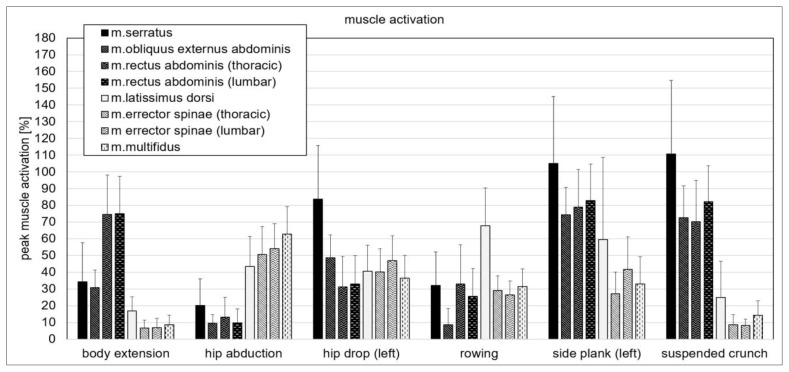
The muscle activation values of the eight tested trunk muscles differed during the sling training exercises (Figure 1).
Figure 1.
Means±SD of peak muscle activation (%) in the eight core muscles tested during the sling training exercises. For exercises with the two difficulty levels, the mean of the hard and easy versions were calculated and included in this figure.
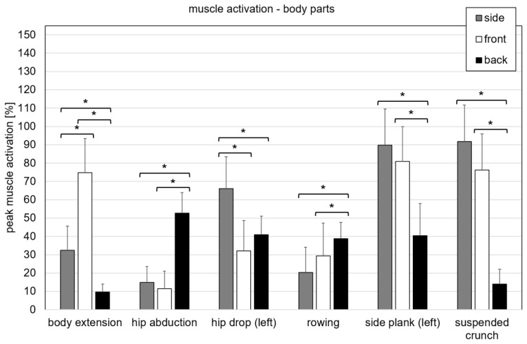
Analysis between the body parts showed significant (p< 0.05) differences within the Friedman analysis. Post-hoc tests demonstrated significant (p< 0.05) differences between the body parts (Figure 2).
Figure 2.
Means±SD of peak muscle activation (%) during the sling training exercises in the three body parts: side (m. serratus, m. obliquus externus abdominis), front (m. rectus abdominis: thoracic and lumbar), and back (m. latissimus dorsi, m. erector spinae: thoracic and lumbar, m. multifidus); *significant difference (p< 0.05).
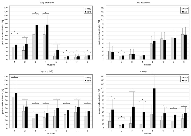
Results showed an influence of the two different difficulty levels in the four exercises from the TRX-OldAge program on muscle activation (Figure 3). ANOVA and Friedman analysis displayed significant differences (p< 0.05) for all four exercises. Post hoc tests showed significant (p< 0.05) differences for all eight muscles during the exercises body extension, hip drop, and rowing, but only for m. obliquus externus during hip abduction (Figure 3) between the easy and the hard versions of the four exercises. Higher muscle activations were always found in the harder versions.
Figure 3.
Means±SD of peak muscle activation (%) during the easy and hard versions of the four sling training exercises of the TRX-OldAge training program (body extension, hip abduction, hip drop (left), and rowing) in the eight tested core muscles: (1) m. serratus; (2) m. obliquus externus abdominis; (3) m. rectus abdominis (thoracic); (4) m. rectus abdominis (lumbar); (5) m. latissimus dorsi; (6) m. erector spinae (thoracic); (7) m. erector spinae (lumbar); (8) m. multifidus; *significant difference (p< 0.05) between the two versions (easy and hard).
DISCUSSION
Previous research on sling training has mostly focused on either push-up or plank sling training exercises in comparison to traditional strengthening exercises (3, 6, 8, 9, 21, 24, 25). The aim of this study was to investigate the level of trunk muscle activation during six different dynamic sling training exercises in healthy, young adults to allow the development of an evidence-based sling training program aimed at improving trunk strength and stability for future studies.
As hypothesized, the six sling exercises differed regarding muscle activation with significant differences between the three body parts (front, side, back). The high muscle activation of the (front) trunk flexor muscles measured during body extension, side plank, and suspended crunch indicates that these exercises could be implemented to increase abdominal strength and therefore trunk stability (5). In contrast to the high activation levels of the abdominal muscles, the back muscles exceeded half their peak reference trial values only during the hip abduction exercise. Exercises with potentially higher muscle activation of the m. latissimus dorsi, m. erector spinae (thoracic), m. erector spinae (lumbar), and m. multifidus could be power pull, low and L and I deltoid fly, or torso rotation. For the side body muscles tested (m. serratus, m. obliquus externus abdominis), the hip drop, side plank, and suspended crunch exercises achieved muscle activations of 60% and higher. In particular, the side plank and the suspended crunch seem to require high activation in the m. obliquus externus abdominis, another important stabilizer of the trunk.
In a recent study by Mok et al. (23), muscle activations of the trunk muscles were examined in young, healthy adults during four sling training exercises (hamstring curl, hip abduction plank, chest press, and 45 degrees rowing). Despite some overlap regarding the exercises and the major muscle groups examined, a comparison between this study and theirs might be difficult as Mok et al. (23) only analyzed isometric muscle activity during the hold positions of the sling training exercises, while this study’s aim was to determine peak muscle activation throughout an entire exercise cycle. Mok et al.’s (23) results provide very relevant information concerning isometric muscle activation during the sling training exercises they examined. However, during sling training, one usually implements dynamic repetitions within the training program, and therefore the dynamic phases prior to and after the potential hold positions should not be neglected. With respect to practical aspects during the planning and implementation of a sling training program, the professionals should consider such differences.
To compare certain sling exercises at different intensities, the PBA used by Gaedtke and Morat (15) and recommended in the TRX Manual (12) to adjust the intensity of sling training exercises was implemented in this study. Therefore, the base of support was moved closer to or further away from the vertical projection of the TRX mount in the ceiling to vary exercise intensity by altering the body angle. Our study results confirmed our hypothesis for the body extension, hip drop, and rowing exercises as all eight tested trunk muscles showed significantly higher muscle activations in the hard version compared to the easy version. In these exercises, the body angle of the participants notably changed between the levels of difficulty, seeming to cause a relevant shift in the vertical projection of the center of mass further away from the base of support, leading to an increased requirement for trunk muscle activation to execute the task correctly. This confirms the feasibility of using the PBA to increase intensity during sling trainer exercises.
Regarding the PBA, muscle activations during the hip abduction exercise did not differ significantly, except for one muscle. This could be the case as the vertical projection of the center of mass moved slightly closer to the base of support in the harder setting, actually reducing the forces acting on the trunk muscles. However, these shifts were rather small due to the flat (lying) body position. Moreover, one should bear in mind that the purpose of the hip abduction exercise is to train the hip abductors rather than the trunk. Therefore, a harder version of the exercise is not necessarily visible when examining the trunk muscles as was done in this study. The hip abductor muscles were not investigated in this study, so it is not possible to state whether the recommended difficulty level fulfils its purpose for the hip abduction exercise.
While the PBA seems to be effective in adjusting the intensity of a workout, it also shows one limitation of sling training, as the maximal distance between the center of mass and the base of support is limited by anthropometrics. Furthermore, a change in body angle is often accompanied by the muscles involved working at different muscle lengths. Designing a sling training intervention based on different mat zones (15) might be suitable for progressively increasing the intensity, even if the intensities can be used more effectively for older/less active persons than for young/fit/athletic persons. Consequently, a long-term sling training intervention could potentially lead to increased trunk stability, resulting in improved performance on functional tests and everyday life performance, and thus potentially being a helpful alternative aiming to reduce fall risk factors in older adults.
As methodological limitations of this study, the potential cross-talk between electrodes and the placement of the surface electrodes (selected with reference to former literature) should be noted. The positions of electrodes were placed using the anatomic landmarks and the muscle maps by Konrad (19). Unfortunately, there was no ultrasound available to check the exact differentiation, particularly between m. erector spinae thoracic, m. erector spinae lumbar and m. multifidus. Regarding peak EMG activity for normalization, the highest muscle activation during either the isometric seated MVC measurements or during the dynamic reference exercises (on the cot) were used. For a higher comparability, similar body postures during the reference EMG measurements (in regard to the sling training exercises) should be taken into account for future studies. Another aspect is that the exercise execution speed was not objectively controlled in this study. In addition, these results are for very active and fit persons, and thus the generalizability of the results to less active persons and other age groups might be limited. Moreover, there are still several more sling training exercises that have not been analyzed concerning trunk muscle activation.
In summary, the sling training exercises investigated in this study seem to be effective for activating important abdominal muscles and provide important complements of previous studies with sling exercises. As assumed, changing the body angle during sling training exercises is shown to be a feasible way of adjusting the intensity of sling training. This could potentially be used in longitudinal sling training studies to assure a controlled, progressively increasing training intervention, preferably with older/less active adults.
Supplementary Material
ACKNOWLEDGEMENTS
The authors thank all the participants for taking part in this study. This work was supported by the ‘Stifterverband fuer die deutsche Wissenschaft’ as part of an excellent teaching concept (by the first author) in the master study program ‘Sport and Movement Gerontology’ at the German Sport University Cologne. First results of this study have been presented by the first author at the European Congress of Sport Science (ECSS) in July 2018 in Dublin.
REFERENCES
- 1.Akuthota V, Ferreiro A, Moore T, Fredericson M. Core Stability Exercise Principles. Curr Sports Med Rep. 2008;7(1):39–44. doi: 10.1097/01.CSMR.0000308663.13278.69. [DOI] [PubMed] [Google Scholar]
- 2.Akuthota V, Nadler SF. Core strengthening. Arch Phys Med Rehabil. 2004;85(3 Suppl 1):S86–92. doi: 10.1053/j.apmr.2003.12.005. [DOI] [PubMed] [Google Scholar]
- 3.Beach TAC, Howarth SJ, Callaghan JP. Muscular contribution to low-back loading and stiffness during standard and suspended push-ups. Hum Mov Sci. 2008;27(3):457–472. doi: 10.1016/j.humov.2007.12.002. [DOI] [PubMed] [Google Scholar]
- 4.Behm DG, Drinkwater EJ, Willardson JM, Cowley PM. The use of instability to train the core musculature. Appl Physiol Nutr Metab. 2010;35(1):91–108. doi: 10.1139/H09-127. [DOI] [PubMed] [Google Scholar]
- 5.Behm DG, Leonard AM, Young WB, Bonsey WAC, MacKinnon SN. Trunk Muscle Electromyographic Activity With Unstable and Unilateral Exercises. J Strength Cond Res. 2005;19(1):193–201. doi: 10.1519/1533-4287(2005)19<193:TMEAWU>2.0.CO;2. [DOI] [PubMed] [Google Scholar]
- 6.Borreani S, Calatayud J, Colado JC, Moya-Nájera D, Triplett NT, Martin F. Muscle activation during push-ups performed under stable and unstable conditions. J Exerc Sci Fit. 2015;13(2):94–98. doi: 10.1016/j.jesf.2015.07.002. [DOI] [PMC free article] [PubMed] [Google Scholar]
- 7.Brumitt J. Core assessment and training. Champaign, Illinois: Human Kinetics; 2010. [Google Scholar]
- 8.Byrne JM, Bishop NS, Caines AM, Crane KA, Feaver AM, Pearcey GEP. Effect of using a suspension training system on muscle activation during the performance of a front plank exercise. J Strength Cond Res. 2014;28(11):3049–3055. doi: 10.1519/JSC.0000000000000510. [DOI] [PubMed] [Google Scholar]
- 9.Calatayud J, Borreani S, Colado JC, Martin F, Rogers ME. Muscle activity levels in upper-body push exercises with different loads and stability conditions. Phys Sportsmed. 2014;42(4):106–119. doi: 10.3810/psm.2014.11.2097. [DOI] [PubMed] [Google Scholar]
- 10.Cugliari G, Boccia G. Core Muscle Activation in Suspension Training Exercises. J Hum Kinet. 2017;56(1):61–71. doi: 10.1515/hukin-2017-0023. [DOI] [PMC free article] [PubMed] [Google Scholar]
- 11.Dannelly BD, Otey SC, Croy T, Harrison B, Rynders CA, Hertel JN, Weltman A. The effectiveness of traditional and sling exercise strength training in women. J Strength Cond Res. 2011;25(2):464–471. doi: 10.1519/JSC.0b013e318202e473. [DOI] [PubMed] [Google Scholar]
- 12.Fitness Anywhere. TRX® Suspension Training® Course Manual. San Francisco: Fitness Anywhere Inc; 2009. [Google Scholar]
- 13.Fong SSM, Tam YT, Macfarlane DJ, Ng SS, Bae YH, Chan EW, Guo X. Core Muscle Activity during TRX Suspension Exercises with and without Kinesiology Taping in Adults with Chronic Low Back Pain: Implications for Rehabilitation. Evid Based Complement Alternat Med. 2015;2015 doi: 10.1155/2015/910168. 910168. [DOI] [PMC free article] [PubMed] [Google Scholar]
- 14.Fowles JR. What I always wanted to know about instability training. Appl Physiol Nutr Metab. 2010;35(1):89–90. doi: 10.1139/H09-134. [DOI] [PubMed] [Google Scholar]
- 15.Gaedtke A, Morat T. TRX Suspension Training: A New Functional Training Approach for Older Adults - Development, Training Control and Feasibility. Int J Exerc Sci. 2015;8(3):224–233. [PMC free article] [PubMed] [Google Scholar]
- 16.Harris S, Ruffin E, Brewer W, Ortiz A. Muscle activation patterns during suspension training exercises. Int J Sports Phys Ther. 2017;12(1):42–52. [PMC free article] [PubMed] [Google Scholar]
- 17.Hicks GE, Simonsick EM, Harris TB, Newman AB, Weiner DK, Nevitt MA, Tylavsky FA. Cross-sectional associations between trunk muscle composition, back pain, and physical function in the health, aging and body composition study. J Gerontol A Biol Sci. 2005;60(7):882–887. doi: 10.1093/gerona/60.7.882. [DOI] [PubMed] [Google Scholar]
- 18.Kibler WB, Press J, Sciascia A. The role of core stability in athletic function. Sports Med. 2006;36(3):189–198. doi: 10.2165/00007256-200636030-00001. [DOI] [PubMed] [Google Scholar]
- 19.Konrad P. The ABC of EMG: A Practical Introduction to Kinesiological Electromyography. Scottsdale, Arizona: Noraxon Inc; 2006. [Google Scholar]
- 20.Lehman GJ, McGill SM. The importance of normalization in the interpretation of surface electromyography: a proof of principle. J Manipulative Physiol Ther. 1999;22(7):444–446. doi: 10.1016/s0161-4754(99)70032-1. [DOI] [PubMed] [Google Scholar]
- 21.Maeo S, Chou T, Yamamoto M, Kanehisa H. Muscular activities during sling- and ground-based push-up exercise. BMC Res Notes. 2014;7(1):192–198. doi: 10.1186/1756-0500-7-192. [DOI] [PMC free article] [PubMed] [Google Scholar]
- 22.McGill SM. Low back stability: from formal description to issues for performance and rehabilitation. Exerc Sport Sci Rev. 2001;29(1):26–31. doi: 10.1097/00003677-200101000-00006. [DOI] [PubMed] [Google Scholar]
- 23.Mok NW, Yeung EW, Cho JC, Hui SC, Liu KC, Pang CH. Core muscle activity during suspension exercises. J Sci Med Sport. 2015;18(2):189–194. doi: 10.1016/j.jsams.2014.01.002. [DOI] [PubMed] [Google Scholar]
- 24.Snarr RL, Esco MR. Electromyographical comparison of plank variations performed with and without instability devices. J Strength Cond Res. 2014;28(11):3298–3305. doi: 10.1519/JSC.0000000000000521. [DOI] [PubMed] [Google Scholar]
- 25.Snarr RL, Esco MR, Witte E, Jenkins C, Brannan R. Electromyographic activity of rectus abdominis during a suspension push-up compared to traditional exercises. J Exerc Physiol Online. 2013;16(3):1–8. [Google Scholar]
- 26.Sternlicht E, Rugg S. Electromyographic analysis of abdominal muscle activity using portable abdominal exercise devices and a traditional crunch. J Strength Cond Res. 2003;17(3):463–468. doi: 10.1519/1533-4287(2003)017<0463:eaoama>2.0.co;2. [DOI] [PubMed] [Google Scholar]
- 27.Vera-Garcia FJ, Grenier SG, McGill SM. Abdominal muscle response during curl-ups on both stable and labile surfaces. Phys Ther. 2000;80(6):564–569. [PubMed] [Google Scholar]
- 28.Willardson JM. Core stability training: applications to sports conditioning programs. J Strength Cond Res. 2007;21(3):979–985. doi: 10.1519/R-20255.1. [DOI] [PubMed] [Google Scholar]
- 29.Willson JD, Dougherty CP, Ireland ML, Davis IM. Core stability and its relationship to lower extremity function and injury. J Am Acad Orthop Surg. 2005;13(5):316–325. doi: 10.5435/00124635-200509000-00005. [DOI] [PubMed] [Google Scholar]
- 30.Yu B, Gabriel D, Noble L, An KN. Estimate of the Optimum Cutoff Frequency for the Butterworth Low-Pass Digital Filter. J Appl Biomech. 1999;15(3):318–329. [Google Scholar]
Associated Data
This section collects any data citations, data availability statements, or supplementary materials included in this article.


