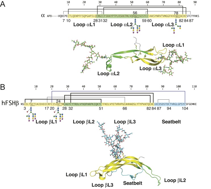
Figure 1.
FSH subunit primary structures. (A) FSHα primary and tertiary structures. (B) FSHβ primary and tertiary structures. Amino acid sequences of FSHα and FSHβ subunits indicated by single-letter amino acid code. Cystine knot loops in primary and tertiary structures are indicated by yellow highlighting for loops L1 and L3 and green for loop L2. The seatbelt loop is colored cyan. Lines above the subunit sequences connecting Cys residues (C) indicate disulfide bonds. Cys residue numbers are shown below. Cys knot disulfides are indicated by thick lines. N-glycosylation site sequons of the type NXT (N, Asn; X, any residue except Pro; T, Thr) are underlined, and oligosaccharide structures are shown below each subunit. Key to oligosaccharide structures can be found in Fig. 3. FSH subunit three-dimensional structures were extracted from pdb file 4ay9 using PyMol. Known FSH glycans were added to the FSH polypeptide backbone using GLYCAM.

