

Abstract
Background: Trapezium dislocations are rare injuries. Methods: A PubMed search of the term “trapezium dislocation” was conducted. Publications reporting a complete trapezium dislocation were included in the review. Results: The PubMed search resulted in 168 results. Fourteen publications reporting on 16 complete trapezium dislocations met inclusion criteria. A case of delayed diagnosis of a trapezium dislocation is presented. The literature is reviewed for pertinent clinically relevant information with respect to trapezium dislocations. A systematic method for radiographic analysis of trapezium dislocations and classification are described, and a treatment algorithm is presented. Conclusions: Trapezium dislocations are infrequent injuries with few cases reported in the literature. Given the rarity of this injury, diagnosis and appropriate treatment may be delayed due to difficulty in recognition. Using the described method of radiographic analysis, delayed diagnosis may be avoided with implementation of timely treatment.
Keywords: dislocation, trapezium, thumb, carpus, CMC
Introduction
Trapezium dislocations are relatively uncommon carpal injuries.5,10,22 Mechanisms of injury are generally high energy in nature given the robust periarticular support of the many articulations of the trapezium.1 Approximately 16 cases of complete trapezium dislocations in 14 reports have been described in the literature.2-4,7,9,10,13-15,17-20,22 The most commonly reported directions of dislocation are volar or dorsoradial, hypothesized to occur due to hyperflexion and axial load to the first ray, although the exact mechanisms determining the direction of dislocation are not fully understood. Fracture-dislocations of the scaphotrapezial and first carpometacarpal articulations have been described, but no classification system has been proposed for complete trapezium dislocations and those not associated with trapezial fracture.14,21 Recognition of these infrequent injuries can be difficult to the untrained eye, as Gilula’s 3 arcs do not account for the position of the trapezium as originally described.6 Outcomes after treatment for trapezium dislocations are generally satisfactory, although loss of pinch and grip strength is common.4 Herein, we report a case of a complete dorsoradial trapezium dislocation, review the literature of this uncommon injury, describe a method for radiographic analysis and identification, propose a new trapezium dislocation classification scheme, and discuss treatment options and outcomes for this injury.
Methods
A PubMed search was conducted using the term “trapezium dislocation,” resulting in 168 publications. These articles were reviewed for cases involving complete dislocation of the trapezium. Fourteen publications met criteria for inclusion in this review.
Results
Sixteen trapezium dislocations in 14 publications were found (Table 1).2-4,7,9,10,13-15,17-20,22 Four dislocations (25%) were dorsal,2,9,18 and 12 (75%) were volar.3,4,7,10,13-15,17,19,20,22 Twelve patients (75%) presented on the day of injury and were diagnosed with a trapezium dislocation at presentation.3,4,10,11,13,15,17-20,22 Three patients (18.75%) presented for evaluation within 1 week of injury (range: 2-5 days).2,7,9 One patient (6.25%) did not seek treatment until 14 weeks after injury due to unresolved hand pain at which time he was diagnosed with a trapezium dislocation.15 Seven (43.75%) patients were treated with open reduction and percutaneous pinning.2-4,17-19 One (6.25%) patient was treated with open reduction alone.12 Three patients (18.75%) were treated with closed reduction and percutaneous pinning.9,10, 22 Two (12.5%) patients were treated with closed reduction and casting/splinting.13,20 Three patients (18.75%) were treated with trapeziectomy.7,15 All patients’ functional outcomes were acceptable. Seven cases (43.75%) reported complications with 3 patients experiencing posttraumatic arthritis, 3 patients reporting thumb stiffness, and 1 patient reporting occasional aching pain with heavy activity.4,7,17,18,19,22 One patient with thumb stiffness required tenolysis of the flexor pollicis longus 1 year after initial surgery.19
Table 1.
Review of Publications Reporting Trapezium Dislocations.
| Publication | Dislocation type | Time to treatment | Treatment | Outcomes | Complications |
|---|---|---|---|---|---|
| Boe2 | Dorsal | 5 days | Open reduction percutaneous pinning | Normal mobility and strength at 2.5 years | None |
| Brewood et al3 | Volar | 1 day | Open reduction percutaneous pinning | Painless thumb movement and return to work at 7 weeks | None |
| Clarke and Raphael4 | Volar | 1 day | Open reduction percutaneous pinning | Full thumb range of motion at 7 months | Scaphotrapeziotrapezoidal arthrosis |
| Goldberg et al7 | Volar | 3 days | Trapeziectomy | No complaints at 8 months | Decreased thumb opposition, abduction, IP flexion compared with opposite side |
| Ichikawa and Inoue9 | Dorsal | 2 days | Closed reduction percutaneous pinning | Full range of motion and no pain of the thumb at 3 months | None |
| Kenyon et al10 | Volar | 1 day | Closed reduction percutaneous pinning | Return to full manual work at 8 weeks | None |
| Kopp et al11 | Volar | 1 day | Open reduction | Pain free at 6 months | None |
| Mckie et al13 | Volar | 1 day | Closed reduction and casting | Full thumb range of motion at a “few” weeks | None |
| Peterson et al15 | Case 1: Volar | 1 day | Trapeziectomy | Full painless thumb ROM by 4 months | None |
| Case 2: Volar | 14 weeks | Trapeziectomy | Painless ROM by 1 month | None | |
| Seimon et al17 | Volar | 1 day | Open reduction percutaneous pinning | Returned to work at 8 weeks | Decreased opposition; obliteration of the CMC joint at 11 months |
| Sherlock et al18 | Case 1: Dorsal | 1 day | Open reduction percutaneous pinning | Pain free at 6 months | Complaints of thumb stiffness at early follow-up |
| Case 2: Dorsal | 1 day | Open reduction percutaneous pinning | Pain free at 6 weeks | Trapeziotrapezoidal sclerosis | |
| Siegel and Hertzberg19 | Volar | 1 day | Open reduction percutaneous pinning | Decreased IP flexion, thumb adduction and opposition | Flexor pollicis longus adhesions requiring tenolysis 1 year after surgery |
| Vente and de Ruiter20 | Volar | 1 day | Closed reduction and splinting × 5 weeks | Full painless thumb ROM at 13 months | None |
| Wintman et al22 | Volar | 1 day | Closed reduction percutaneous pinning | Full ROM and return of grip strength by 1 year | Occasional ache in the affected hand after heavy work |
Note. IP = interphalangeal; ROM = range of motion; CMC = carpometacarpal.
Case Presentation
A 49-year-old male laborer with no significant medical history presented to our office 3 weeks after sustaining a right hand injury in an automobile rollover accident. The patient had been evaluated and treated on the day of the accident at another facility. Right hand radiographs obtained at that time were reported to have been read as normal with no evidence of fracture or dislocation, but unfortunately these radiographs were not available to the authors. Subsequently, a surgeon placed a Kirschner wire across the first metacarpophalangeal (MCP) joint for unknown reasons. The patient presented to our facility complaining of pain located at the radial aspect of his right hand at the base of the first metacarpal and pain elicited with attempted grip. On physical exam, the patient had swelling about the base of the first metacarpal, pain with attempted motion of the thumb, and an inability to pinch or grip secondary to pain. The flexor pollicis longus and extensor pollicis longus remained intact along with full sensation to the thumb. New radiographs were obtained, demonstrating a complete dorsoradial dislocation of the trapezium (Figure 1).
Figure 1.

Anteroposterior (a), oblique (b), and lateral (c) radiographs of normal right hand demonstrating a dorsoradial trapezium dislocation.
The patient was taken to the operating room for further surgical management. An attempt at closed reduction was not successful. Therefore, the trapezium was approached dorsally, just radial to the first dorsal extensor compartment. The capsule was incised, revealing an intact trapezium. Several attempts of reduction were undertaken without success. A trapeziectomy was then performed in conjunction with an abductor pollicis longus to flexor carpi radialis suture suspensionplasty. The Kirschner wire was removed from the first MCP joint. Postoperative fluoroscopy images demonstrated adequate trapeziectomy with first metacarpal suspension.
The thumb carpometacarpal joint was immobilized for 4 weeks followed by initiation of hand therapy. Three months postoperatively, the patient had complete resolution of pain, performed grip and pinch without difficulty, and reported no difficulty performing activities of daily living. Unfortunately, the patient was subsequently lost to follow-up and unable to be contacted.
Discussions
Radiographic Analysis—An Extension of Gilula’s Arcs
In 1979, Gilula originally described his method of carpal analysis by emphasizing the 3 normal arcs (Figure 2), principles of parallelism, and overlapping articular surfaces.6 Disruptions of these arcs and/or overlapping articular surfaces are found in most carpal injuries; however, they cannot detect trapezial injuries.6
Figure 2.
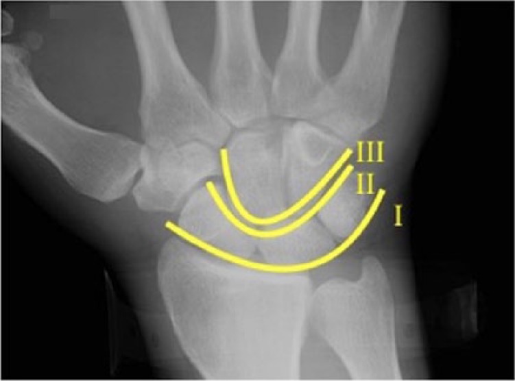
Anteroposterior radiograph of the right wrist demonstrating Gilula’s 3 carpal arcs.
To promote accurate and prompt recognition of trapezial injuries, an extension of Gilula’s first arc can be utilized.10 This is enabled by the relatively constant relationship of the trapezium with the scaphoid, first metacarpal, and trapezoid. In particular, the consistent relationship of the trapezium to the scaphoid is apparent on anteroposterior and lateral radiographs. To detect trapezial injuries, Gilula’s originally described first arc (the most proximal arc visualized on the anteroposterior wrist radiograph), can be extended radially to end just distal to the trapezium (Figure 3).6 Disruption at the border of Gilula’s extended first arc between the scaphoid and trapezium demonstrates a scaphotrapezial dislocation with translation in the radial-ulnar direction (Figure 4). Disruption of both the scaphotrapezial and first carpometacarpal articulation may occur and is termed a complete trapezium dislocation.
Figure 3.

Anteroposterior radiograph of a normal right wrist demonstrating Gilula’s extended first arc traversing the proximal border of the triquetrum, lunate, scaphoid, and trapezium.
Figure 4.
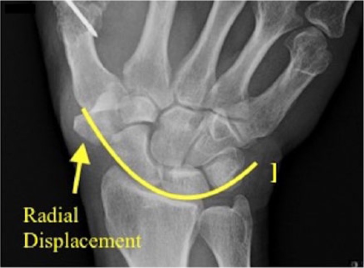
Anteroposterior radiograph of the right wrist demonstrating disruption of Gilula’s extended first arc resulting from radial displacement of the trapezium.
Gilula’s originally described arcs were all based on the anteroposterior radiograph.6 As with Gilula’s extended first arc, disruption of the normal arc contour will identify a malpositioned trapezium in the radial or ulnar direction, but will not provide information regarding the volar-dorsal plane. On the lateral view, a line parallel to the volar border of the scaphoid normally traverses the volar scaphoid and trapezial border (Figure 5). If this line does not traverse the volar trapezial border, it is indicative of trapezium displacement in the volar or dorsal direction (Figure 6).
Figure 5.

Lateral radiograph of a normal right wrist demonstrating the volar scaphotrapezial line.
Figure 6.

Lateral radiograph of a right wrist demonstrating disruption of the volar scaphotrapezial line (a) resulting from a complete dorsal trapezium dislocation with the absence of the trapezium from its articulations with the first metacarpal and scaphoid (b).
With this additional formal analysis, relative disruption of Gilula’s extended arc and displacement of the volar scaphotrapezial line will describe the trapezial displacement in radial-ulnar and volar-dorsal planes. This is particularly useful as the 2 most common dislocation directions are dorsoradial and volar.10,4
Systematic analysis of the wrist is necessary given the intricate anatomy of the carpal rows. In 1979, Gilula originally described his method of systematic analysis emphasizing parallelism, the 3 normal arcs, and articular overlap.6 Prior to his description, much had been written in the literature about carpal injury “aunt minnie” appearances, but no systematic approach had been provided. Gilula’s original descriptions analyzed many carpal relationships; however, it stopped short of addressing the trapezium.6 Fortunately, these same principles can be applied to the trapezium’s relationship to the scaphoid and first metacarpal to provide a simple method for recognition of injury and placement into a classification scheme.
Prompt recognition of these injuries is necessary to prevent adverse outcomes and avoidable surgeries.4 Upon suspicion of a carpal injury, anteroposterior, lateral, and oblique radiographs of the hand and wrist should be obtained and analyzed as previously described. A Robert’s view—an anteroposterior view of the trapezium with the hand in full pronation—or computed tomographic scan may also be considered.8,16
A Classification Scheme for Isolated Dislocations of the Trapezium
Dislocations of the trapezium have been anatomically described in the dorsoradial and volar directions, although no formal classification scheme has been described. Classification schemata are valuable because they clarify discussion between providers, may direct treatment, and help predict outcomes; it is difficult to improve patient care without clear descriptions of the problem. The mechanisms of dislocation and fracture-dislocation have been hypothesized to be the result of axial compression and hyperflexion of the first ray, although no cadaveric studies have confirmed this mechanism.18 These injuries require critical radiographic analysis of carpal anatomy to prevent misdiagnosis, delayed diagnosis, and promote expeditious treatment. Volar dislocation is the most common direction given the robust palmar scaphotrapezial and radial ligament complex.1,18 With this anatomy in mind, and reviewing the available cases of trapezial dislocation in the literature, we propose a new classification system (Supplementary Figure 1a-1d).2-4,7,9,10,13-15,17-20,22
Fracture-dislocations have also been reported.14,19 In 1988, Walker et al published a classification of trapezium fractures.21 In 2015, Kose et al expanded on this classification to describe fracture-dislocations of the thumb carpometacarpal joint involving the trapezium, but did not take into account complete dislocation of the trapezium from its articulations with the trapezoid and scaphoid.12 The classification proposed in this article expands on these classifications to take into account the most commonly reported trapezium dislocations.
Treatment
A treatment algorithm can be utilized once the trapezium dislocation has been identified (Supplementary Figure 2). After recognition of the direction of trapezium dislocation using radiographic analysis, acute dislocations of the trapezium should be urgently reduced with the aid of an assistant or fingertrap providing distraction force on the first carpometacarpal joint.4 In the event that a trapezium dislocation is irreducible, an attempt at open reduction is warranted.10 As in our case, a delay in treatment of 3 weeks significantly decrease the probability of successful closed or open reduction, necessitating the salvage procedure of trapeziectomy with or without suture suspensionplasty or ligament reconstruction and tendon interposition, depending on surgeon preference.
If concentric reduction of the trapezium is obtained, stabilization is necessary to maintain alignment of the adjacent carpal articulations by means of splinting, casting, or percutaneous fixation.4 Three reports have demonstrated excellent patient outcomes with closed reduction and percutaneous pinning.9,10,22 Seven cases demonstrated excellent outcomes with open reduction and percutaneous fixation.2-4,17-19 Two cases were treated with closed reduction and cast/splint immobilization with acceptable outcomes, although 1 patient reported an occasional ach with heavy work.13,20 One patient was treated with open reduction alone, reporting excellent function at 6 months.11
Initial excision of the dorsally dislocated trapezium was advocated in early reports of this injury.7,15 The risk of avascular necrosis was used as rationale for this procedure for initial management but has not been demonstrated clinically. Successful patient outcomes have been reported for patients treated with initial trapeziectomy.7,15 Currently, trapeziectomy is considered a salvage procedure in cases where open reduction is not feasible.4
Outcomes after reduction of complete trapezium dislocations are generally satisfactory.2-4,7,9,10,13-15,17-20,22 Potential deficits on physical exam include decreased key pinch strength, wrist range of motion, and grip strength.4,7,17,18,19,22 No cases of avascular necrosis after dislocation have been described. Seven cases reported complications with 3 of these cases reporting development of arthritis, 1 case with flexor pollicis adhesions, and 3 cases of thumb stiffness.4,7,17,18,19,22 However, all cases of trapezium dislocation demonstrated acceptable return function at final follow-up.2-4,7,9,10,13-15,17-20,22
Conclusions
Complete trapezium dislocations are uncommon injuries that often require operative intervention to maintain a stable reduction. The majority of trapezium dislocations reported were in the volar direction, recognized within 5 days of injury, and treated with either closed or open reduction with percutaneous fixation, or trapeziectomy. As demonstrated by our case presentation, a delay in recognition may require the more invasive procedure of trapeziectomy with or without ligament reconstruction or tendon interposition for initial treatment, as open or closed reduction may no longer be feasible. All patients reported resolution of pain within the first 8 months postoperatively, and several patients were able to return to work within 8 weeks. Loss of range of motion and stiffness of thumb were the most common complications, with 1 patient requiring tenolysis of the flexor pollicis longus.
There are a number of limitations to the proposed classification system. Without a larger number of cases, it is difficult to determine the natural history of these injuries. Furthermore, without a prospective database capturing such dislocations, it is challenging to determine how the classification will guide treatment. Despite this, there is value in advancing a common language for discussing these injuries, so that we may improve care for our patients in the future. Further investigation is needed.
Supplementary Material
Footnotes
Supplemental material is available in the online version of the article.
Ethical Approval: This study was approved by our institutional review board.
Statement of Human and Animal Rights: This article does not contain any studies with human or animal subjects
Statement of Informed Consent: Informed consent was obtained from all individual participants in this study
Declaration of Conflicting Interests: The author(s) declared no potential conflicts of interest with respect to the research, authorship, and/or publication of this article.
Funding: The author(s) received no financial support for the research, authorship, and/or publication of this article.
References
- 1. Bettinger PC, Berger RA. Functional ligamentous anatomy of the trapezium and trapeziometacarpal joint (gross and arthroscopic). Hand Clin. 2001;17(2):151-168, vii. [PubMed] [Google Scholar]
- 2. Boe S. Dislocation of the trapezium (multangulum majus). A case report. Acta Orthop Scand. 1979;50(1):85-86. [DOI] [PubMed] [Google Scholar]
- 3. Brewood AF. Complete dislocation of the trapezium: a case report. Injury. 1985;16(5):303-304. [DOI] [PubMed] [Google Scholar]
- 4. Clarke SE, Raphael JR. Combined dislocation of the trapezium and the trapezoid: a case report with review of the literature. Hand (N Y). 2010;5(1):111-115. doi: 10.1007/s11552-009-9216-5. [DOI] [PMC free article] [PubMed] [Google Scholar]
- 5. Dunn AW. Fractures and dislocations of the carpus. Surg Clin North Am. 1972;52(6):1513-1538. [DOI] [PubMed] [Google Scholar]
- 6. Gilula LA. Carpal injuries: analytic approach and case exercises. AJR Am J Roentgenol. 1979;133(3):503-517. doi: 10.2214/ajr.133.3.503. [DOI] [PubMed] [Google Scholar]
- 7. Goldberg I, Amit S, Bahar A, Seelenfreund M. Complete dislocation of the trapezium (multangulum majus). J Hand Surg. 1981;6(2):193-195. [DOI] [PubMed] [Google Scholar]
- 8. Horch R. A new method for treating isolated fractures of the os trapezium. Arch Orthop Trauma Surg. 1998;117(3):180-182. [DOI] [PubMed] [Google Scholar]
- 9. Ichikawa T, Inoue G. Complete dislocation of the trapezium. Case report. Scand J Plast Reconstr Surg Hand Surg. 1999;33(3):335-337. [DOI] [PubMed] [Google Scholar]
- 10. Kenyon RM, Kelly EG, Padinjarathala B. Traumatic isolated trapezium dislocation without fracture: a case report and review of the literature. Case Rep Orthop. 2016;2016:1798941. doi: 10.1155/2016/1798941. [DOI] [PMC free article] [PubMed] [Google Scholar]
- 11. Kopp JR. Isolated palmar dislocation of the trapezoid. J Hand Surg Am. 1985;10(1):91-93. [DOI] [PubMed] [Google Scholar]
- 12. Kose O, Keskinbora M, Guler F. Carpometacarpal dislocation of the thumb associated with fracture of the trapezium. J Orthop Traumatol. 2015;16(2):161-165. doi: 10.1007/s10195-014-0288-9. [DOI] [PMC free article] [PubMed] [Google Scholar]
- 13. McKie LD, Rocke LG, Taylor TC. Isolated dislocation of the trapezium. Arch Emerg Med. 1988;5(1):38-40. [DOI] [PMC free article] [PubMed] [Google Scholar]
- 14. Mumtaz MU, Drabu NA. Open complete dislocation of trapezium with a vertically split fracture: a case report. Cases J. 2009;2:9092. doi: 10.1186/1757-1626-2-9092. [DOI] [PMC free article] [PubMed] [Google Scholar]
- 15. Peterson CL. Dislocation of the multangulum majus or trapezium (and its treatment in 2 cases with extirpation). Arch Chir Neerl. 1950;2(4):369-376. [PubMed] [Google Scholar]
- 16. Ramoutar DN, Katevu C, Titchener AG, et al. Trapezium fracture—a common technique to fix a rare injury: a case report. Cases J. 2009;2:8304. doi: 10.4076/1757-1626-2-8304. [DOI] [PMC free article] [PubMed] [Google Scholar]
- 17. Seimon LP. Compound dislocation of a trapezium. A case report. J Bone Joint Surg Am. 1972;54(6):1297-1300. [PubMed] [Google Scholar]
- 18. Sherlock DA. Traumatic dorsoradial dislocation of the trapezium. J Hand Surg Am. 1987;12(2):262-265. [DOI] [PubMed] [Google Scholar]
- 19. Siegel MW, Hertzberg H. Complete dislocation of the greater multangular (trapezium). A case report. J Bone Joint Surg Am. 1969;51(4):769-772. [PubMed] [Google Scholar]
- 20. Vente JP, de Ruiter K. Complete dislocation of the trapezium multangulum majus. Neth J Surg. 1983;35(1):33-35. [PubMed] [Google Scholar]
- 21. Walker JL, Greene TL, Lunseth PA. Fractures of the body of the trapezium. J Orthop Trauma. 1988;2(1):22-28. [DOI] [PubMed] [Google Scholar]
- 22. Wintman BI, Fowler JL, Baratz ME. Traumatic dislocation of trapezium: case report and review of the literature. Am J Orthop (Belle Mead NJ). 2000;29(3):229-232. [PubMed] [Google Scholar]
Associated Data
This section collects any data citations, data availability statements, or supplementary materials included in this article.