Abstract
Introduction:
Periorbital hyperpigmentation (POH) is one of the common conditions seen in outpatient department. Despite of its huge prevalence, clinical data regarding its etiology and associations are still insufficient.
Materials and Methods:
We conducted a clinico-investigational study in 50 patients of periorbital pigmentation. A detailed clinical history was recorded, clinical examination and laboratory investigation including complete blood count, vitamin B12 level, and thyroid profile are done.
Results:
The mean age of the patients presenting with periorbital hyperpigmentation was 29.5 years, out of 50 patients 42 (84%) were females and 8 (16%) were males. About 14% patients give positive family history of POH, history of atopy was positive in 30% of patients. History of various other habits like lack of adequate sleep, prolonged exposure to computers, rubbing eyes, and application of various cosmetics were also found to be positive in these patients. The other associated clinical findings were freckles (12%), telengectesia (2%), erythema (2%), and melasma (2%). In maximum (90%) number of patients, both upper and lower eyelids were involved and pigmentation involving >1 cm of eyelid margin was seen in 62% of patients. Laboratory investigations showed anemia in 10% of patients and low serum vitamin B12 in 12%; however, none of the patients has deranged thyroid profile.
Conclusion:
POH has a multifactorial etiology and role of correcting various faulty habits is important factor in its management. Presence of anemia and low serum vitamin B12 levels also points toward need of detailed laboratory evaluation in these patients.
Keywords: Etiological factors, periorbital pigmentation, vitamin B12
Introduction
Periorbital hyperpigmentation (POH) is one of the common conditions seen in our out-patient department. It presents as brown to dark color pigmentation over bilateral periorbital area, which can also extend to eyebrows, malar regions, temporal regions, and lateral nasal root. This condition is aesthetically bothering to the patients and has been found to be associated with significant impairment in quality of life.[1,2] The condition is seen in both sex, with female preponderance.[3] It is more commonly seen in patients of darker skin type.[4]
The etiology is multifactorial, including genetic/hereditary, postinflammatory hyperpigmentation, dermal melanin deposition, secondary to atopic or allergic contact dermatitis, anemia, stress, faulty habits, periorbital edema, superficial location of vasculature, and shadowing due to skin laxity, nutritional deficiencies, and chronic illness.[5,6]
Despite its high prevalence, there are limited data regarding its etiology and associations. We conducted a cross-sectional study in 50 subjects of periorbital pigmentation to outline the clinical and etiological profile of POH.
Materials and Methods
A hospital-based cross-sectional study was conducted in 50 patients of periorbital pigmentation attending the out-patient department of Sucheta Kriplani hospital, from November 2014 to September 2016. Patients of age >18 years and Fitzpatrick skin type I to IV were enrolled in the study, pregnant and lactating females were excluded.
After enrollment, a detail history was recorded which included, family history of POH, history of atopy, prolonged exposure to bright screens (computer and television), topical cosmetics applications, diet (veg/non-veg), sleep patterns, and thyroid disorder. The colour and extension of pigmentation from lid margin was recorded (<0.5 cm from lid margins, 0.5–1 cm from lid margins, >1 cm from lid margins). Other cutaneous findings and nail and mucosal pigmentation were also recorded. In all the patients, complete blood count, serum vitamin B12 levels, and thyroid function test were done.
Results and Observations
Fifty patients ranging between 18 and 51 years of age were recruited in the study. The mean age of patients was 29.5 years. Out of 50 patients, 42 (84%) were females and 8 (16%) were males. Majority of our patients were housewives (n = 23). POH was asymptomatic in majority of the patients 29 (58%); however, 16 (32%) patients reported itching over the periorbital area.
In many patients, there was positive history of various risk factors associated with POH. Sleep deprivation was noted in only 4 (8%) patients, rest of our patients has adequate sleep of 7–8 h. In 9 (18%) patients, there was history of prolonged exposure (>8 h) to bright screen (computer, television). Family history of POH was positive in 7 (14%) patients and history of atopy was positive in 30% of patients. In 15 (30%) female patients, there was positive history of application of kajal/eyeliner on regular bases.
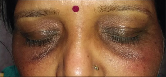
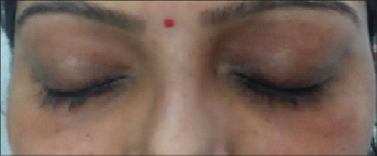
In the clinical evaluation, both upper and lower eyelids were involved in maximum number of patients, 45 (90%). The pigmentation was involving >1 cm of eyelid margin in 60% of patients [Figure 1], 0.5–1 cm of eyelid margin in 36% [Figure 2], and <1 cm of eyelid margin in 4% cases [Figure 3]. The other associated clinical findings were freckles (12%), telangiectesia (2%), erythema (2%), and melasma (2%).
Figure 1.

Periorbital pigmentation extending >1 cm from eyelid margins
Figure 2.

Periorbital pigmentation extending 0.5–1 cm from eyelid margins
Figure 3.

Periorbital pigmentation extending <1 cm from eyelid margins
Laboratory investigations showed anemia [hemoglobin (Hb) <10 mg/dL] in 5 (10%) of patients and low-serum vitamin B12 (<200 pg/mL) in 6 (12%); out of these six patients, only two had anemia (hemoglobin = 9.9 and 9.6 mg/dL), in rest of the patients hemoglobin levels were normal. Thyroid function test was turned out to be normal in all of the patients.
Discussion
POH is also known as periorbital melanosis, infraorbital melanosis, under eye circle, or dark eye circle. POH can be primary or secondary; when it is primary, it is termed as ICHOR (idiopathic cutaneous hyperchromia of orbital region). Secondary causes can be many, for example, postinflammatory hyperpigmentation secondary to atopic and allergic contact dermatitis, periorbital edema, excessive subcutaneous vascularity, shadowing due to skin laxity, and tear trough associated with aging. Ranu et al.[6] classified POH into four types: constitutional (most common type), postinflammatory, vascular, and shadow effect and others. On the basis of wood's lamp examination and ultrasonogram, Huang YL et al.[7] also categorized POH into pigmented (brown), vascular (blue to purple), structural, and mixed type.
As shown in previous studies, our study also found more number of female patients than males (M:F = 5:1). One possible reason for this female preponderance could be that females are more conscious for their appearance. Genetic basis of this condition has been proposed in past; Goodman and Belcher[5] reported familial cases of periorbital pigmentation with autosomal dominant trait; Sheth et al.,[3] and Ranu et al.[6] had also reported positive family history in their patients of POH. In our study, we found a positive family history of POH in 14% (n = 7) of patients, thus contributing to the genetic basis of this condition.
Conditions such as smoking, sleep deprivation, and prolong exposure to bright screen can contribute to palpebral hyperchromia due to the stasis of blood vessels. These factors were present in our study also and similar findings has been reported in previous study; this strengthens correlation of these factors with the presence of POH.[3,8]
Sixteen (32%) of our patients had history of itching over periorbital area, out of which eight gave history of atopy. Patients with history of atopy have habit of frequent rubbing or itching over periorbital area; this can cause local inflammation and increases pigmentation. In present study, history of atopy was present in 30% of patients; this finding is in concordance with the study by Sheth et al.[3] and Ranu et al.[6] We also found history of various eye cosmetic application (kajal/eyeliner) in significant number 30% (n = 15) of our patients. Various allergens in the eye cosmetics can cause allergic contact dermatitis and results in postinflammatory hyperpigmentation over periorbital area.
Anemia was found in only 10% of our patients which was not statistically significant. Sheth et al.[3] has reported iron deficiency anemia in significant number of patients in their study. Facial pallor due to anemia makes the periorbital region look comparatively darker, and due to low hemoglobin, there is lack of enough oxygen supply in the periorbital area leading to POH.
We could not found any association of hyperthyroidism/hypothyroidism in these patients. Deranged thyroid profile in patients with POH has been reported[3]; however, the association was not significant.
Deficiency of Vitamin B12 has been known to be responsible for increase pigmentation; however, its role in the etiopathogenesis of POH has not been studied in past. We found that low vitamin B12 was present in 12% of our patients, out of which only two had anemia; this finding suggest that low vitamin B12 can be present in patients who do not have any clinical signs of anemia, thus emphasizing role of screening for vitamin B12 levels in these patients. We hypothesized that correction of vitamin B12 deficiency in these patients can improve the periorbital pigmentation. However, more number of studies with large sample size is required to substantiate this finding.
Conclusion
POH has a multifactorial etiology, with various associations. Correction of underlying factors and laboratory abnormalities is essential for the management of this condition. Although many topical depigmenting creams and various peeling agents have been tried, results are variable. Our study shows role of genetic factors, contact allergic dermatitis to eye cosmetics, atopic diathesis, and association with vitamin B12 deficiency in POH. Addressing modifiable causative factors, namely, minimizing cosmetics, rubbing, friction, and vitamin B12 supplementation can act as adjunctive modalities in managing POH.
Financial support and sponsorship
Nil.
Conflicts of interest
There are no conflicts of interest.
References
- 1.Freitag FM, Cestari TF. What causes dark circles under the eyes? J Cosmet Dermatol. 2007;6:211–5. doi: 10.1111/j.1473-2165.2007.00324.x. [DOI] [PubMed] [Google Scholar]
- 2.Gendler EC. Treatment of Periorbital Hyperpigmentation. Aesthet Surg J. 2005;25:618–24. doi: 10.1016/j.asj.2005.09.018. [DOI] [PubMed] [Google Scholar]
- 3.Sheth PB, Shah HA, Dave JN. Periorbital hyperpigmentation. A study of its prevalence, common causative factors and its association with personal habits and other disorders. Indian J Dermatol. 2014;59:151–7. doi: 10.4103/0019-5154.127675. [DOI] [PMC free article] [PubMed] [Google Scholar]
- 4.Pearl E, Grimes MD. Aesthetic and Cosmetic Surgery of Darker Skin Type. 1st ed. Philadelphia (USA): Lippincott; 2008. [Google Scholar]
- 5.Goodman RM, Belcher RW. Periorbital hyperpigmentation. An overlooked genetic disorder of pigmentation. Arch Dermatol. 1969;100:169–74. doi: 10.1001/archderm.100.2.169. [DOI] [PubMed] [Google Scholar]
- 6.Ranu H, Thng S, Goh BK, Burger A, Goh CL. Periorbital hyperpigmentation in Asians: An epidemiologic study and a proposed classification. Dermatol Surg. 2011;37:1297–303. doi: 10.1111/j.1524-4725.2011.02065.x. [DOI] [PubMed] [Google Scholar]
- 7.Huang YL, Chang SL, Ma L, Lee MC, Hu S. Clinical analysis and classification of dark eye circle. Int J Dermatol. 2014;53:164–70. doi: 10.1111/j.1365-4632.2012.05701.x. [DOI] [PubMed] [Google Scholar]
- 8.Sarkar R, Ranjan R, Garg S, Garg VK, Sonthalia S, Bansal S. Periorbital hyperpigmentation: A comprehensive review. J Clin Aesthet Dermatol. 2016;9:49–55. [PMC free article] [PubMed] [Google Scholar]


