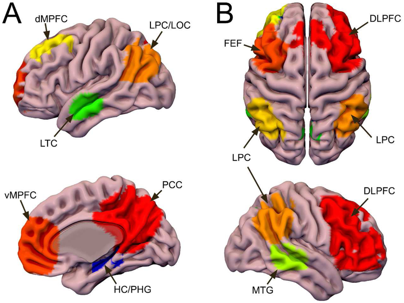
Figure 1. DMN and ECN Functional Templates.
DMN and ECN regions identified using ICA on both task- and rs-fMRI. The task-fMRI components were masked by the corresponding rs-fMRI components to create a template of regions showing activation/deactivation during the task and connectivity at rest. A: The DMN functional template includes the posterior cingulate cortex/precuneus (PCC/pC), ventromedial prefrontal cortex (vMPFC), dorsomedial prefrontal cortex (dMPFC), lateral temporal cortices (LTC), lateral parietal/occipital cortices (LPC/LOC), and portions of the hippocampus and parahippocampal gyrus (HC/PHG). B: The ECN functional template includes bilateral dorsolateral prefrontal cortex (DLPFC), frontal eye fields (FEF), lateral parietal cortices (LPC), and middle temporal gyri (MTG). Both: These regions were used to extract time-series for functional analyses and as seeds for probabilistic tractography. Surf Ice (https://www.nitrc.org/projects/surfice/) was used to create this display of the DMN template on the surface of the MNI152 T1 brain.

