Abstract
Case summary
A 14-year-old neutered female Burmese cat was referred for investigation of a caudal oropharyngeal mass. CT showed a thin walled cyst-like structure filling and expanding from the right tympanic bulla. Histopathology showed fragments of mildly dysplastic squamous epithelium and aggregates of keratin. These findings were considered consistent with a diagnosis of cholesteatoma.
Relevance and novel information
To the best of our knowledge, this is the first reported case of a cholesteatoma in a cat. Cholesteatoma should be considered a differential diagnosis for cats presenting with a caudal oropharyngeal mass, a history of chronic ear disease or a history of previous, surgically managed middle ear disease. Advanced imaging and biopsies should be considered important in the diagnosis of these lesions.
Keywords: Cholesteatoma, oropharyngeal mass, ear, otitis, ventral bulla osteotomy
Case description
A 14-year-old neutered female Burmese cat was referred for investigation of a caudal oropharyngeal mass.
Three weeks prior to referral the cat presented to the primary care veterinarian for lethargy, altered vocalisation, snorting when eating and masticating with one side of the mouth. A temperature of 39.4°C was noted, as well as gingival recession on the upper-left carnassial tooth. The teeth were scaled, polished and three extractions were performed. At this time, ventral deviation of the soft palate was noted on the right-hand side. Following dental treatment, the clinical signs resolved. A right-sided nasopharyngeal polyp had been removed by traction via the pharynx when the cat was 8 months old, followed by a ventral bulla osteotomy (VBO).
On presentation, physical examination revealed a firm, immobile right-sided caudal oropharyngeal mass, a mild right-sided head tilt and a body condition score of 3.5/9. Auscultation revealed mild stridor with the point of maximum intensity over the larynx. The remainder of the examination was unremarkable. Laryngoscopy and pharyngoscopy immediately after induction of anaesthesia, but prior to intubation, revealed a hypoplastic soft palate with a markedly curved caudal free border that was situated rostral to the epiglottis. The mass had an intact mucosa over its surface, which extended into the pharynx.
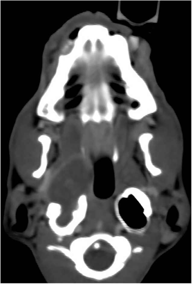
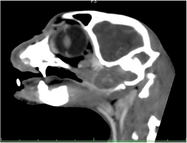
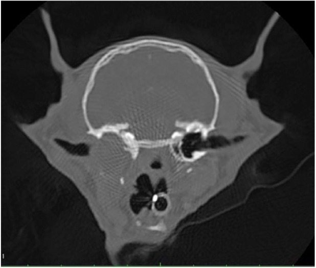
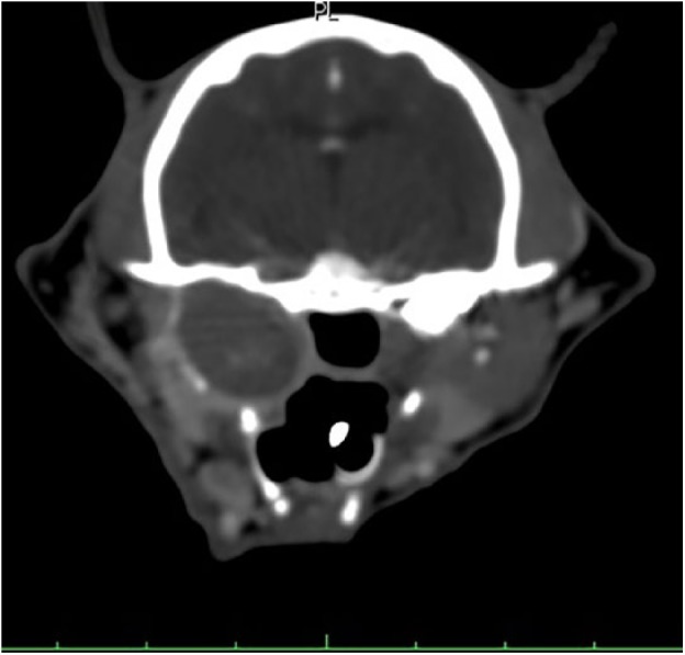
CT (General Electric Brightspeed 16, Model: 2335179-2) showed a thin walled cyst-like structure filling and expanding from the right tympanic bulla. This extended rostrally through the site of the previous VBO, into the right retropharyngeal space, bulging medially into the caudal nasopharynx and oral cavity (Figure 1). The mass measured 13 mm (dorsoventral) by 13 mm (lateromedial) by 27 mm (rostrocaudal). A relatively consistent attenuation (HU68) was noted throughout the mass, apart from a small central region of increased attenuation (HU85). A CT scan following administration of contrast revealed enhancement of the thin wall of the structure but not of the contents (Figures 1–4). Mild enlargement of the right mandibular lymph node, widespread bronchitis and right middle lung lobe atelectasis were also seen.
Figure 1.

Dorsal plane reconstruction of the post-contrast study (soft tissue window) demonstrating the thin rim of contrast enhancement surrounding the mass as it extends through the rostral wall of the right tympanic bulla
Figure 2.

Sagittal plane reconstruction of the post-contrast study (soft tissue window) through the plane of the right tympanic bulla, showing loss of the rostral bulla wall due to rostral extension of the mass
Figure 3.

Transverse image of the post-contrast study (bone window) demonstrating the loss of the right bony bulla wall
Figure 4.

Transverse image of the post-contrast study (soft tissue window) demonstrating the mass bulging medially into the nasopharynx and oral cavity
TruCut needle biopsies were taken from the mass via the oral cavity, which yielded pale/tan homogeneous, friable tissue. On otoscopic examination of the right ear, material similar to that of the biopsies could be seen in the deeper parts of the horizontal canal extending into the middle ear cavity, and grab biopsies were taken via the external ear canal. The cat was discharged with meloxicam (Metacam 0.1 mg/kg PO q24h; Boehringer Ingelheim).
Histopathological examination of the samples showed sections consisting of amorphous necrotic cellular debris with foci of suppurative inflammation. Small clumps of bacteria were also noted, along with rare fragments of mildly dysplastic squamous epithelium and aggregates of keratin. These findings were considered consistent with a diagnosis of cholesteatoma. No cytology was performed on submitted samples.
Treatment options, including a ventral bulla osteotomy or a lateral bulla osteotomy, with or without a total ear canal ablation, were discussed with the owner. The owner elected to not pursue surgical removal of the cholesteatoma. A course of potentiated amoxicillin (12.5 mg/kg PO q12h) for 14 days was started based on the histopathological findings of suppurative inflammation in the middle ear.
The cat lived for a further 3 years, during which time it was treated over 30 times by its primary care veterinarian with antibiotics for recurrent episodes of suppurative otitis. The cat was eventually euthanased after developing neurological signs including knuckling and loss of sensation on all four limbs, inability to ambulate, anorexia and facial rubbing, but it was not reported whether these signs were lateralised. A post-mortem examination was not permitted.
Discussion
To our knowledge this is the first reported case of a cholesteatoma in a cat.
The term cholesteatoma is well described in the literature but is potentially a misnomer as these lesions are not neoplastic in nature and do not contain cholesterol.1 A cholesteatoma describes a benign keratinising squamous cell cyst. Histologically, they are made up of a keratinised stratified squamous-lined cyst, filled with squamous debris.2–6 Visually, they often appear as cysts with a pearly white content.7 Cholesteatomas grow invasively, destroying neighbouring structures as keratin debris accumulates within the cyst’s lining.
Cholesteatomas are defined as either congenital or acquired.8 In human medicine, cholesteatoma is a rare clinical finding with an incidence of three and 9.2 per 100,000 population in children and adults, respectively.9 In the veterinary literature there is ambiguity with the terms cholesteatoma and cholesterol granuloma. At times they are used synonymously, particularly in equine medicine.6 However, cholesterol granulomas consist of cholesterol clefts embedded in granulation tissue and the suspected aetiology is different from cholesteatomas.10,11
Proposed theories for the formation of a cholesteatoma include invagination of the tympanic membrane into the middle ear and displacement of squamous epithelium from the external ear canal into the middle ear.12 In a gerbil model, it was shown that the tympanic membrane was the origin of the cholesteatoma epithelium. Attempts to induce cholesteatomas in cats through ligation of the Eustachian tube or ligation of the external auditory canal have been unsuccessful.13–15 All current theories suggest that chronic inflammation and bacterial infection are important predisposing factors for the development and growth of cholesteatomas.16,17 The presence of bacteria on the histopathological evaluation of the lesion in this report suggests that chronic infection could have played a role in the formation of this cholesteatoma.
Historically, canine cholesteatomas have been report-ed to be secondary complications of aural surgery, particularly total ear canal ablation with lateral bulla osteotomy (TECA-LBO), in which inflamed or infected epithelium could be left behind after surgery.16 In the absence of surgery, ongoing infection and inflammation have also been shown to be a primary cause of cholesteatomas such as in otitis externa and media. In this case the presence of the middle ear polyp, its effect on the middle ear and external ear canal, or the surgical procedure (ventral bulla osteotomy) to manage this could have contributed to the formation of the cholesteatoma.
In this case, there were few clinical signs related to the mass. However, given the location and the destructive, invasive nature of cholesteatomas, it is possible for affected animals to present with a range of clinical signs. In a case series of 20 dogs the observed clinical signs on presentation included head tilt, asymmetrical facial palsy, ataxia, nystagmus, circling and asymmetrical atrophy of the temporalis and masseter muscles. Given the proximity of the vestibulocochlear nerve and the facial nerve the majority of signs are likely attributed to their involvement. Four of the dogs in the case series had pain on opening their mouth or were unable to open their mouth fully. Respiratory noise and effort was also reported in the study.4 Mass lesions in the nasopharyngeal region have been associated with respiratory compromise and noise due to an obstructive effect.18 In this case, the previously performed VBO may have allowed the mass to extend ventrally out of the previous bulla osteotomy site and therefore cause less destruction laterally or medially relative to the bulla. This may have impacted on the clinical signs present in this case.
Diagnosis of this condition may be delayed as epidermoid cysts can often be present for a prolonged period of time before there is accumulation of sufficient keratin within them, or the surrounding inflammation and infection is significant enough to cause clinical signs.19 Bacterial infection of the central nervous system has been linked to an aural cholesteatoma that eroded through the petrous portion of the temporal bone, eventually causing infection of the vestibulocochlear ganglion.20 In this case it is unclear if the neurological signs seen prior to euthanasia were caused by the cholesteatoma as no further investigation was undertaken at the time and a necropsy was not performed. Meningitis has been reported in two dogs that had bone lysis into the cranial fossa; one of these was also found to be septic.4
Initial investigation into this case involved the use of contrast CT imaging to identify the location and nature of the lesion. In both human and veterinary medicine CT scans have been shown to be the imaging modality of choice.21–23 Changes visible on the CT include osteoproliferation, osteolysis of the bulla, expansion of the bulla and bone lysis in the petrosal portion of the temporal bone. Local lymphadenomegaly may also be seen, as was noted in this case.4 Changes can also be seen in the ipsilateral temporomandibular joint with the presence of a periosteal reaction, sclerosis or osteoproliferation.12 MRI is an alternative imaging modality, particularly when neurological signs predominate.12,15,17 Typical MRI findings are material that is isointense compared with brain tissue on T1-weighted images and of mixed intensity on T2-weighted and fluid-attenuated inversion recovery sequences. There is no post-contrast enhancement of the content, but enhancement of the lining of the bulla may be seen.15 MRI is being utilised more in human medicine as a radiation-free form of imaging to detect cholesteatomas, particularly in paediatric patients.19
It may be that many patients with a cholesteatoma may go undiagnosed owing the relative lack of clinical signs early on in the disease, the similarity of these signs to those of chronic otitis externa/otitis media and the lack of investigation into the underlying cause of the clinical signs. Even if not yet clinically significant, early diagnosis and surgical intervention of a cholesteatoma has been shown to improve outcome and prognosis,4,5,20 and early intervention may be curative.
The mainstay of management of patients with cholesteatoma is surgical intervention, often paired with long-term antimicrobial use.4 In dogs the main surgical intervention described is either a TECA-LBO or a VBO, with the former being performed more commonly.4,5 Particular care is needed to ensure as complete an excision as possible, and use of advanced imaging modalities helps with surgical planning and achieving complete removal of abnormal tissue. There was no difference reported in outcome based upon the surgical approach taken. Complete removal of the abnormal epithelium was deemed important, but was often difficult owing to its tight adherence to the abnormal bone. The use of rongeurs, high-speed burrs and a carbon dioxide laser were used to maximise removal.4
However, recurrence of clinical signs or persistence of clinical signs has been reported in around 50% of patients treated. Greci et al5 reported a confirmed recurrence in 4/11 dogs, and a fifth one was suspected but was not investigated. Time to recurrence ranged from 2–13 months. In the series of 20 dogs, 19 of which were surgically managed, nine showed resolution of clinical signs during the follow-up period that ranged from 3–95 months. Signs of middle ear disease, otodynia, head tilt and discomfort opening the mouth were present in the cases that recurred. Advanced disease at the time of first presentation was the main factor associated with recurrence.4,5 Factors that are significantly associated with recurrence after surgery include pain or difficulty with opening the jaw, bulla lysis, neurological disease or bone lysis within the squamous and petrosal portions of the temporal bone evident on CT imaging. Short-term complications of surgery related to damage to the facial nerve were thought to be caused during curettage of the middle ear cavity, but these often resolved.5 The large medial component of the cholesteatoma in this case, which encroached on the oropharynx, meant that repeat VBO may not have provided adequate access to and drainage of this part of the mass and as a result may not have helped to resolve the condition.
Conclusions
Cholesteatoma should be considered a differential diagnosis for cats presenting with a caudal oropharyngeal mass and, as a result, investigation with advanced imaging should be performed. Cats with a history of chronic ear disease or surgical management of middle ear disease that present with deficits of the facial or vestibulocochlear nerve that do not resolve or become chronic should be investigated for potential cholesteatoma.
Footnotes
Accepted: 11 April 2019
Conflict of interest: The authors declared no potential conflicts of interest with respect to the research, authorship, and/or publication of this article.
Funding: The authors received no financial support for the research, authorship, and/or publication of this article.
Ethical approval: This report involved the use of a client-owned animal only, and followed internationally recognised high standards (‘best practice’) of individual veterinary clinical patient care. Ethical Approval from a committee was not therefore needed.
Informed consent: Informed consent was obtained from the owner of the animal described in this report for the procedures undertaken. No animals or humans are identifiable within this publication, and therefore additional Informed Consent for publication was not required.
This paper was handled and processed by the European Editorial Office (ISFM) for publication in JFMS Open Reports
References
- 1. Ferlito A. A review of the definition, terminology and pathology of aural cholesteatoma. J Laryngol Otol 1993; 107: 483–488. [DOI] [PubMed] [Google Scholar]
- 2. Ferlito A, Devaney KO, Rinaldo A, et al. Clinicopathological consultation. Ear cholesteatoma versus cholesterol granuloma. Ann Otol Rhinol Laryngol 1997; 106: 79–85. [DOI] [PubMed] [Google Scholar]
- 3. Rosenberg RA, Hammerschlag PE, Cohen NL, et al. Cholesteatoma vs. cholesterol granuloma of the petrous apex. Otolaryngol Head Neck Surg 1986; 94: 322–327. [DOI] [PubMed] [Google Scholar]
- 4. Hardie EM, Linder KE, Pease AP. Aural cholesteatoma in twenty dogs. Vet Surg 2008; 37: 763–770. [DOI] [PubMed] [Google Scholar]
- 5. Greci V, Travetti O, Di Giancamillo M, et al. Middle ear cholesteatoma in 11 dogs. Can Vet J 2011; 52: 631–636. [PMC free article] [PubMed] [Google Scholar]
- 6. Maxie MG, Youssef S. Nervous system. In: Maxie MG. (ed). Jubb, Kennedy, and Palmer’s pathology of domestic animals. 5th ed. St Louis, MO: Elsevier, 2007, pp 281–457. [Google Scholar]
- 7. Chang P, Fagan PA, Atlas MD, et al. Imaging destructive lesions of the petrous apex. Laryngoscope 1998; 108: 599–604. [DOI] [PubMed] [Google Scholar]
- 8. Smith PG, Leonetti JP, Kletzker GR. Differential clinical and radiographic features of cholesterol granulomas and cholesteatomas of the petrous apex. Ann Otol Rhinol Laryngol 1988; 97: 599–604. [DOI] [PubMed] [Google Scholar]
- 9. Olszweska E, Wagner M, Bernal-Sprekelsen M, et al. Etiopathogenesis of cholesteatoma. Eur Arch Otorhinolaryngol 2004; 261: 6–24. [DOI] [PubMed] [Google Scholar]
- 10. Ilha MRS, Wisell C. Cholesterol granuloma associated with otitis media in a cat. J Vet Diagn Invest 2013; 25: 515–518. [DOI] [PubMed] [Google Scholar]
- 11. Fluehmann G, Konar M, Jaggy A, et al. Cerebral cholesterol granuloma in a cat. J Vet Intern Med 2006; 20: 1241–1244. [DOI] [PubMed] [Google Scholar]
- 12. Travetti O, Guidice C, Greci V, et al. Computed tomography features of middle ear cholesteatoma in dogs. Vet Radiol Ultrasound 2010; 51: 374–379. [DOI] [PubMed] [Google Scholar]
- 13. Chole R, Kodama K. Comparative histology of the tympanic membranes and its relationship to cholesteatoma. Ann Otol Rhinol Laryngol 1989; 98: 761–766. [DOI] [PubMed] [Google Scholar]
- 14. McGinn MD, Chole RA, Henry KR. Cholesteatoma induction. Consequences of external auditory canal ligation in gerbils, cats, hamsters, guinea pigs, mice and rats. Acta Otolaryngol 1984; 97: 297–304. [DOI] [PubMed] [Google Scholar]
- 15. Yamamoto-Fukuda T, Hishikawa Y, Shibata Y, et al. Pathogenesis of middle ear cholesteatoma: a new model of experimentally induced cholesteatoma in Mongolian gerbils. Am J Pathol 2010; 176: 2602–2606. [DOI] [PMC free article] [PubMed] [Google Scholar]
- 16. Risselada M. Diagnosis and management of cholesteatomas in dogs. Vet Clin North Am Small Anim Pract 2016; 46: 623–634. [DOI] [PubMed] [Google Scholar]
- 17. Kuo CL, Shiao AS, Yung M, et al. Updates and knowledge gaps in cholesteatoma research. Biomed Res Int 2015; 2015. DOI: 10.1155/2015/854024. [DOI] [PMC free article] [PubMed] [Google Scholar]
- 18. Ellison GW, Donnell RL, Daniel GB. Nasopharyngeal epidermal cyst in a dog. J Am Vet Med Assoc 1995; 207: 1590–1592. [PubMed] [Google Scholar]
- 19. Hansen S, Sorensen CH, Stage J, et al. Massive cholesteatoma of the frontal sinus: case report and review of the literature. Auris Nasus Larynx 2007; 34: 387–392. [DOI] [PubMed] [Google Scholar]
- 20. Østevik L, Rudlang K, Jahr TH, et al. Bilateral tympanokeratomas (cholesteatomas) with bilateral otitis media, unilateral otitis interna and acoustic neuritis in a dog. Acta Vet Scand 2018; 60: 31–38. [DOI] [PMC free article] [PubMed] [Google Scholar]
- 21. Love NE, Kramer RW, Spodnick GJ, et al. Radiographic and computed tomographic evaluation of otitis media in the dog. Vet Radiol Ultrasound 1995; 36: 375–379. [Google Scholar]
- 22. Russo M, Covelli EM, Meomartino L, et al. Computed tomographic anatomy of the canine inner and middle ear. Vet Radiol Ultrasound 2002; 43: 22–26. [DOI] [PubMed] [Google Scholar]
- 23. Garosi LS, Dennis R, Schwarz T. Review of diagnostic imaging of ear diseases in the dog and cat. Vet Radiol Ultrasound 2003; 44: 137–146. [DOI] [PubMed] [Google Scholar]


