Abstract
Stem cells are considered as the principal source of differentiated cells. In the past few years, the research on the stem cell in various fields had shown success, but the stem cell-based therapies in the dentistry had confined to a particular extent. The present case report was aimed to evaluate the efficacy of stem cells from human exfoliated deciduous teeth (SHED) in the management of a large periapical lesion. A 12-year-old girl reported with a chief complaint of pain in the lower right back tooth region since 5 days. Intraoral examination revealed a deep occlusal cavity in relation to tooth#46 with tenderness on percussion. Radiograph examination revealed periapical radiolucency measuring 1.8×1.0cm in size with perforation at the floor of the pulp chamber in relation to 46. A deciduous tooth from the same child was collected to isolate stem cells. After access opening for tooth #46, pulp was extirpated and a thick mucoperiosteal flap was raised. This was followed by homing of SHED into the periapical area through the window created in the buccal cortical plate and into the root canals of tooth #46 until the orifice. The access cavity was sealed with glass ionomer cement. The patient was subjected to evaluation at regular intervals i.e., two weeks, four months, twelve months, and twenty-four months. The case treated demonstrated complete resolution of periapical radiolucency in the fourth-month review with a positive response to electric pulp testing. This clinical application report concludes that SHED can be effective in treating the periapical lesions in permanent teeth.
Keywords: Periapical Diseases , Permanent Dentition , Dental Pulp , Dental Pulp Test , Stem Cells , Scaffold
Introduction
The stem cells are capable of both self-renewal and differentiation with varying degree of potency and plasticity. Scientific research in the field of stem cells represents a particular interest. Incipient therapeutic strategies have been made possible thanks to great advances in stem cell biology, with the aim of regenerating tissues injured by disease.[1-3]
Dental pulp of the deciduous teeth which are about to exfoliate act as a promising source of mesenchymal stem cells, with the capacity to replicate, renew on their own and divide into millions of cells. Dr. Saongtao Shi a Pedodontist discovered dental pulp stem cells by using the deciduous teeth of his daughter and named them as stem cells from human exfoliated deciduous teeth (SHED).[1]
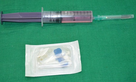
The lesions in the periapical area, which are a sequel of pulpal disease, are most routinely diagnosed during the radiographic examinations. Numerous non-surgical and surgical treatments are available for the treatment of periapical lesions, but due to great advancements in the field of stem cells, new therapeutic strategies have been made possible.[4] A case of periapical lesion that was successfully treated utilizing stem cells from human exfoliated deciduous teeth (SHED) was presented (Figure 1).
Figure1.

Stem cells
Case Report
A 12-year-old girl reported to the Department of Pedodontics and Preventive Dentistry with a chief complaint of pain in the lower right back tooth region since 5 days. Intraoral examination revealed a deep occlusal cavity in relation to tooth#46 with tenderness on percussion. Electric pulp testing to tooth#46 yielded negative response. Radiograph examination revealed periapical radiolucency measuring 1.8×1.0cm in size with perforation at the floor of the pulp chamber in relation to tooth #46 (Figure 2a). Hence, it was diagnosed as acute periapical abscess with perforation in relation to tooth #46. To promote faster healing, we planned for the tissue regeneration technique using SHED.
Figure2.
a: Preoperative radiograph, b: 2nd-week follow-up, c: Fourth-month follow-up, d: one-year follow-up, e: 2nd-year follow-up
After getting approval from the institutional ethical committee and written informed consent from the patient’s parent, a retained normal sound human deciduous tooth from the same child was collected. Enzymatic digestion method was used to isolate the stem cells from the pulp.[5] Under strict aseptic conditions, surgical removal of dental pulp from the tooth was done in the laboratory followed by digestion in collagenase/ dispase, and then characterization and screening were carried out using specific markers. The procedure involving culture media preparation, sample collection, storage, handling, expansion, subculturing, and characterization was performed using methods as described by Vishwanath et al.[6] The isolated SHED cells were immediately transported to the Department of Pedodontics and Preventive Dentistry from the laboratory and were utilized for the treatment of periapical lesion on the same day.
In the first appointment, all the debris from the cavity of tooth #46 and the necrotic pulp tissue was removed followed by irrigation of root canals with normal saline, and then the root canals were dried with absorbent points. Perforation repair was done with type IX glass ionomer cement (GC®, GC-Corp, Asia) and the access cavity was sealed with zinc oxide-eugenol cement (Prime dental product). In the subsequent appointment after 3 days upon the arrival of SHED cells to the Department, tooth #46 root canals were thoroughly irrigated with normal saline and dried with absorbent points. A full thickness mucoperiosteal flap was raised in relation to 46, a window was created through the buccal cortical plate (Figure 3a) to gain access to the periapical area which was followed by placement of scaffold PerioGlas®, homing of SHED into the periapical defect area (Figure 3b) and the mucoperiosteal flap was sutured in place. Then homing of SHED into the canals of 46 until the orifices was done and the access cavity was sealed with type IX glass ionomer cement (GC®, GC-Corp, Asia) with a thickness of 5mm. The patient returned asymptomatic with no signs of pain after two weeks, a modified stainless steel crown with a buccal window to access the tooth for pulp testing in future was cemented in relation to tooth #46 (Figure 2b and 3c). Electric pulp testing was done to evaluate the vitality of tooth #46 through the buccal window created and was negative. Patient failed to report for the 1st, 2nd, and 3rd month recall. At the postoperative fourth-month recall, the intra oral periapical radiograph showed complete resolution of periapical radiolucency that measured 1.8×1.0cm in size preoperatively to tooth #46 (Figure 2c) and responded positively to the electric pulp testing at higher current, i.e. 8. During the 12th month (Figure 2d) and 24th- month recall (Figure 2e) the tooth#46 was asymptomatic and responded positively to electric pulp testing at lower current i.e. 2.
Figure3.

a: Buccal window created, b: Homing of stem cells, c: Cemented modified stainless-steel crown
Discussion
The diagnosis of the periapical radiolucencies is generally made through the routine radiographic examinations or following toothache. These radiolucencies can be classified into periapical granulomas, radicular cysts, or abscesses. The incidence of cysts among periapical lesions is reported to be 6% to 55%.[4]
Scaffolds play a vital role as a delivery vehicle in stem cell-based therapy, which can be used for local repair. Since the activation and attachment of the stem cells is the most important action in the stem cell-based therapies, these scaffolds play a vital role in the attachment of the stem cells as well as bonding between the scaffold and host bone.[7-8] PerioGlas®, a bioactive ceramic, which was used as a scaffold in the present case report, has good osteostimulative and osteoconductive properties with additional advantages such as resorbability and ease of application.[9]
In the present case report, SHED’s were used for the treatment of periapical lesion to tooth #46, clinical and radiographic success was achieved in 4 months interval. Until now, there was no exact mechanism explained in the literature regarding the healing of the periapical lesions that are treated using SHED, as SHED does not have the capacity to differentiate directly into osteoblasts. But the probable success in the present case report may be attributed to the SHED cells capability that are homed into the area to induce new bone formation by forming an osteoinductive template to recruit host osteogenic cells leading to deposition of bone.[10] The osteoinductive capacity of SHED is proven in the present case, as there was radiographic evidence of formation of the bone in the periapical area of the treated tooth. Similar osteogenic regeneration for young permanent teeth treated with SHED was reported by Prasad et al.[11]
Root canal treatment was not initiated as the treated tooth responded positively during the fourth, twelfth and twenty-four-month follow-ups to electric pulp test, which may be indicative of re-innervation of pulp tissue inside the root canals. In one human study, immature permanent tooth treated with SHED displayed positive response to electric pulp testing from the 3rd month recall to the 12th month of follow-up.[11] This may be attributed to the neurogenic potential of SHED owing to their neural crest origin. In addition, SHED may have activated the residual pulp and periodontal stem cells to differentiate which may have generated highly vascularized living tissue.[12] In an immunohistochemistry study of a revascularized immature permanent tooth that regained pulp sensibility after regenerative endodontic therapy, neurons and nerve fibres were observed in newly formed tissue in the canal space.[13]
Dental pulp stem cells can be differentiated into both endothelial cells that would support revascularization [14-15] and neurons, which would support the reinnervation of the regenerated pulp tissue.[16] The triad properties i.e high proliferation rate, odontogenic, and osteogenic differentiation of SHED makes it a unique modality for treating teeth with periapical pathology. However, further research can be directed towards the evaluation of the bona fide feature of bone/marrow structures of SHED-induced regenerated bone, by bone biopsy.
Conclusion
The present case report depicts the potential capability of SHED in revascularization of permanent molar, but further clinical research with more samples is needed to evaluate and ascertain the success of SHED in regenerative endodontics.
Footnotes
Conflict of Interest: The authors declare that they have no conflict of interest.
References
- 1.Singh H, Bhaskar DJ, Rehman R, Jain CD, Khan M. Stem cells: An Emerging future in dentistry. Int J Adv Health Sci. 2014; 1: 17–23. [Google Scholar]
- 2.Marawar PP, Mani A, Sachdev S, Sodhi NK, Anju A. Stem cells in dentistry: An overview. Pravara Med Rev. 2012; 4: 11–15. [Google Scholar]
- 3.Potdar PD, Jethmalani YD. Human dental pulp stem cells: Applications in future regenerative medicine. World J Stem Cells. 2015; 7: 839–851. doi: 10.4252/wjsc.v7.i5.839. [DOI] [PMC free article] [PubMed] [Google Scholar]
- 4.Fernandes M, Ataide ID. Nonsurgical management of periapical lesions. J Conserv Dent. 2010;13:240–245. doi: 10.4103/0972-0707.73384. [DOI] [PMC free article] [PubMed] [Google Scholar]
- 5.Laino G, d'Aquino R, Graziano A, Lanza V, Carinci F, Naro F, et al. A new population of human adult dental pulp stem cells: a useful source of livingautologous fibrous bone tissue (LAB) J Bone Miner Res. 2005; 20: 1394–1402. doi: 10.1359/JBMR.050325. [DOI] [PubMed] [Google Scholar]
- 6.Vishwanath VR, Nadig RR, Nadig R, Prasanna JS, Karthik J, Pai VS. Differentiation of isolated and characterized human dental pulp stem cells and stem cells from human exfoliated deciduous teeth: An in vitro study. J Conserv Dent. 2013; 16: 423–428. doi: 10.4103/0972-0707.117509. [DOI] [PMC free article] [PubMed] [Google Scholar]
- 7.Shiehzadeh V, Aghmasheh F, Shiehzadeh F, Joulae M, Kosarieh E, Shiehzadeh F. Healing of large periapical lesions following delivery of dental stem cells with an injectable scaffold: new method and three case reports. Indian J Dent Res. 2014; 25: 248–253. doi: 10.4103/0970-9290.135937. [DOI] [PubMed] [Google Scholar]
- 8.Gerhardt LC, Boccaccini AR. Bioactive Glass and Glass-Ceramic Scaffolds for Bone Tissue Engineering. Materials (Basel) 2010; 3: 3867–3910. doi: 10.3390/ma3073867. [DOI] [PMC free article] [PubMed] [Google Scholar]
- 9.Brunelli G, Carinci F, Girardi A, Palmieri A, Caccianiga GL, Sollazzo V. Perioglass® and its osteogenic potential. Europ J Inflamm. 2012; 10:43–47. [Google Scholar]
- 10.Miura M, Gronthos S, Zhao M, Lu B, Fisher LW, Robey PG, et al. SHED: stem cells from human exfoliated deciduous teeth. Proc Natl Acad Sci U S A. 2003; 100: 5807–5812. doi: 10.1073/pnas.0937635100. [DOI] [PMC free article] [PubMed] [Google Scholar]
- 11.Prasad MGS, Ramakrishna J, Babu DN. Allogeneic stem cells derived from human exfoliated deciduous teeth (SHED) for the management of periapical lesions in permanent teeth: Two case reports of a novel biologic alternative treatment. J Dent Res Dent Clin Dent Prospects. 2017; 11: 117–122. doi: 10.15171/joddd.2017.021. [DOI] [PMC free article] [PubMed] [Google Scholar]
- 12.Namour M, Theys S. Pulp revascularization of immature permanent teeth: a review of the literature and a proposal of a new clinical protocol. Scientific World Journal. 2014; 2014: 737503. doi: 10.1155/2014/737503. [DOI] [PMC free article] [PubMed] [Google Scholar]
- 13.Lei L, Chen Y, Zhou R, Huang X, Cai Z. Histologic and Immunohistochemical Findings of a Human Immature Permanent Tooth with Apical Periodontitis after Regenerative Endodontic Treatment. J Endod. 2015; 41: 1172–1179. doi: 10.1016/j.joen.2015.03.012. [DOI] [PubMed] [Google Scholar]
- 14.d'Aquino R, Graziano A, Sampaolesi M, Laino G, Pirozzi G, de Rosa A, et al. Human postnatal dental pulp cells co-differentiate into osteoblasts and endotheliocytes: a pivotal synergy leading to adult bone tissue formation. Cell Death Differ. 2007; 14: 1162–1171. doi: 10.1038/sj.cdd.4402121. [DOI] [PubMed] [Google Scholar]
- 15.Nakashima M, Iohara K, Sugiyama M. Human dental pulp stem cells with highly angiogenic and neurogenic potential for possible use in pulp regeneration. Cytokine Growth Factor Rev. 2009; 20: 435–440. doi: 10.1016/j.cytogfr.2009.10.012. [DOI] [PubMed] [Google Scholar]
- 16.Arthur A, Rychkov G, Shi S, Koblar SA, Gronthos S. Adult human dental pulp stem cells differentiate toward functionally active neuronsunder appropriate environmental cues. Stem Cells. 2008; 26: 1787–1795. doi: 10.1634/stemcells.2007-0979. [DOI] [PubMed] [Google Scholar]


