

Abstract
Objective:
To compare disc and nerve root findings, image quality, and pain between supine lumbar MRI positions with straightened v s flexed hips and knees.
Methods:
In this prospective pilot study, 14 adults with sciatica or suspected lumbar radiculopathy underwent MRI supine with their hips and knees flexed and then straightened. For each position, two experienced radiologists assessed disc contour, location/size of disc herniation, nerve root affection, image quality, image evaluation difficulty, and sagittal angles between the vertebral bodies at each disc level L3-S1. Patients scored pain (0–10) after MRI in each position. We compared MRI assessments and mean pain scores (t-test, log-transformation) between the two positions.
Results:
We found no clear difference in disc bulges, disc herniation, nerve root affection, image quality, or image evaluation difficulty between MRI with straightened v s flexed knees/hips. Herniation size differed ≤ 0.6 mm between the two positions. Sagittal angles between neighboring vertebral bodies differed ≤3.8°. Mean pain score after MRI with straightened v s flexed knees/hips was 4.64 v s 3.29 (p = 0.005).
Conclusion:
In this pilot study, supine lumbar MRI with straightened vs flexed hips/knees showed similar disc and nerve root findings. The straightened position appeared more painful.
Advances in knowledge:
In previous studies, spondylolisthesis increased on supine MRI with straightened v s flexed lower limbs, but corresponding data on disc findings were lacking. In this pilot study, supine lumbar MRI with straightened rather than flexed hips and knees was more painful and did not improve the diagnosis of disc or nerve root findings.
Introduction
In patients with sciatica or suspected lumbar radiculopathy, MRI may show normal or borderline findings with no clear nerve root compromise or disc herniation. Lumbar radicular pain can indeed occur without morphological impingement. However, in some cases, MRI in a different position might have shown different findings, since the patient’s position (supine, sitting, erect; lumbar flexion/extension) can affect lumbar MRI findings.1–6 Different positions may not be important on an initial scan, but may—if MRI findings and symptoms diverge—be used to clarify the clinical relevance of the findings or detect relevant additional findings (e.g. disc herniation,1,2 facet cysts,7 spondylolysis8). Lumbar MRI in various supine positions has revealed less bulging discs on extension and flexion views than on neutral views,9 increased axial dural sac area if a lumbar pillow is added10 and increased spondylolisthesis on MRI with straightened vs flexed lower limbs.8
Patients with sciatica usually undergo MRI in the supine position with flexed hips and knees. We hypothesized that straightened hips and knees might provoke lumbar extension and pain, and might reveal disc and nerve root findings not verified on supine MRI with flexed hips and knees. We found no previous study regarding this specific hypothesis, and decided to perform a pilot study. The purpose of this pilot study was to compare disc and nerve root findings, image quality and pain between supine lumbar MRI positions with straightened v s flexed hips and legs.
Methods and Materials
The regional research ethics committee approved this prospective Norwegian study. All patients gave their informed consent prior to inclusion. The study included 14 patients (regarded sufficient for a pilot study) recruited by clinicians and referred for lumbar MRI during separate periods in 2014 and 2016. Inclusion criteria were age above 18 years and MRI referral indicating low back pain radiating to the leg or lumbar radiculopathy. Exclusion criteria (not observed) were serious disease making MRI difficult, contraindications for MRI, metal implants likely to cause MRI artefacts, prior lumbar surgery or malformation, and pain making MRI with extended hips and legs impossible.
All patients underwent a standard clinical lumbar MRI on a 1.5 T scanner (Symphony Vision, Siemens Medical Solutions, Erlangen, Germany) with sagittal T 1- and T 2 weighted and axial T 2 weighted images in the supine position with their hips and knees flexed and with a standard foam wedge beneath their knees (Figure 1). They then had the foam wedge removed, straightened their hips and knees, and had their two T 2 weighted sequences repeated with unchanged MRI parameters. The patients scored their current pain on a numeric scale from 0 (no pain) to 10 (worst pain possible) prior to MRI, after MRI with flexed hips/knees, and after MRI with straightened hips/knees.
Figure 1.
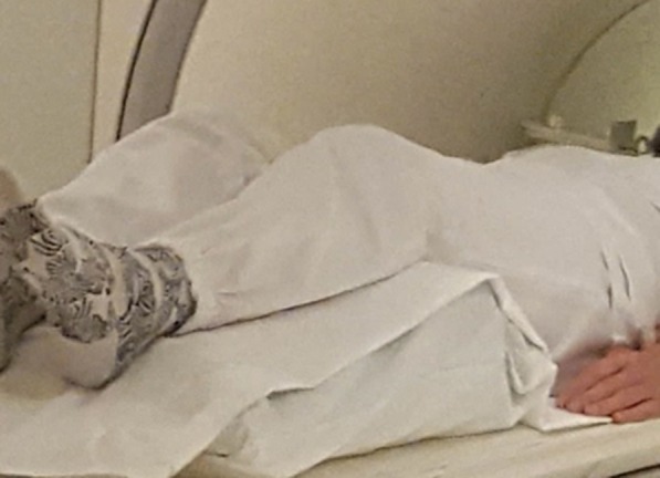
Supine lumbar MRI position with flexed hips and knees and a standard foam wedge beneath the knees.
Two radiologists with >10 years of experience in spine imaging independently evaluated findings at each disc level L3-S1 on a clinical Picture Archiving and Communication System unit using recommended criteria.11 For each position (straightened or flexed hips and knees), they evaluated disc contour (normal, bulge, herniation), the location of any disc herniation (central only, left side, right side, both sides) as well as its axial size (<1/3, 1/3–2/3, > 2/3 of the spinal canal, and largest size in millimetres). They further assessed nerve root affection in the recesses (no contact, in contact, displaced not compressed, compressed) and foramina (normal or no fat around the root, indicating root affection). They also rated image quality (5-point scale) and image evaluation difficulty (yes/no). Image quality was rated as 1 (non-diagnostic), 2 (severe blurring, restricted evaluation), 3 (slight blurring, restricted evaluation), 4 (slight blurring, unrestricted evaluation possible) or 5 (excellent, no artefacts). At each disc level, the radiologists measured the angle between the posterior borders of the upper and lower vertebrae and the angle between the upper and lower endplates.
The radiologists compared disc and nerve root findings between MRI with straightened vs flexed hips/knees by directly comparing the images, as recommended.12 In all cases of disagreement, the two radiologists negotiated a conclusive rating. For measurements, the conclusive value was the mean of both radiologists’ values. For each patient and disc level, we calculated differences in ratings/values between MRI with straightened vs flexed hips/knees. We used paired sample t tests on log-transformed data (which were normally distributed) to compare mean values between the two positions.
Results
Table 1 shows MRI findings and pain scores for the 14 patients (mean age 52 years, range 37–65 years; nine females). Data were complete for all variables in all patients. No patient in this study focussing on disc and nerve root findings had MRI findings indicating tumour, infection, spondylolysis or facet cysts. Figure 2 illustrates findings and measurements made in one of the patients.
Table 1.
MRI findings and pain scores in the study sample
| Patient no | Pain scores a | MRI disc findings (axial herniation size in flexed/straightened position) |
| 1 Female 45 years | 2, 2, 2 | Bulge L3/L4, L4/L5 and L5/S1 without nerve root contact |
| 2 Female 58 years | 4, 3, 6 | Herniation central L3/L4 (7.0/7.0 mm) in contact with right L4 root; bulge L4/L5 without nerve root contact |
| 3 Female 61 years | 3, 3, 4 | Bulge L3/L4 and L4/L5 without nerve root contact |
| 4 Female 65 years | 0, 0, 5 | Herniation central L3/L4 (3.2/3.2 mm) in contact with right L4 root; bulge L4/L5 compressing right and in contact with left L5 root; herniation central L5/S1 (2.4/2.8 mm) in contact with both S1 roots |
| 5 Female 52 years | 6, 7, 9 | Bulge L4/L5 compressing both L5 roots; herniation central L5/S1 (3.9/3.9 mm) without nerve root contact |
| 6 Female 37 years | 6, 7, 6 | Bulge L4/L5 in contact with both L5 nerve roots |
| 7 Female 52 years | 2, 2, 3 | Herniation central L4/L5 (3.6/3.6 mm) in contact with both L5 nerve roots; herniation central L5/S1 (4.8/4.8 mm) without root contact |
| 8 Male 42 years | 5, 2, 3 | Bulge L3/L4 in contact with right L4 root; bulge L4/L5 without nerve rot contact |
| 9 Male 58 years | 1, 1, 2 | Bulge L3/L4 in contact with both L4 roots; herniation L4/L5 right foramen (4.7/5.0 mm) displacing but not compressing right L5 root and in contact with left L5 root |
| 10 Male 50 years | 5, 1, 3 | Herniation right side (4.4/4.8 mm) L5/S1 in contact with right S1 root |
| 11 Male 59 years | 5, 5, 8 | Herniation central L4/L5 (2.8/3.2 mm) and L5/S1 (3.6/3.5 mm) without nerve root contact |
| 12 Female 60 years | 0, 4, 5 | Bulge L3/L4 and L4/L5 without nerve root contact, herniation right and left side L5/S1 (3.4/3.4 mm) without nerve root contact |
| 13 Female 48 years | 4, 4, 4 | Herniation central L4/L5 (3.8/3.8 mm) and central sequestrated L5/S1 (5.8/5.4 mm) without nerve root contact |
| 14 Male 47 years | 5, 5, 5 | Bulge L3/L4 in contact with both L4 roots; herniation left side L4/L5 (4.6/4.9 mm) in contact with left L5 root; herniation both sides L5/S1 (6.2/5.6 mm) compressing right and in contact with left S1 root |
Patients’ scores of their current pain on a scale from 0 (no pain) to 10 (worst pain possible) prior to MRI, after MRI with flexed hips/knees, and after MRI with straightened hips/knees, respectively
Figure 2.
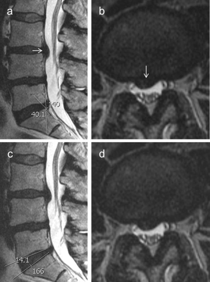
MRI findings in Patient No 2. A disc herniation at L3/L4 (arrows) in contact with the right L4 nerve root is unchanged between supine positions with flexed (a, b) and straightened (c, d) hips and knees. The figure also illustrates angles measured at each disc level between the posterior borders of the upper and lower vertebrae (a) and between the upper and lower endplates (c).
There was no difference in disc bulges (n = 14), disc herniation (n = 15), nerve root findings (n = 23, one dislocation, four compressions), or image evaluation difficulty (all “no”) between MRI with straightened vs flexed knees/hips. Image quality was rated 4 for one scan in the flexed position (due to slight blurring because of motion artefacts, not affecting image evaluation) and otherwise 5 (excellent).
Mean axial herniation size in straightened vs flexed position was 4.33 vs 4.28 mm (p = 0.29). No disc herniation differed more than 0.6 mm in axial size between the two positions. Mean score for current pain immediately prior to the MRI examination was 3.43. Mean pain score after MRI in straightened position vs after MRI in flexed position was 4.64 vs 3.29 (p = 0.005).
The mean sagittal angles (degrees) on MRI with straightened/flexed knees and hips were between the posterior vertebral body borders 7.9/7.3 at L3/L4, 13.1/12.8 at L4/L5 and 36.9/36.3 at L5/S1 and between the upper and lower end plates 6.1/5.9 at L3/L4, 6.8/6.8 at L4/L5 and 13.7/13.0 at L5/S1. No sagittal angle differed more than 3.8° between the two positions in any patient or level.
Discussion
In this study, supine lumbar MRI with straightened hips and knees revealed the same disc and nerve root findings as supine lumbar MRI with flexed hips and knees. Mean pain scores were higher in the straightened position. The results supported our hypothesis that a position with straightened hips/legs might provoke pain, but not that it might cause changed disc and nerve root findings.
The change in sagittal axes at the disc level (≤3.8 degrees) was likely too small to affect these findings, despite a mean change of only 5.7 degrees in lumbar lordosis L1/S1 led to increased degree of spondylolisthesis in the study by Daghighi et al.8 Alternatively, the studied disc and nerve root findings were too minor to show appreciable change. More marked findings might perhaps have changed. However, a small change in a marked MRI finding with a clear clinical relevance is unlikely to provide further clinically useful information. Results from a sample with marked findings would not help to clarify the clinical relevance of smaller borderline findings, which was our goal.
The reasons for increased pain in the position with straightened hips and legs are unclear. Increased lumbar sagittal angles (extension) may induce facet joint-related pain, but the very small increase in sagittal angles in this study was unlikely to affect the facet joints. It is a common clinical experience that patients with lumbar radicular pain may be more comfortable with a pillow beneath their knees when resting. This was not related to a decrease in morphological nerve root affection in our study.
Strengths of the study include prospective design, well-defined eligibility and evaluation criteria, and independent image evaluations by two experienced radiologists. The main limitation is small sample size. Furthermore, we did not randomize the order of the two positions (we prioritized first the clinically indicated MRI in flexed position), and some changes in pain during the scanning may not be due to changed position. We also did not investigate whether the type of pain changed, e.g. from radicular to facet type. Still, the lack of any clear differences in MRI findings in this pilot study does not motivate a further larger study on similarly selected patients imaged in the same two positions.
To provide potential clinically relevant findings, a further study might apply other eligibility criteria (e.g. diverging clinical and MRI findings on initial imaging) and other positions (e.g. supine position with straightened legs as well as a lumbar pillow to increase lumbar extension). For clinical practice, these preliminary results suggest that MRI with just straightened rather than flexed hips/knees may provoke pain without adding much to the diagnosis of disc herniation and nerve root compromise. The results are reassuring for maintaining usual practice of imaging with flexed hips and knees.
Conclusion
This pilot study (n = 14) revealed similar disc and nerve root findings regardless of whether patients underwent supine lumbar MRI with straightened or with flexed hips and knees. The straightened position appeared more painful.
Contributor Information
Ansgar Espeland, Email: ansgar.espeland@helse-bergen.no.
Nina Dalen, Email: .
REFERENCES
- 1. Lao LF , Zhong GB , Li QY , Liu ZD , Lao LF . Kinetic magnetic resonance imaging analysis of spinal degeneration: a systematic review . Orthop Surg 2014. ; 6 : 294 – 9 . doi: 10.1111/os.12137 [DOI] [PMC free article] [PubMed] [Google Scholar]
- 2. Zamani AA , Moriarty T , Hsu L , Winalski CS , Schaffer JL , Isbister H , et al. . Functional MRI of the lumbar spine in erect position in a superconducting open-configuration Mr system: preliminary results . J Magn Reson Imaging 1998. ; 8 : 1329 – 33 . doi: 10.1002/jmri.1880080622 [DOI] [PubMed] [Google Scholar]
- 3. Tarantino U , Fanucci E , Iundusi R , Celi M , Altobelli S , Gasbarra E , et al. . Lumbar spine MRI in upright position for diagnosing acute and chronic low back pain: statistical analysis of morphological changes . J Orthopaed Traumatol 2013. ; 14 : 15 – 22 . doi: 10.1007/s10195-012-0213-z [DOI] [PMC free article] [PubMed] [Google Scholar]
- 4. Weishaupt D , Schmid MR , Zanetti M , Boos N , Romanowski B , Kissling RO , et al. . Positional MR imaging of the lumbar spine: does it demonstrate nerve root compromise not visible at conventional MR imaging? Radiol 2000. ; 215 : 247 – 53 . doi: 10.1148/radiology.215.1.r00ap06247 [DOI] [PubMed] [Google Scholar]
- 5. Zou J , Yang H , Miyazaki M , Wei F , Hong SW , Yoon SH , et al. . Missed lumbar disc herniations diagnosed with kinetic magnetic resonance imaging . Spine 2008. ; 33 : E140 – E144 . doi: 10.1097/BRS.0b013e3181657f7e [DOI] [PubMed] [Google Scholar]
- 6. Madsen R , Jensen TS , Pope M , Sørensen JS , Bendix T . The effect of body position and axial load on spinal canal morphology: an MRI study of central spinal stenosis . Spine 2008. ; 33 : 61 – 7 . doi: 10.1097/BRS.0b013e31815e395f [DOI] [PubMed] [Google Scholar]
- 7. Niggemann P , Kuchta J , Hoeffer J , Grosskurth D , Beyer HK , Delank KS . Juxtafacet cysts of the lumbar spine: a positional MRI study . Skeletal Radiol 2012. ; 41 : 313 – 20 . doi: 10.1007/s00256-011-1186-3 [DOI] [PubMed] [Google Scholar]
- 8. Daghighi MH , Poureisa M , Arablou F , Fouladi DF . Supine spinal magnetic resonance imaging with straightened lower extremities in spondylolisthesis: a comparison with the conventional technique . Eur J Radiol 2015. ; 84 : 921 – 6 . doi: 10.1016/j.ejrad.2015.01.023 [DOI] [PubMed] [Google Scholar]
- 9. Parent EC , Videman T , Battié MC . The effect of lumbar flexion and extension on disc contour abnormality measured quantitatively on magnetic resonance imaging . Spine 2006. ; 31 : 2836 – 42 . doi: 10.1097/01.brs.0000245834.30646.aa [DOI] [PubMed] [Google Scholar]
- 10. Hansen BB , Hansen P , Grindsted J , Rasti Z , Bliddal H , Riis RGC , et al. . Conventional supine MRI with a lumbar Pillow-An alternative to weight-bearing MRI for diagnosing spinal stenosis?: a cross-sectional study . Spine 2017. ; 42 : 662 – 9 . doi: 10.1097/BRS.0000000000001889 [DOI] [PubMed] [Google Scholar]
- 11. Fardon DF , Williams AL , Dohring EJ , Murtagh FR , Gabriel Rothman SL , Sze GK . Lumbar disc nomenclature: version 2.0: recommendations of the combined task forces of the North American spine Society, the American Society of spine radiology, and the American Society of Neuroradiology . Spine 2014. ; 39 : E1448 – 65 . doi: 10.1097/BRS.0b013e3182a8866d [DOI] [PubMed] [Google Scholar]
- 12. Berg L , Gjertsen O , Hellum C , Neckelmann G , Johnsen LG , Eide GE , et al. . Reliability of change in lumbar MRI findings over time in patients with and without disc prosthesis--comparing two different image evaluation methods . Skeletal Radiol 2012. ; 41 : 1547 – 57 . doi: 10.1007/s00256-012-1394-5 [DOI] [PubMed] [Google Scholar]