Abstract
Background
Due to increasing popularity in Sweden during the last decade, alpacas are frequently encountered by practising veterinarians and pathologists. Knowledge regarding their health and diseases under Swedish conditions is, however, limited.
Objectives
To improve knowledge about the health of alpacas in Sweden by collecting information on diseases and health status.
Design
A retrospective study was made of 93 necropsies conducted on alpacas in Sweden during the period 2001–2013.
Setting
Data were obtained from the two major veterinary pathology centres in Sweden. The alpacas were hobby or farm animals and they were submitted by veterinarians in local practices or at a national animal healthcare organisation.
Results
The digestive system was most frequently affected (29 per cent), with parasitic gastroenteritis (17 per cent) and hepatic disease being especially prevalent (15 per cent fascioliasis and 7 per cent hepatitis). Cardiovascular conditions (9 per cent), systemic diseases (7 per cent) and perinatal deaths were also common, including abortions (10 per cent) and fatal septicaemia (4 per cent). Wasting/emaciation was a frequent finding (26 per cent). Other diagnoses included dermatitis (8 per cent), diseases of the central nervous system (8 per cent), traumatic injuries (7 per cent), neoplasia (5 per cent), pneumonia (5 per cent) and nephritis (3 per cent).
Conclusions
This study identified areas of concern regarding diagnostic and pathological procedures, for which specific measures have been recommended. One particular cause for concern was the number of deaths from emaciation in weanling alpacas during late winter or early spring. For adult alpacas, infectious and non-infectious causes of death were approximately equally frequent. Many of the diseases were considered clinically acute but pathology often showed them to be chronic conditions that had eventually deteriorated and presented as acute cases in the late stages. This study revealed similarities in the health/disease status reported in other European countries and in North America. The results can be used by alpaca keepers and veterinary practitioners to improve management, diagnosis and treatment of alpacas.
Keywords: alpacas, camelids, diseases, pathology
Introduction
Importation of South American camelids, especially alpacas (Vicugna pacos), into Sweden started during the 1990s. Since then their popularity has grown steadily and interest for the alpaca especially has grown during the last decade. Alpacas are mainly kept for their fine quality fibre, but also as companion animals and tourist attractions.1 One advantage of these animals compared with cattle is that they can graze marginal land without damaging the soil structure due to their relatively low bodyweight and the unusual structure of their digits. In spring 2013, the Swedish Alpaca Association estimated the number of alpacas in Sweden to be between 1500 and 2000 individuals. Most herds consist of about 10 animals but there are larger herds of up to 100 animals.1 Since 2009, a part of the Swedish Animal Healthcare Programme (Farm and Animal Health) has been dedicated to camelids and offers several services including veterinary consulting, test for endoparasites and free necropsies (http://www.gardochdjurhalsan.se/sv/andra-djurslag/kameldjur/om-gard-djurhalsan-kameldjur/, accessed May 18, 2017). It has 111 member farms and currently cares for an estimated 1200 alpacas (K de Verdier, personal communication, February 2017).
Alpacas are kept in an extensive grazing system at pasture on the slopes of the Andes, that is, at 4300 m above sea level, where many of the indigenous people depend on alpaca husbandry for their livelihood. At these altitudes, maximum and minimum daytime temperatures do not vary much throughout the year. As a guide, climate data for Juliaca airport (Peru) at an elevation of 3825 m, between 1961 and 1990, show the maximum daytime temperatures to be 16°C–18°C and the minimum night-time temperatures to be −7.5°C to 3.5°C. The wet season occurs between December and March (monthly rainfall 85–135 mm) and the dry season from June to August (monthly rainfall 3.1–5.8 mm) (http://www.nationalparks-worldwide.info/sam/peru/regions/andes/andes-climate.html, accessed November 19, 2017). The pasture is meagre (scarce in quality or quantity) during the dry season and consists mainly of the harsh Peruvian feather grass called ‘Ichu’ which is endemic to the area. No extra feed or mineral supplements are usually provided. Water is available from glacier-fed springs or from precipitation. Alpacas are often kept together with llamas and may be herded together in small, open enclosures during the night for protection, being allowed to range freely on the pasture during the day. There are also some alpaca herds in South America that are managed more intensively, including improved nutrition and genetic selection programmes, with a view to improving fibre quality.2
In contrast, alpacas in Sweden are kept on lowland pasture at lower altitudes than their counterparts in the Andes. They have access to mineral blocks and a continuous supply of water; supplementary feeding and shelter is provided during cold weather. The climate varies throughout the country, but in Uppsala (Sweden) maximum daytime temperatures during the summer are around 20°C and minimum night-time temperatures are around 12°C. Corresponding temperatures in winter are 0°C and −5°C. Monthly precipitation varies from 25 mm (February) to 78 mm (August) (https://weather-and-climate.com/average-monthly-Rainfall-Temperature-Sunshine, Uppsala, Sweden, accessed November 19, 2017).
The environment in Scandinavia is new to these animals, which poses several challenges in terms of infectious diseases, nutrition and adaptation to the climate. In Sweden, camelids are kept outdoors in smaller pastures than in their native habitat in the Andes. Often the same pastures are used all year round, which increases the parasite burden. During cold winters they may be kept indoors, which predisposes them to infections and parasitism due to closer contact between animals and to manure. Owners and veterinarians may be unfamiliar with the health issues of these animals, with their behaviour when sick, or with the most effective treatments. A postal survey conducted among Swedish alpaca owners in 2008 made this very clear1; only 10 per cent were satisfied with the help they received and 50 per cent thought veterinarians lacked sufficient knowledge about alpacas. Such a response shows the need for veterinary education, which in turn requires scientifically based facts about these animals’ needs and diseases in the environment where they are kept. Lack of knowledge precludes the development of effective treatment and preventive strategies.
The aim of this study was to identify diseases and causes of death in alpacas in Sweden from necropsy data during a 13-year period. The intention was to provide a knowledge base for Swedish veterinarians and to compare Swedish data with available information from other European countries and from North America. This information is vital for the provision of adequate veterinary care for the growing population of alpacas in Sweden and should serve as a basis for improving alpaca welfare in Sweden.
Materials and methods
Information was assembled from reports of 93 necropsies conducted between February 2001 and October 2013 at the two major pathology centres in Sweden, the National Veterinary Institute (SVA) in Uppsala and Eurofins (formerly AnalyCen) in Kristianstad and Skara. The animals were a mixture of hobby and farm animals, submitted by veterinarians in local practices or at a national animal healthcare organisation. One alpaca was born in Germany, one in Chile and all others in Sweden. Information from the remittance report for each necropsy and archived information (mail correspondence, letters from the owners, etc) were also collected. The information retrieved included: age, sex, body condition, means of death (euthanased or found dead), history, main clinical signs, clinical diagnoses, treatments, pathology diagnosis, macroscopic and histopathological findings, and results of laboratory tests performed. Results are reported as frequency (n) and per cent (%).
Results
Age and sex distribution
A total of 93 reports came from the two pathology centres: 50 from SVA and 43 from Eurofins. Both the alpaca population and the number of necropsies increased during the 13-year period (figure 1). The animals originated from most parts of Sweden, from both large and small herds. The largest age group comprised individuals between one and five years of age (n=30; 32 per cent), followed by newborns up to five months (n=17; 18 per cent) (table 1). The majority of all necropsy cases (67 per cent) were female but the proportion varied between age groups (figure 2). Twenty-eight per cent were males and in 5 per cent of cases, the sex was not specified.
Figure 1.
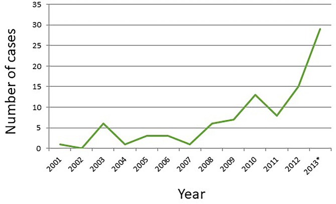
Alpaca necropsy cases at National Veterinary Institute (SVA) and Eurofins between February 2001 and October 2013, completed by year.
Table 1.
Alpaca necropsy cases reported by age for 93 cases
| Category | n | % |
| Age | ||
| Prenatal | 10 | 11 |
| Crias/calves | ||
| 0–24 hours | 5 | 5 |
| 1–10 days | 3 | 3 |
| 2–3 weeks | 3 | 3 |
| 1–4 months | 6 | 6 |
| Juveniles | ||
| 5–12 months | 11 | 12 |
| Adults | ||
| 1–5 years | 30 | 32 |
| 6–9 years | 13 | 14 |
| ≥10 years | 8 | 9 |
| Unknown adult age | 4 | 4 |
| Total | 93 | 100 |
Figure 2.
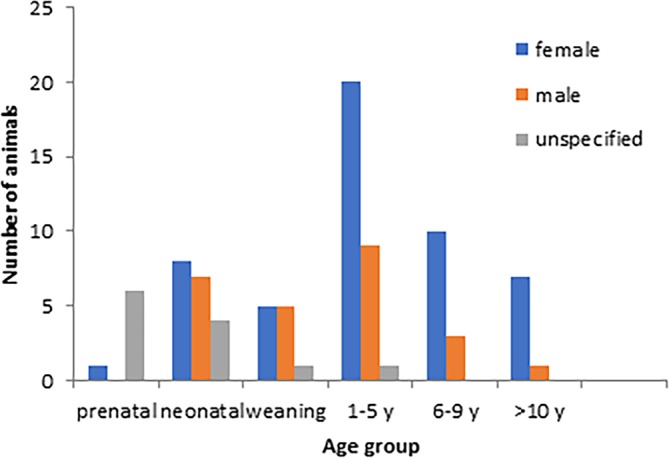
Sex distribution among alpacas in different age groups.
Clinical signs of disease
Wasting (n=24, 26 per cent), sudden death (n=16, 17 per cent), weakness (n=14, 15 per cent) and inappetence (n=10, 11 per cent) were the most commonly reported clinical signs (table 2). Emaciation was most frequent in weaning animals and animals aged one to five years, whereas there was a tendency for neonatal animals to be found dead without previous signs of illness.
Table 2.
A total of 113 clinical signs were reported for 93 alpacas by age group
| Neonatal | Weaning | 1–5 years | 6–9 years | ≥10 years | Unspecified adults | Total | |
| Fever | 1 | – | 1 | 1 | – | – | 3 |
| Swollen lymph nodes | – | – | – | – | 1 | – | 1 |
| Inappetence | – | 1 | 3 | 1 | 4 | 1 | 10 |
| Emaciation | 2 | 7 | 9 | 2 | 3 | 1 | 24 |
| Weakness | 5 | 1 | 5 | 1 | 1 | 1 | 14 |
| Diarrhoea | 1 | – | 1 | – | – | – | 2 |
| Colic | – | 1 | 2 | 1 | – | – | 4 |
| Recumbency | – | 1 | – | 1 | 1 | – | 3 |
| Collapse | – | – | – | 1 | – | – | 1 |
| Anaemia | – | – | 1 | – | – | – | 1 |
| Respiratory | 2 | – | 1 | 2 | – | – | 5 |
| Dyspnoea | |||||||
| Coughing | 1 | – | 1 | – | – | – | 2 |
| Unspecified | – | – | 2 | – | 1 | – | 3 |
| Neurological | |||||||
| Staggering | 2 | – | – | – | – | – | 2 |
| Seizures | – | – | – | – | – | 1 | 1 |
| Paresis | – | – | 2 | 0 | – | – | 2 |
| Blindness | – | – | 2 | – | – | – | 2 |
| Behavioural changes | – | 2 | 1 | – | – | – | 3 |
| Unspecified | 0 | – | 2 | – | – | – | 2 |
| Anuria | – | 1 | – | – | – | – | 1 |
| Lameness | – | 1 | 2 | – | – | – | 3 |
| Heart murmur | – | – | 1 | – | 1 | – | 2 |
| Dermatitis | – | – | 4 | 1 | 1 | – | 6 |
| Sudden death | 8 | – | 3 | 4 | – | 1 | 16 |
| Totals | 22 | 15 | 43 | 15 | 13 | 5 | 113 |
More than one clinical sign was reported in some animals.
Diagnoses premortem and recorded treatment
In 23.6 per cent (n=22) of cases, a premortem diagnosis was provided such as colic, dermatitis, pneumonia, malformation, fracture, lameness and nerve damage. Thirty-three alpacas (35 per cent) had received treatment for their illness before death. The most common treatment was antibiotics (n=17; 52 per cent) (penicillin, streptomycin, Engemycin [oxytetracycline], clindamycin, sulphadoxine or trimethoprim), followed by B vitamins (n=13; 39 per cent), non-steroidal anti-inflammatory drugs (n=9; 27 per cent), selenium (n=3; 9 per cent), glucose (n=3; 9 per cent), cortisone (n=2; 6 per cent), analgesics (n=2; 6 per cent), vitamins A and D (n=2; 6 per cent), coccidiostats (n=2: 6 per cent) and anthelmintics (n=2; 6 per cent). More than one treatment may have been administered to an individual animal.
Cause of death and main pathologies
Excluding perinatal deaths (n=5), the majority of the alpacas (n=54) were found dead; in one, the mode of death was not reported and the remainder were euthanased (n=23). The causes of euthanasia of 20 alpacas are shown in the online supplementary table. The causes of euthanasia in each of three other animals were: (1) emaciation due to a tooth abscess and mandibular osteomyelitis, (2) dyspnoea related to malformation of the nasal cavity and (3) chronic mastitis and sepsis. The most common pathologies affected primarily the digestive system (n=27; 29 per cent) and the circulatory system (n=8; 9 per cent). Infectious diseases were the major cause of disease and death among alpacas up to four months of age. These included one case of enteritis caused by Escherichia coli, a systemic infection by Streptococcus species and four cases of septicaemia with suppurative processes in the brain with one of these also in the lungs (aetiology not identified). Non-infectious problems dominated among the weanling animals, which showed progressive loss of body condition resulting in emaciation during the late winter and early spring.
Pathologies of specific organ systems and analyses
The online supplementary table details the pathology findings and their relation to clinical observations in a selection of cases. In many cases, more than one pathology was described in the same animal. In 10 (11 per cent) alpacas no lesions were observed; eight of these were aborted, one was a cria born weak, which died two hours after birth, and the other one was another cria found dead in the field, its mother could not be identified. Histopathology was conducted in 74 (80 per cent) alpacas.
vetreco-2017-000239supp001.pdf (41.5KB, pdf)
For the diseases affecting the digestive system, the majority were chronic (23/27; 86 per cent), showing no signs of imminent fatal disease until their chronic conditions suddenly became acute shortly before death. The majority of enteritis cases among adults were attributed to parasitic infestation (7/9; 78 per cent). Intestinal parasites were incidental findings in more than 1 out of 10 alpacas submitted for necropsy and included: Eimeria species (n=4; 4 per cent), Nematodirus species (n=2; 2 per cent), Trichostrongyloidea (2; 2 per cet), Trichuris species (1; 1 per cent), Ostertagia species (1; 1 per cent) and Haemonchus species (n=1; 1 per cent). Eimeria species (E macusaniensis, E punoensis and E alpacae) were commonly found together with nematodes. Other frequent diagnoses in the digestive system included hepatitis or cholangiohepatitis with some cases associated with liver fluke (Dicrocoelium dendriticum, Fasciola hepatica), hepatic lipidosis, gastric ulcers or acute mechanical gastrointestinal conditions.
Among the cardiac disease cases (n=8; 9 per cent), three (3 per cent) had a history of treatment but in none of these was cardiac disease mentioned as a differential diagnosis. The cardiac diseases included acute or chronic myocarditis or endocarditis (n=4), cardiomyopathy (n=2), myocardial degeneration (n=1) and cardiac malformation (n=1). Sarcocystis species in the myocardium was an incidental finding in three alpacas.
Four alpacas (4 per cent) were diagnosed with pneumonia. In two of these (2 per cent) an infectious cause was identified. One (1 per cent) had a suppurative/necrotising bronchopneumonia caused by Corynebacterium pseudotuberculosis. The other was found dead at pasture and had a Pasteurella species infection.
Diseases of the urinary system included three animals with interstitial nephritis (3 per cent). In one of them, a nine-month-old male, a 3 mm calcium carbonate urolith was found obstructing a ureter. The obstruction had caused dilation of the renal pelvis, chronic cystitis, interstitial nephritis and anuria.
Ten (11 per cent) of the carcasses were aborted fetuses, the majority from full-term or close to full-term pregnancy. One abortion was due to necrotising placentitis only visibly histologically, and one to C pseudotuberculosis abscesses in the gravid uterine horn. None of the other fetuses had signs of trauma, infection, inflammation, malformation or other diagnostic findings. Tests for Schmallenberg virus (n=3), Brucella species (n=6), Toxoplasma gondii (n=2), Listeria species (n=1), Sarcocystis species (n=1), Neospora species (n=1), Coxiella burnetii (n=3), equine herpesvirus type 1 (n=1) and general bacteriological culture (n=2) were negative.
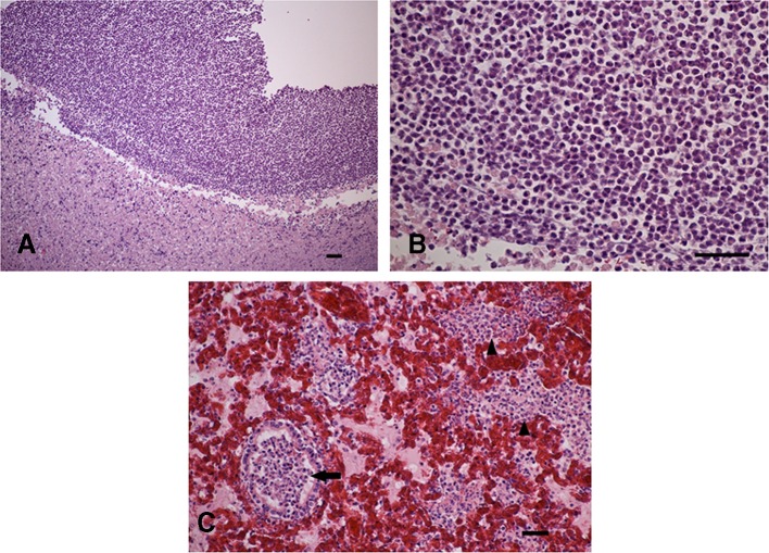
Diseases affecting the CNS included a case in which the submitting veterinarian had suspected listeriosis or polioencephalomalacia and pathology revealed cerebrocortical necrosis typical for polioencephalomalacia caused by thiamine deficiency in ruminants. Other diseases involving the CNS included four cases of suppurative processes in the brain of neonatal alpacas (one of these had also purulent pneumonia, figure 3A–C) and two cases of non-suppurative encephalitis. One of these, a two-year-old female, showed progressively worsening general condition and blindness, histologically, perivascular infiltrations of mononuclear cells and deposits of amorphous, eosinophilic, hyaline material around pathogenic organisms (Splendore-Hoeppli phenomenon) (figure 4), a suspected immune-related process with a previous infection was observed but the agent was not identified.
Figure 3.
Histological sections from a one-month-old alpaca with brain abscess and purulent pneumonia. (A) Brain abscess, delimited area of purulent necrosis of brain substance, (B) abundance of neutrophils, (C) purulent pneumonia, exudate with abundant neutrophils in lumen of bronchiole (arrow), alveoli filled with neutrophilic exudate (arrowheads), hyperaemia. Haematoxylin and eosin (H&E) stained, paraffin embedded section. Size bar=100 µm.
Figure 4.
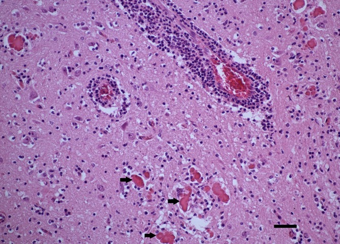
Histological section of brain of two-year-old alpaca with non-purulent encephalitis. Perivascular cuff, deposits of eosinophilic hyaline material consistent with Splendore-Hoeppli material (arrows). Haematoxylin and eosin (H&E) stained, paraffin embedded section. Size bar=100 µm.
Seven cases (8 per cent) involved dermatitis, four of which had dermatitis as the sole pathology. Chorioptic mange was an incidental finding in two cases.
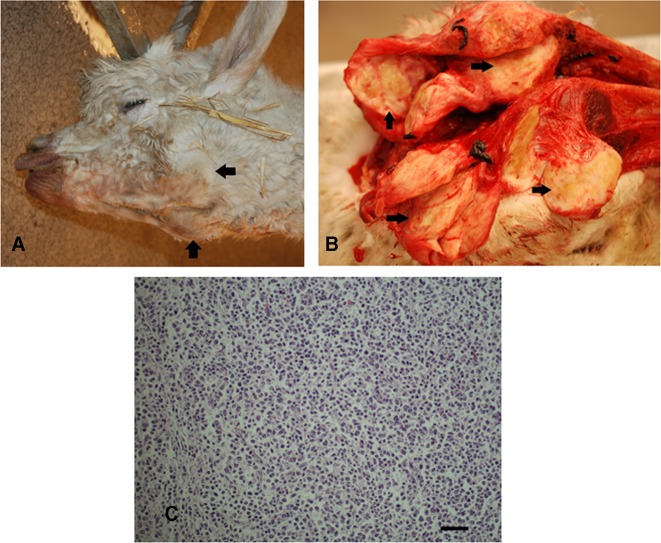
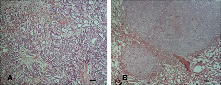
Neoplasias were diagnosed in five animals (5 per cent), four alpacas of wide age range (from 1.5 to 10 years of age) had malignant lymphomas, one case is illustrated in figure 5A–C. A 14-year-old female alpaca had cholangiocellular carcinoma (figure 6A) with metastasis in the lungs (figure 6B) and lymph nodes.
Figure 5.
Multicentric malignant lymphoma in a 10-year-old alpaca. (A) Enlarged mandibular head lymph nodes (arrow), (B) cut surface of enlarged head lymph nodes (arrows), replacement of normal lymph node architecture by homogenous pale, firm tissue, (C) histopathology of malignant lymphoma, neoplastic proliferation of lymphocytes, characterised by densely packed round to slightly elongated, pleomorphic lymphocytes in a fine fibrous stroma, relatively high mitotic activity. Haematoxylin and eosin (H&E) stained, paraffin embedded section. Size bar=100 µm. (Photographs by Katinka Belak [A, B] and Dolores Gavier-Widén [C]).
Figure 6.
Histological photographs of cholangiocarcinoma in a 14-year-old alpaca. (A) Cholangiocarcinoma carcinoma in the liver, ductules and acini formed by cuboidal or columnar epithelial carcinomatous cells, (B) lung exhibiting metastasis of cholangiocarcinoma. Haematoxylin and eosin (H&E) stained, paraffin embedded section. Size bar=100 µm.
Some of the identified traumatic injuries were caused by predation by wolves, fighting wounds and slipping on ice.
In 24 cases (25.8 per cent), from nine farms, the major finding was wasting. A further 24 alpacas (25.8 per cent) were classified as being in poor bodily condition. Wasting was particularly frequent in weanling alpacas, 5–12 months of age (7/11; 63.6 per cent). Both sexes and all age groups were affected. Poor body condition/emaciation was often present in association with diseases, such as enteritis, malignant lymphoma, gastric ulcer, multiple abscesses, mandibular osteomyelitis and cholangiohepatitis. In two six-month-old alpacas, emaciation was the only diagnosis. Parasites were identified in five emaciated alpacas: Eimeria species (E punoensis and E alpacae), Ostertagia ostertagi, Camelostrongylus mentulatus, Trichuris species and Nematodirus species.
Bacteriological examination was conducted in 37 (39.8 per cent) alpacas. Tests for surveillance of infections were all negative: tuberculosis (n=7), paratuberculosis (n=7), brucellosis (n=8), bluetongue (n=1).
As a result of our findings, we have been able to formulate some guidelines for personnel involved in the care of alpacas (table 3).
Table 3.
Advice on health management and diagnostics for alpaca owners, practising veterinarians and pathologists based on our results
| Advice for alpaca owners |
|
| Advice for practising veterinarians |
|
| Advice for pathologists |
|
Discussion
The purpose of this study was to determine the most common causes of pathology leading to diseases or death in Swedish alpacas. The camelid health programme within Farm and Animal Health in Sweden largely contributed to the increased number of necropsies since its implementation in 2009.
Several difficulties were identified while processing the information in the reports; for example, the interpretation of the referring veterinarian’s thoughts on a diagnosis based on clinical findings and blood analyses. Defining chronically versus acutely diseased animals caused some difficulties. A large proportion of the cases presented clinically as acute (54 per cent) but were later identified as suffering from chronic disease that had become acute, for example, urolithiasis, malnutrition and chronic myocarditis, enteritis, nephritis and hepatitis or cholangiohepatitis. Thus, the majority only showed signs of disease for a day or a few days before they died.
In reports on camelid health, neonatal deaths, intestinal diseases, parasitism, nutritional disorders and skin diseases are considered to be the major concerns around the world. The necropsy findings in this study correspond relatively well to those in camelids previously reported from Canada,3 England and Wales,4 and Germany.5 Hepatic lipidosis and/or malnutrition were frequent in severely ill, inappetent or wasting Swedish alpacas of all ages. Increased use of simple diagnostic tests or a blood analysis could, in many cases, have given valuable information and guided treatment and prognosis. The clinical development of hepatic lipidosis, progressing to inappetence, should not be underestimated.6
Emaciation as the cause of death was seen in 9 per cent of camelid deaths in a report from Germany5 but was not reported from Canada.3 In the present study, wasting was the major presenting sign, occurring in 25.8 per cent (n=24) of the alpacas. Similarly, in a recent review from England and Wales,4 wasting (observed in 20.3 per cent of the submission) and diarrhoea (observed in 18.4 per cent of the submissions) causing malabsorption were the main clinical histories reported in the laboratory submissions. Differences in the incidence of wasting between studies may be due to several factors, for example, if it is reported as a clinical sign or as a necropsy finding. Also, wasting may be related to gastrointestinal disease, inappetence related to other diseases or other disturbances. Thus, direct comparisons between studies cannot be made.
Abortions were often of undiagnosed cause (undiagnosed in eight alpacas, 80 per cent); similarly, another study reported that infections were not found in 20 of the 21 abortion submissions and a clear cause for the abortion was not stated.3 Systemic bacterial infections and malformations were common causes of death in crias during their first week of life. In another report, bacterial infection was usually related to omphalitis,3 however, omphalitis was not diagnosed in our study. Digestive system disease (eg, enteritis, liver disease and gastric ulceration) was frequent in adult alpacas, and was also the most common diagnosis in other reports.3
Dermatitis was diagnosed in 7.5 per cent (n=7) of the cases; four alpacas were euthanased due to chronic dermatitis although the causative agent was not identified. In a study on South American camelids in the UK, skin disease was reported in 9.1 per cent of the surveyed population; the frequency was highest in alpacas. There was a broad range of skin lesions and 15 different diagnoses had been made by the veterinarians: ectoparasite infestation and zinc deficiency were the most frequent presumptive causes.7 In the present study, in only two cases was a cause (chorioptic mange) identified. While the zinc status was not known in the alpacas in our study, it is recommended that this parameter should be monitored.
Sarcocystis species were found in four (4.3 per cent) of the alpacas. The life cycle of Sarcocystis involves a definitive host (often a predator) and an intermediate host (a prey, often herbivores). Since the alpacas are sometimes kept in habitats inhabited by wolves and wolf predation occurs, here reported in six (6.4 per cent) of the alpacas, it could be speculated that wolves act as definitive host for Sarcocystis species in this environment. However, the definitive host species of Sarcocystis species in Sweden has not been identified.
Several animals died due to illnesses that could not be treated with antibiotics and anti-inflammatory drugs, for example, severe, chronic cases of nephritis. These animals must be identified by the field practitioner and euthanased if surgery or intensive care at an animal hospital is not an option. Many of the alpacas sent in to necropsy had died suddenly after only a few days of illness according to the clinicians. However, pathology investigations revealed chronic conditions in many of these cases. It is therefore important that veterinary practitioners keep in mind the stoic nature of these animals and act appropriately as an alpaca with signs of illness should not be left untreated. Improved diagnostic procedures that can be used in the field will likely play a major part in the effort to improve animal welfare for sick alpacas. In a postal survey of camelid owners performed in the UK in 1992–1993, a third of the deaths were reported to be of unknown cause and a diagnosis had been made by a veterinarian in less than half of the deceased camelids.8 In the present study the numbers were even less, an antemortem diagnosis was made in in 22 (23.6 per cent) of the cases. The fact that the diagnoses found in the referrals were vague and often based only on clinical signs explains the inconsistency observed between the clinical diagnoses and the pathology diagnoses. This also reinforces the need for improved diagnostic procedures in the field.
The results of this study have profound implications both for alpaca owners and for practising veterinarians who may be tasked with treating alpacas in the field. Since many of the animals in this study were relatively young, the largest group being between one and five years of age, euthanasia is not an easy decision to take unless the veterinarian is certain the condition either has a bad prognosis or is too severe to treat from an animal welfare aspect. There were animals in this study with long periods of wasting (sometimes leading to emaciation and death), inappetence or even such severe signs as hindlimb paresis, which had not been examined by a veterinarian. Information needs to be provided to camelid owners regarding the need for veterinary assessment in order to facilitate early diagnosis and treatment. Thus, owner education is also important to improve animal welfare.
In conclusion, the most common causes of diseases leading to death in Swedish alpacas were identified from necropsy reports. It is anticipated that the information collated in this report will provide valuable insight into the prevalence of subclinical conditions affecting the overall health of alpacas in Sweden, such as endoparasitism and ectoparasitism, metabolic imbalances and nutritional deficiencies. This information is valuable for owners for husbandry purposes and for veterinarians, both to provide prophylaxis for better herd health and as an aid to diagnosing and treating clinically sick alpacas.
Footnotes
Contributors: CB summarised and analysed the data. RB, JMM and KdV designed the study, supervised the data compilation, contributed with interpretation of results and drafted the manuscript. LNH, NK, KBel and KBer acquired the data by conducting the pathology work. DG-W supervised the pathology work, contributed to the design and to the writing of the manuscript, took the histological photographs and is the corresponding author.
Funding: The authors have not declared a specific grant for this research from any funding agency in the public, commercial or not-for-profit sectors.
Competing interests: None declared.
Provenance and peer review: Not commissioned; externally peer reviewed.
Data sharing statement: No additional data are available.
References
- 1. Verdier, K DE, Bornstein S. Questionnaire study 2008: Alpacas in Sweden – a new challenge. Svensk veterinärtidning 2010;62:19–23. [Google Scholar]
- 2. Postigo JC, Young KR, Crews KA. Change and Continuity in a Pastoralist Community in the High Peruvian Andes. Hum Ecol 2008;36:535–51. 10.1007/s10745-008-9186-1 [DOI] [Google Scholar]
- 3. Shapiro JL, Watson P, McEwen B, et al. . Highlights of camelid diagnoses from necropsy submissions to the Animal Health Laboratory, University of Guelph, from 1998 to 2004. Can Vet J 2005;46:317–8. [PMC free article] [PubMed] [Google Scholar]
- 4. Twomey DF, Wu G, Nicholson R, et al. . Review of laboratory submissions from New World camelids in England and Wales (2000-2011). Vet J 2014;200:51–9. 10.1016/j.tvjl.2014.01.021 [DOI] [PubMed] [Google Scholar]
- 5. Gunsser I, Kiesling C. Breeding and/or handling problems? Causes of death in South American camelids. South American Camelids Research: Proceedings of the 4th European Symposium on South American Camelids and DECAMA European Seminar, Göttingen, 7-9 October 2004, M. GERKEN and C. REINIERI: Wageningen Academic Publishers, 2006. [Google Scholar]
- 6. Tornquist SJ, Van Saun RJ, Smith BB, et al. . Hepatic lipidosis in llamas and alpacas: 31 cases (1991-1997). J Am Vet Med Assoc 1999;214:1368–72. [PubMed] [Google Scholar]
- 7. D’Alterio GL, Knowles TG, Eknaes EI, et al. . Postal survey of the population of South American camelids in the United Kingdom in 2000/01. Vet Rec 2006;158:86–90. 10.1136/vr.158.3.86 [DOI] [PubMed] [Google Scholar]
- 8. Davis R, Keeble E, Wright A, et al. . South American camelids in the United Kingdom: population statistics, mortality rates and causes of death. Vet Rec 1998;142:162–6. 10.1136/vr.142.7.162 [DOI] [PubMed] [Google Scholar]
Associated Data
This section collects any data citations, data availability statements, or supplementary materials included in this article.
Supplementary Materials
vetreco-2017-000239supp001.pdf (41.5KB, pdf)


