Abstract
Purpose
This study aims to investigate the feasibility of robot-assisted simulated strabismus surgery using the new da Vinci Xi Surgical System and to report what we believe is the first use of a surgical robot in experimental eye muscle surgery.
Methods
Robot-assisted strabismus surgeries were performed on a strabismus eye model using the robotic da Vinci Xi Surgical System. On the lateral rectus of each eye, we performed a procedure including, successively, a 4-mm plication followed by a 4-mm recession of the muscle to end with a 4-mm resection. Operative time from conjunctival opening to closing and successful completion of the different steps with or without complications or unexpected events were assessed.
Results
Robot-assisted strabismus procedures were successfully performed on six eyes. The feasibility of robot-assisted simulated strabismus surgery is confirmed. The da Vinci Xi system provided the appropriate dexterity and operative field visualization necessary to perform conjunctival and Tenon's capsule opening and closing, muscle identification, suturing, desinsertion, sectioning, and resuturing. The mean duration to complete the whole procedure was 27 minutes (range, 22–35). There were no complications or unexpected intraoperative events.
Conclusions
Experimental robot-assisted strabismus surgery is technically feasible using the new robotic da Vinci Xi Surgical System. This is, to our knowledge, the first use of a surgical robot in ocular muscle surgery.
Translational Relevance
Further experimentation will allow the advantages of robot-assisted microsurgery to be identified while underlining the improvements and innovations necessary for clinical use.
Keywords: da Vinci, muscle, robot, strabismus, surgery
Introduction
Strabismus is a common ophthalmologic disorder affecting 3% of preschool children1 and approximately 4% of adults.2,3 Although strabismus surgery is still most commonly performed on children, Astle et al.4 recently reported a significant increase in the number of strabismus operations performed on adults. Conversely, the number of ophthalmic surgeons competent in this field is decreasing.5 Thus, waiting time and access to surgery can be a problem. Horizontal rectus muscle surgery has many indications and is the most common strabismus surgery performed.3 Procedures include recession, resection, plication, tenotomy, transposition, and adjustable suture surgery.
Robot-assisted surgery has expanded over the last 20 years in macrosurgical specialties (urological, general, digestive, gynecological, cardiovascular), but microsurgical specialties, including ophthalmology, remain largely ignored. There are several reasons for this discrepancy, including the following: (1) the current rhythm of eye surgeons, which could be qualified as a “robotic” as they routinely perform a large number of similar procedures per day (i.e., cataract surgeries, intravitreal injections, and so forth); (2) the need for direct visualization of the ocular surface, the periocular and intraocular tissues that eye surgeons already have; (3) the robotic da Vinci Surgical System (Intuitive Surgical Inc., Sunnyvale, CA) and the ARES (Auris Surgical Endoscopy System) robot (Auris Surgical Robotics, San Carlos, CA)—the only two surgical robots approved by the U.S. Food and Drug Administration for human surgery—not being specifically designed for microsurgical specialties such as ocular surgery; and (4) the cost of robotic systems and the potentially steep learning curve for traditional surgeons. There are nonetheless many potential advantages to the use of robotics in ophthalmology, including increased precision and maneuverability of movements, scalability of motion, tremor filtration, better ergonomics, access to deep spaces or organs, task automation, and surgical training.6,7 In addition, the use of robotics and telesurgery might be a good option in areas that lack ophthalmologic infrastructures and/or skilled surgeons. As a result, robotics may improve patient care and could well become clinically relevant for strabismus surgery in the future.
The robotic da Vinci Surgical System is the most widespread platform used in human surgery. Four models have been launched since it received U.S. Food and Drug Administration approval in 2000: S, Si, Si HD, and Xi. Surgeons can control the tools and camera from a remote workstation. The system can filter tremors and provides 3D visualization of the operative field. We first used the Si HD to perform robot-assisted ocular surface surgery in experimental8,9 and clinical settings.10,11 We recently demonstrated the feasibility of all of the main steps of simulated cataract surgery using the new robotic da Vinci Xi Surgical System in combination with a phacoemulsification system.12 While attempts of da Vinci–assisted posterior-segment surgeries (i.e., pars plana vitrectomy, intraocular foreign body removal) have been reported,13 to our knowledge, investigations concerning orbital or eye muscle surgery do not exist in the literature.
This study aims to investigate the feasibility of robot-assisted simulated strabismus surgery using the robotic da Vinci Xi Surgical System and to report, to our knowledge, the first use of a surgical robot in experimental eye muscle surgery.
Materials and Methods
The Robot
The da Vinci Xi Surgical System consists of three components: a mobile instrument cart with four articulated arms, a vision cart, and a surgeon console used to control the robotic arms.12 The mobile cart contains the articulated robotic arms, three of which carry surgical instruments, and a fourth that manipulates the digital stereoscopic camera, allowing the surgeon to visualize the surgical field. The camera provides 3D vision with progressive magnification up to 15 times. It can be placed on any of the arms, and it autofocuses. Each of the arms has multiple joints that allow for three-dimensional movement of the surgical instruments. The surgeon's console is equipped with an optical viewing system, two telemanipulation handles, and five pedals. The optical viewing system offers a three-dimensional, high-definition view of the operating field and displays text messages and icons that reflect the status of the system in real time. The field of view is 80 degrees, and the frame rate is 59.94 frames per second. The two telemanipulation handles allow for the remote manipulation of the four articulated robotic arms. Master-slave controls replicate the surgeon's hand motions, filtering tremor and offering the possibility of using an adjustable motion-scaling ratio. The following robotic tools were used for the surgical procedures: micro bipolar forceps, Black Diamond micro forceps, and Potts scissors (Intuitive Surgical Inc., Sunnyvale, CA).
Dry Lab Strabismus Surgery Model
The strabismus eye dry lab system (Simulated Ocular Surgery; Philips Studio, Bristol, UK) was used to simulate strabismus surgery. The eye is a hollow sphere with a scleral shell, which is 0.5 mm thick. Four rectus muscles are glued to the sclera. In addition, there is a layer of conjunctiva and Tenon's capsule. The artificial anterior segment includes a cornea that provides a view of the anterior chamber. The eyes were placed on a model head.
Simulated Surgical Procedures
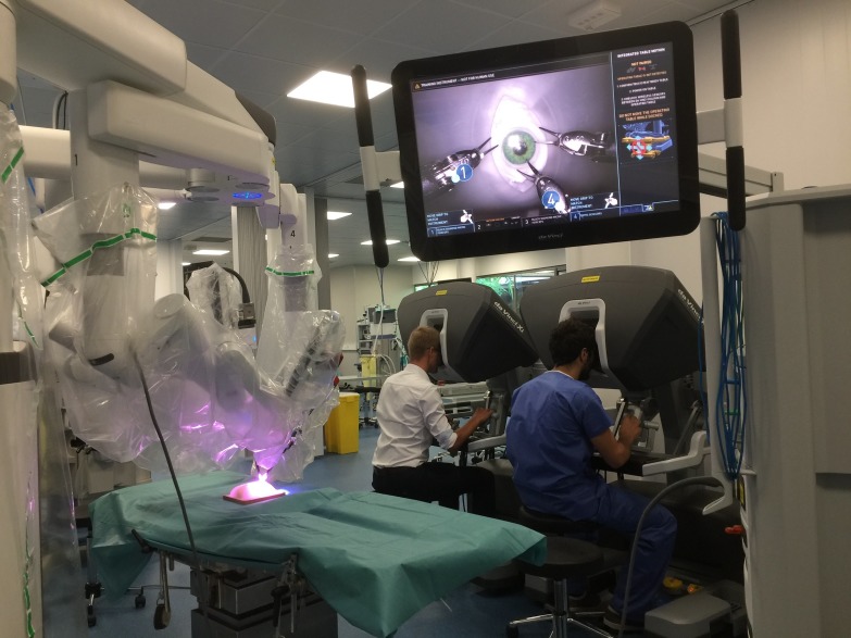
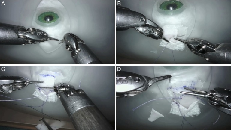
An ophthalmic surgeon (TB) with prior experience in robotic microsurgery and certified by the Robotic Assisted Microsurgical and Endoscopic Society performed the surgical procedures. Surgical movements were scaled to 1.5:1. The eye model was installed under the da Vinci Xi system arms. The camera was installed vertically above the eye on arm number 2, and other instruments were placed around the eye in a triangular fashion (Fig. 1). Procedures were performed on a right eye lateral rectus. The opening of the conjunctiva was made using the fornix-based approach (approximately 6–7 mm behind the limbus) with the Potts scissors (arm number 4) and the micro bipolar forceps (arm number 1) (Fig. 2A). The Tenon's capsule was then dissected to reach the lateral rectus. The Tenon's capsule and conjunctiva were retracted temporally using an 8/0 polyglactin thread to facilitate exposure of the lateral rectus.
Figure 1.
Installation. On the left side are the surgical tables with the strabismus dry-lab training system installed under the robotic da Vinci Xi Surgical System above it. On the right side and in the background are the surgeon's remote consoles. The video screen (top) shows the operative field with the three robotic arms above the eye model.
Figure 2.
Surgical sequences. (A) The conjunctival opening was made with the Potts scissors (arm number 4) and the micro bipolar forceps (arm number 1). (B) The rectus muscle was identified and sutured with 5/0 polyglactin. (C) Desinsertion of the muscle from the sclera was made with Potts scissors (arm number 4). (D) Resuturing of the muscle to the globe was made using the Black Diamond micro forceps (arm number 3) and the micro bipolar forceps (arm number 1) with 5/0 polyglactin. The muscle insertion was secured with two partial-thickness scleral passes.
For each eye, we performed successively a 4-mm plication followed by a 4-mm recession of the muscle, ending with a 4-mm resection (Supplementary Video S1). The 4 mm was measured using a caliper and surgical felt pen. The first step was the 4-mm plication. Two single-armed 5/0 polyglactin sutures were placed at each edge of the muscle 4 mm from the insertion (Fig. 2B). The needles were then passed immediately anterior to the insertion of the muscle, and the sutures were used to pull the posterior portion of the muscle forward and fold it on itself. In a second step, a 4-mm recession was performed. Desinsertion of the muscle from the sclera was performed with Potts scissors (Fig. 2C). Resuturing of the muscle posterior to its original insertion site to the globe was performed using the Black Diamond micro forceps (arm number 3) and the micro bipolar forceps (arm number 1). The muscle was reattached to the sclera with two 5/0 polyglactin sutures (Fig. 2D). In the third step, resection was performed. Sutures were placed 4 mm posterior to the insertion site of the muscle. The muscle was detached from its insertion, and the distal portion of the muscle was excised. The sutures were then passed through the original insertion site of the muscle allowing the shortened muscle to be pulled up to the original site. Lastly, the Tenon's capsule and conjunctiva were closed with five 8/0 polyglactin stitches.
The primary endpoint of this study is the feasibility of a robot-assisted simulated strabismus surgery using the da Vinci Surgical System. For each complete procedure (plication, recession, resection), operative time (from the creation of the conjunctival opening to the end of the conjunctival closing) and successful completion of the different steps with or without complications or unexpected events were assessed. The Ophthalmology Surgical Competency Assessment Rubric for Strabismus Surgery in resident training (OSCAR: strabismus), a standardized tool described by Golnik et al.,14 was used to assess the completion of the procedures. The absence of conflicts between the instruments themselves and between the instruments and the model head was also studied.
Results
Six complete procedures were performed at the IRCAD center (Strasbourg, France). We confirmed the feasibility of robot-assisted simulated strabismus surgery. The robotic da Vinci Xi Surgical System provided the dexterity and operative field visualization necessary to perform the main steps of strabismus surgery as defined by OSCAR: conjunctival and Tenon's capsule opening and closing, lateral rectus identification, suturing, desinsertion, sectioning, and resuturing. The time required for the docking of the robot and the installation of the arms, camera, instruments, and the dry lab model was 45 minutes. The mean operative time to complete the whole procedure was 27 minutes (range, 22–35). All of the scheduled steps were performed. We did not observe any conflicts between the instruments themselves or with the head model. There were no cases of scleral or conjunctiva damage during the procedures.
Discussion
Horizontal rectus surgery is the most frequent strabismus surgery performed in the United States.3 The rate of complications is low, and the psychosocial and economic benefits of surgery for adult and pediatric patients has now been well documented.15 However, the relative lack of skilled surgeons makes access to surgery difficult, and the question of the role of robotics in strabismus surgery, as in many other ophthalmologic surgeries, is legitimate.
The da Vinci is a master-slave surgical robot that enables restitution of surgeons' movements with improved accuracy (7 degrees of freedom) through motion scaling and tremor filtering. Although not specifically designed for eye surgery, there have been several investigations of the potential applicability of the da Vinci system in both anterior- and posterior-segment eye surgeries since 2006.10,11,13 However, the current kinematics of the robotic arms, even in the latest version, and the absence of specific ophthalmologic instruments were found to be hurdles for further clinical research in intraocular surgery. Beyond the field of ophthalmology, the da Vinci system has been used by neurosurgeons to investigate the feasibility of minimally invasive skull base surgery using a supraorbital trans-eyebrow keyhole approach.16 Robot-assisted muscle surgery has also been reported for many large muscles, such as latissimus dorsi, diaphragm, Heller, bladder, vagina, and pelvic muscles.17–19
We report here the feasibility of robot-assisted simulated strabismus surgery using the Xi da Vinci Surgical System. All steps including lateral rectus plication, recession, and resection were completed successfully on a strabismus eye model. This is, to our knowledge, the first use of the da Vinci system and, more generally, of a surgical robot in eye muscle surgery.
The robotic instruments were well adapted to the different steps of surgery. With the exception of the measurement of the lateral rectus muscle (4 mm) for plication, resection, and recession using a caliper, no human assistance was needed for the procedures. Unlike robot-assisted simulated cataract surgery,12 no other specific microsurgical instruments had to be used in combination with the robotic system. Tissue handling (i.e., muscle, Tenon's, conjunctiva, sclera) and knot tying using 8/0 and 5/0 polyglactin were easy with Black Diamond micro forceps and micro bipolar forceps, two tools that have been specially designed for microsurgery. All the steps of ICO-OSCAR strabismus were performed except for the forced duction test (step 2) and muscle hooking (step 5). Steps 1 (draping), 12 (hemostasis), and 14 (knowledge of instruments) were not applicable to this experimental setting. Communication between members of the surgical team (step 17) was carried out using the microphone on the surgeon console. We did not observe the needle or thread breakage frequently seen with da Vinci–assisted procedures using 9/0 or 10/0 nylon.9 Manipulation of the instruments around the eyeball was easy in comparison to previously experimented intraocular procedures.9,12 Access to the orbit proved very easy without helper-induced potential awkwardness, although the absence of muscle tension in our model did not require the use of a hook on the muscle. This is interesting as helper interference or awkwardness can be observed as strabismus surgery often requires that four hands intervene in a limited amount of space. On the other hand, the setup time necessary for installation of the robot, the camera, and the instruments largely exceeded the operative time. Other drawbacks are the current cost of the robotic system and its maintenance and the need for trained staff and additional disposable instruments. However, the absence of conflict between the instruments and the model head suggests that deep-set orbits are not a problem when the initial positioning of the robotic tools is correct. Finally, we appreciated the 8-mm camera of the Xi model that allows better visual definition and clarity than the previous Si HD model. It provides both magnification and 3D image quality close to those one can find in modern surgical microscopes. Thus, visualization of the operative field was perfectly adequate for strabismus surgery, notably allowing perfect visualization of the sclera to avoid perforation. The autofocus system on the image is very practical and time saving. In all, single-procedure time (recession or plication or resection) is estimated to be about 10 minutes, which is nearly the same time observed in classical surgeries.
Simulation can be used not only to teach or enhance strabismus surgical training but also to investigate new surgical techniques, such as robot-assisted strabismus surgery. There are currently no widely accepted simulators for strabismus surgery, although some residency programs use a wet lab with animal tissue.20 Cadaveric ocular muscles are difficult to manipulate, and specimens are not readily available. As an alternative, nonbiological strabismus surgery simulator systems or dry-lab models have been developed.5 Due to the consistent quality of synthetic materials, strabismus dry-lab models are more easily replicated than the biological model using pig eyes and cold cuts.8,20 One such model, the Simulated Ocular Surgery strabismus eye, was recently introduced to improve strabismus skills.21 The strabismus eye mimics the appearance and the feel of a human eye well. The material has the same needle resistance as human sclera. It has a conjunctival layer with elastic properties similar to human conjunctiva. Under this layer, there is a more fibrous Tenon's capsule layer. Muscles have the same dimensions, degree of elasticity, and needle-feel resistance as real muscles. Three drawbacks did appear, however: the dissection of the Tenon's capsule and identification of the muscle was too easy since there is no adherence between the capsule and conjunctiva on one hand and the capsule and muscles on the other. Secondly, the model is not completely realistic as there is no need to perform hemostasis, which must be done when anterior ciliary vessels bleed. Finally, this model is static. Although not the perfect simulation model, this strabismus eye is, in our experience, a recommendable model for use to practice and/or assess new surgical techniques. In addition to becoming an educational model, the robot could also be used in vivo and pave the way for telesurgery in response to the lack of strabologists.
In conclusion, we demonstrate, we believe for the first time, the feasibility of robot-assisted simulated strabismus surgery. The new da Vinci Xi Surgical System possesses the necessary dexterity to perform nearly all the steps of ocular muscle surgeries. We believe that strabismus surgery is potentially the best indicator among all the ocular procedures that have been previously reported with this surgical robot. We also expect that the next steps in research will lead to the development of microsurgical robots with small cameras to allow subconjunctival access. In this context of preparation for future use, experimental investigations are needed to define specifications for all types of ocular procedures, including strabismus surgery. In addition, research could stimulate the development of surgical robots dedicated to ocular surgery. These improvements will certainly allow strabismus surgeons to separate what is considered today to be a fantasy (robot-assisted strabismus surgery) from what will become our next reality: mini-invasive, image-guided surgery.
Supplementary Material
Acknowledgments
The authors thank Richard Bastier, Henri and Jean-Frédéric Chibret, Arnaud Danse, Janel Hooven, Armel Jaunasse, Julien Lacaux, Eric Leplat, Bernard Stoloff for their advice, proofreading, and technical support.
Disclosure: T. Bourcier, None; J. Chammas, None; D. Gaucher, None; P. Liverneaux, None; J. Marescaux, None; C. Speeg-Schatz, None; D. Mutter, None; A. Sauer, None
References
- 1.McKean-Cowdin R, Cotter SA, Tarczy-Hornoch K, et al. Prevalence of amblyopia or strabismus in Asian and non-Hispanic white preschool children: multi-ethnic pediatric eye disease study. Ophthalmology. 2013;120:2117–2124. doi: 10.1016/j.ophtha.2013.03.001. [DOI] [PMC free article] [PubMed] [Google Scholar]
- 2.Martinez-Thompson JM, Diehl NN, Holmes JM, Mohney BG. Incidence, types, and lifetime risk of adult-onset strabismus. Ophthalmology. 2014;121:877–882. doi: 10.1016/j.ophtha.2013.10.030. [DOI] [PMC free article] [PubMed] [Google Scholar]
- 3.Repka MX, Lum F, Burugapalli B. Strabismus, strabismus surgery, and reoperation rate in the United States: analysis from the IRIS Registry. Ophthalmology. 2018;125:1646–1653. doi: 10.1016/j.ophtha.2018.04.024. [DOI] [PubMed] [Google Scholar]
- 4.Astle AT, Foulsham T, Foss AJ, McGraw PV. Is the frequency of adult strabismus surgery increasing? Ophthalmic Physiol Opt. 2016;36:487–493. doi: 10.1111/opo.12306. [DOI] [PubMed] [Google Scholar]
- 5.Adebayo T, Abendroth M, Elera GG, et al. Developing and validating a simple and cost-effective strabismus surgery simulator. J AAPOS. 2018;22:85–88. doi: 10.1016/j.jaapos.2017.11.006. [DOI] [PubMed] [Google Scholar]
- 6.Nuzzi R, Brusasco L. State of the art of robotic surgery related to vision: brain and eye applications of newly available devices. Eye Brain. 2018;10:13–24. doi: 10.2147/EB.S148644. [DOI] [PMC free article] [PubMed] [Google Scholar]
- 7.de Smet MD, Naus GJL, Faridpooya K, Mura M. Robotic-assisted surgery in ophthalmology. Curr Opin Ophthalmol. 2018;29:248–253. doi: 10.1097/ICU.0000000000000476. [DOI] [PubMed] [Google Scholar]
- 8.Bourcier T, Nardin M, Sauer A, et al. Robot-assisted pterygium surgery: feasibility study in a nonliving porcine model. Trans Vis Sci Tech. 2015;4:9. doi: 10.1167/tvst.4.1.9. [DOI] [PMC free article] [PubMed] [Google Scholar]
- 9.Chammas J, Sauer A, Pizzuto J, et al. Da Vinci Xi robot-assisted penetrating keratoplasty. Trans Vis Sci Tech. 2017;6:21. doi: 10.1167/tvst.6.3.21. [DOI] [PMC free article] [PubMed] [Google Scholar]
- 10.Bourcier T, Becmeur PH, Mutter D. Robotically assisted amniotic membrane transplant surgery. JAMA Ophthalmol. 2015;133:213–214. doi: 10.1001/jamaophthalmol.2014.4453. [DOI] [PubMed] [Google Scholar]
- 11.Bourcier T, Chammas J, Becmeur PH, et al. Robotically assisted pterygium surgery: first human case. Cornea. 2015;34:1329–1330. doi: 10.1097/ICO.0000000000000561. [DOI] [PubMed] [Google Scholar]
- 12.Bourcier T, Chammas J, Becmeur PH, et al. Robot-assisted simulated cataract surgery. J Cataract Refract Surg. 2017;43:552–557. doi: 10.1016/j.jcrs.2017.02.020. [DOI] [PubMed] [Google Scholar]
- 13.Bourla DH, Hubschman JP, Culjat M, Tsirbas A, Gupta A, Schwartz SD. Feasibility study of intraocular robotic surgery with the da Vinci surgical system. Retina. 2008;28:154–158. doi: 10.1097/IAE.0b013e318068de46. [DOI] [PubMed] [Google Scholar]
- 14.Golnik KC, Motley WW, Atilla H, et al. The ophthalmology surgical competency assessment rubric for strabismus surgery. J AAPOS. 2012;16:318–321. doi: 10.1016/j.jaapos.2012.04.005. [DOI] [PubMed] [Google Scholar]
- 15.MacKenzie K, Hancox J, McBain H, Ezra DG, Adams G, Newman S. Psychosocial interventions for improving quality of life outcomes in adults undergoing strabismus surgery. Cochrane Database Syst Rev. 2016;12 doi: 10.1002/14651858.CD010092.pub4. :CD010092. [DOI] [PMC free article] [PubMed] [Google Scholar]
- 16.Hong WC, Tsai JC, Chang SD, Sorger JM. Robotic skull base surgery via supraorbital keyhole approach: a cadaveric study. Neurosurgery. 2013;72:33–38. doi: 10.1227/NEU.0b013e318270d9de. [DOI] [PubMed] [Google Scholar]
- 17.Mottrie A, Carpentier P, Schatteman P, et al. Robot-assisted laparoscopic radical cystectomy: initial experience on 27 consecutive patients. J Robot Surg. 2007;1:197–201. doi: 10.1007/s11701-007-0035-9. [DOI] [PMC free article] [PubMed] [Google Scholar]
- 18.Chung JH, You HJ, Kim HS, Lee BI, Park SH, Yoon ES. A novel technique for robot assisted latissimus dorsi flap harvest. J Plast Reconstr Aesthet Surg. 2015;68:966–972. doi: 10.1016/j.bjps.2015.03.021. [DOI] [PubMed] [Google Scholar]
- 19.Davila HH, Brown K, Dara P, Bruce L, Goodman L, Gallo T. Robotic-assisted laparoscopic apical suspension: description of the spiral technique [published online ahead of print October 3, 2018] J Robot Surg. doi: 10.1007/s11701-018-0879-1. [DOI] [PubMed]
- 20.White CA, Wrzosek JA, Chesnutt DA, Enyedi LB, Cabrera MT. A novel method for teaching key steps of strabismus surgery in the wet lab. J AAPOS. 2015;19:468–470. doi: 10.1016/j.jaapos.2015.05.020. [DOI] [PubMed] [Google Scholar]
- 21.Gertsch KR, Kitzmann A, Larson SA, et al. Description and validation of a structured simulation curriculum for strabismus surgery. J AAPOS. 2015;19:3–5. doi: 10.1016/j.jaapos.2014.09.007. [DOI] [PubMed] [Google Scholar]
Associated Data
This section collects any data citations, data availability statements, or supplementary materials included in this article.


