. 2019 May 24;8:e45342. doi: 10.7554/eLife.45342

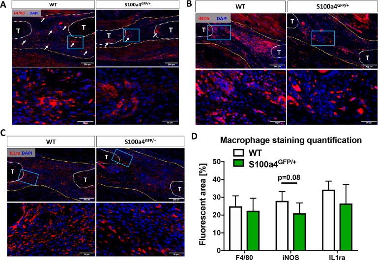
Figure 4. S100a4 haploinsufficiency alters the macrophage response to tendon injury.

(A) F4/80 staining demonstrates decreased macrophage content in the healing tendon of S100a4GFP/+ repairs at D14. White arrows identify concentrated areas of macrophages. (B) Expression of the M1 macrophage marker iNOS is markedly reduced in S100a4GFP/+ repairs at D14. (C) Expression of the M2 macrophage marker IL1ra is not different between WT and S100a4GFP/+ repairs at D14. Tendon ends are outlined in white, scar tissue is outlined in yellow, blue boxes indicate location of higher magnification images (n = 4 per group). (D) The percent area of F4/80+, iNOS+ and IL1ra+ staining, normalized to tissue area was quantified (n = 4) (un-paired t-test).

Figure 4.

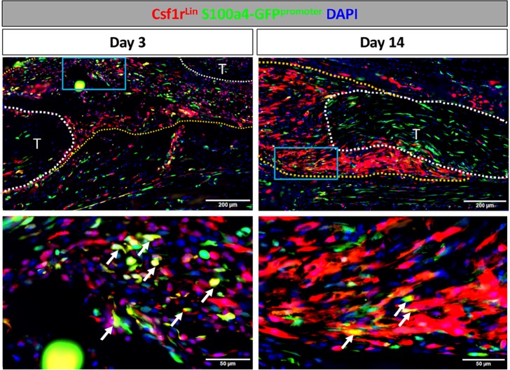
Figure 4—figure supplement 1. S100a4 is expressed by macrophages during early tendon healing.

Figure 4—figure supplement 1.

To identify S100a4+ macrophages, Csf1r-iCre; Rosa-Ai9; S100a4-GFPpromoter+ mice were induced with tamoxifen. Tmx was given on D0-2 for mice harvested on D3, and D0-2 and every 48 hr thereafter until harvest for samples harvested at D14. On D3 several macrophages actively express S100a4 (white arrows). By D14 the presence of Csfr1Lin+ cells increased, but very few cells actively expressed S100a4 (white arrows).
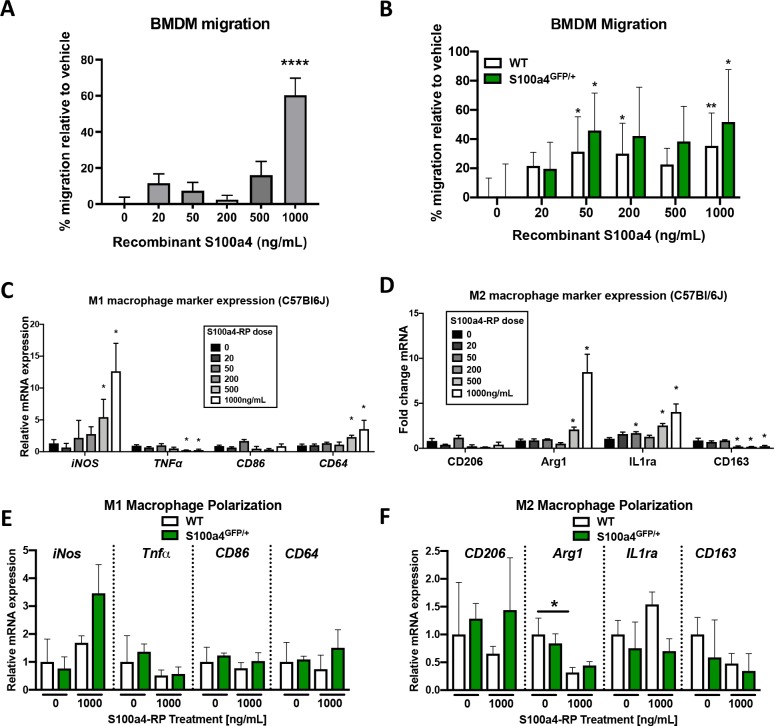
Figure 4—figure supplement 2. S100a4 promotes macrophage migration and alters polarization.

Figure 4—figure supplement 2.

(A) S100a4 promotes migration of C57BL/6J bone marrow derived macrophages (BMDMs). (****) Indicates p<0.0001 vs. vehicle treated cells (1-way ANOVA). (B) No change in migration was observed in vehicle treated WT and S100a4GFP/+ BMDMs, while significant increases in migration were observed in WT and S100a4GFP/+ BMDM treated with 50 ng/mL and 1000 ng/mL S100a4-RP. (*) indicates p<0.05, (**) indicates p<0.01 vs. genotype-matched vehicle treated cells (2-way ANOVA). (C and D) Following treatment with S100a4-RP (20–1000 ng/mL), significant increases in M1 polarization markers (iNos, CD64) were observed relative to vehicle treated C57BL/6J BMDMs, as was a significant decrease in TNFα, and no change in CD86 expression (C). Significant increases in M2 markers Arg2 and IL1ra were seen with S100a4-RP treatment, while a decrease in CD163 expression, and no change in CD206 was also observed in C57BL/6J BMDMs (D). (*) indicates p<0.05 between vehicle and S100a4-RP treatment (1-way ANOVA). (E and F) Expression of M1 (E) and M2 (F) macrophage markers were not significantly different between WT and S100a4GFP/+ BMDMs in vehicle treated or upon treatment with 1000 ng/mL S100a4-RP. (*) indicates p<0.05 vs. genotype-matched vehicle treated cells (2-way ANOVA). (n = 3 per treatment).
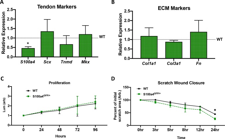
Figure 4—figure supplement 3. Tendon cell S100a4 haploinsufficiency does not alter tenogenic and matrix gene expression or proliferation but enhances migration.

Figure 4—figure supplement 3.

(A) qPCR analysis primary tendon cells from WT and S100a4GFP/+ mice. Expression of S100a4 is significantly reduced (~50%) in S100a4GFP/+ tendon cells, relative to WT. No changes in tenogenic genes Scx, Tnmd and Mkx. (*) indicates p<0.05 vs. expression in WT cells. (B) No changes in expression of matrix genes Col1a1, Col3a1 or Fn are observed between WT and S100a4GFP/+ tendon cells. Data are normalized to WT expression and β-actin. (un-paired t-test). (C) No changes in proliferation were observed between WT and S100a4GFP/+ tendon cells (2-way ANOVA). (D). Cell migration was assessed by measuring closure of a scratch wound. Data are plotted as % of initial scratch area. Closure was significantly increased in S100a4GFP/+ tendon cells at 24 hr (2-way ANOVA).