Abstract
Introduction:
Congenital granular cell epulis is a rare and benign lesion in newborn. There are some papers of the entity; however, there are very few reports focusing on its macroscopic view.
Case Presentation:
A 0-day-old boy was noted to have a mass consisting of multiple nodules on maxillary gingiva, and it was excised. The mass was measured about 2 cm in its greatest diameter. Surface of cross-section was characteristically whitish-yellow and very smooth. Histopathologically, the lesion was composed of a proliferation of large polygonal cells with demarcated cell membrane, granular cytoplasm, and small uniform nuclei. Immunohistochemically, these cells were negative for S100. The diagnosis was concluded as congenital granular cell epulis.
Discussion and Conclusion:
We reported a typical case of congenital granular cell epulis. It is noteworthy that the cross-section observed in this case was very characteristically whitish-yellow and smooth.
Keywords: Congenital granular cell epulis, macroscopic view
Introduction
Congenital granular cell epulis is a localized mass that mostly develops on the maxilla of newborn. This is a rare and benign lesion. There are some papers of the entity, in which histological or immunohistochemical characteristics of the lesion have been mainly reported; however, there are very few reports focusing on its macroscopic view.1,2
Case Presentation
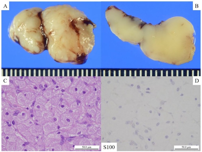
A 0-day-old boy was noted to have a mass consisting of multiple nodules on maxillary gingiva. He was born by normal vaginal delivery at 40 weeks gestation weighing 3115 g. The mass was excised. It consisted of multiple nodules and measured about 2 cm in its greatest diameter (Figure 1A). Surface of cross-section was characteristically whitish-yellow and very smooth (Figure 1B). Histopathologically, the lesion was composed of a proliferation of large polygonal cells with demarcated cell membrane, granular cytoplasm, and small uniform nuclei (Figure 1C). The overlying surface epithelium was not hyperplastic. Immunohistochemically, these cells were negative for S100 (Figure 1D). The final diagnosis was congenital granular cell epulis.
Figure 1.
Gross findings, microscopic findings, and immunohistochemical findings of specimen: (A) the mass consisted of multiple nodules and measured about 2 cm in its greatest diameter; (B) surface of cross-section was characteristically whitish-yellow and very smooth; (C) the lesion was composed of a proliferation of large polygonal cells with demarcated cell membrane, granular cytoplasm, and small uniform nuclei, bar = 50 μm (H&E stain; original magnification: ×400); and (D) immunohistochemically, these cells were negative for S100, bar = 50 μm (original magnification: ×400).
Discussion and Conclusion
Congenital granular cell epulis mostly occurs on the maxilla of newborns which is characterized by sheet-like proliferation of large polygonal cells with demarcated cell membrane, granular cytoplasm, and small uniform nuclei.3 With similar macroscopic findings, granular cell tumor is usually considered as a differential diagnosis. Unlike congenital granular cell epulis, granular cell tumor usually occurs in adults, is immunoreactive for S100 and sometimes shows pseudoepitheliomatous hyperplasia.3,4 Histopathological findings of our case are consistent with congenital granular cell epulis. As the cells are negative for S100, granular cell tumor is ruled out. Congenital granular cell epulis is a rare entity. Although there are some papers on it, we actually seldom encounter this lesion. Our case has typical findings of this rare entity and deserves reporting.
More noteworthy is the macroscopic view of this lesion. Microscopic characteristics, immunohistochemical findings, and/or its origin have been discussed in previous papers; however, there are very few reports focusing on its clear macroscopic view. The cross-section of this lesion was characteristically whitish-yellow and smooth. The cross-section image shown in this article was taken after fixation with 10% neutral buffered formalin. The specimen was fixed properly and sufficiently soon after surgical resection. Although this is a case report and further studies are required, the cross-section surface observed in this case could reflect monotonous sheet-like proliferation of granular cells. We have less information of the color of the lesion before surgery, and it could be different from that of after the fixation. However, this macroscopic view is interesting as a teaching case, and could be an aid for not only clinicians but also pathologists to macroscopically diagnose the entity.
This case is worth reporting, as it shows not only typical histopathological findings but also unique cross-section findings.
Acknowledgments
The authors would like to thank all members of their department for the expert technical assistance and general support they provided.
Footnotes
Declaration of Conflicting Interests:The author(s) declared no potential conflicts of interest with respect to the research, authorship, and/or publication of this article.
Funding:The author(s) received no financial support for the research, authorship, and/or publication of this article.
Author Contributions: KM and SY participated in conception of the idea and writing of the manuscript. KM, SY, MM, and HN were associated with the pathological interpretation of this case. All authors have read and approved the final manuscript.
Availability of Data and Materials: The data set supporting the findings and conclusions of this case report is included within the article.
Informed Consent: Informed consent was obtained from the patient and his family on admission for the publication of this case report and any accompanying images.
ORCID iD: Sohsuke Yamada  https://orcid.org/0000-0003-2662-0024
References
- 1. Katsutoshi K, Matsuzaka K, Akashi Y, et al. Congenital epulis: a case and review of the literature. Bull Tokyo Dental Col. 2018;59:127–132. [DOI] [PubMed] [Google Scholar]
- 2. Zhang B, Tan X, Zhang K, Li Y, Chen J, Zhang F. A study of cell proliferation using immunohistological staining: a case report of congenital granular cell epulis. Int J Pediatr Otorhinolaryngol. 2016;88:58–62. [DOI] [PubMed] [Google Scholar]
- 3. Yıldırım C, Zerener T, Şençimen M, Akgün ÖM, Altuğ HA, Çiçek AF. Congenital gingival granular cell tumor: a case report. J Dent (Shiraz). 2017;18:70–72. [PMC free article] [PubMed] [Google Scholar]
- 4. Bilen BT, Alaybeyoğlu N, Arslan A, Türkmen E, Aslan S, Celik M. Obstructive congenital gingival granular cell tumour. Int J Pediatr Otorhinolaryngol. 2004;68:1567–1571. [DOI] [PubMed] [Google Scholar]


