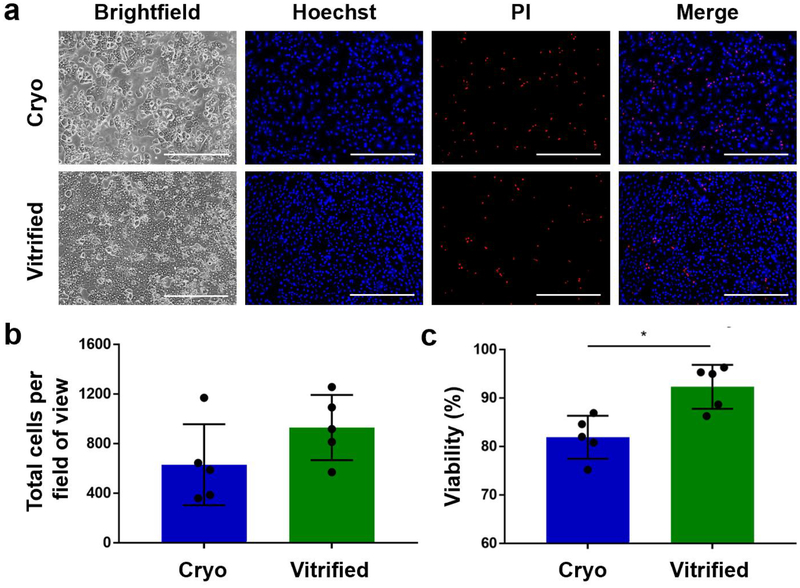
Figure 5.
Viability of cryopreserved (blue) and bulk droplet vitrified (green) hepatocytes after 24 hours monolayer culture. a. Representative images of Hoechst (all hepatocytes) / propidium iodide (PI) (dead hepatocytes) staining of the cultures. b. Total number of attached hepatocytes per field of view. c. Viability of attached hepatocytes. Scale bars: 400μm. Stars: p<0.05 Error bars: SD.

