CASE PRESENTATION
A healthy 24-year-old female presented at the emergency department (ED) after a car accident with ambulance while injured severely after the bus got run over her lower limb. As the trauma team was activated, her primary survey was started:
Ac (Airway and cervical collar): She was awake and could talk. Cervical collar was fixed, oxygenation with face mask was started.
B (Breathing): Her chest rising was symmetrical without any laceration or abrasion. Chest auscultation was clear and there was no tenderness or crepitation on palpation. No tracheal shift was found. She had normal respiratory rate and O2 saturation of 94% at ambient air.
C (Circulation): Two large bore IV lines were inserted and blood samples were obtained. Her vital signs were BP = 60/40 mmHg, PR = 130/min, RR = 12. E-FAST was performed which was negative for free fluid in abdomen, pelvis and thorax, tamponade, and hemopneumothorax. Her pelvis was unstable on examination and pelvic wrapping was performed with sheath. IV fluid therapy with normal saline was started followed by 3 units of packed RBC transfusion. More pack cells and FFP were also requested.
D (Disability): She had Glasgow coma scale of 15/15 with normal size and reactive pupil. No neurologic deficit was found except disability of lower extremities due to crush injury.
E (Exposure): She had no midline spinal tenderness with normal sphincter anal tone, but there was a laceration in the perineum which extended to the vagina.
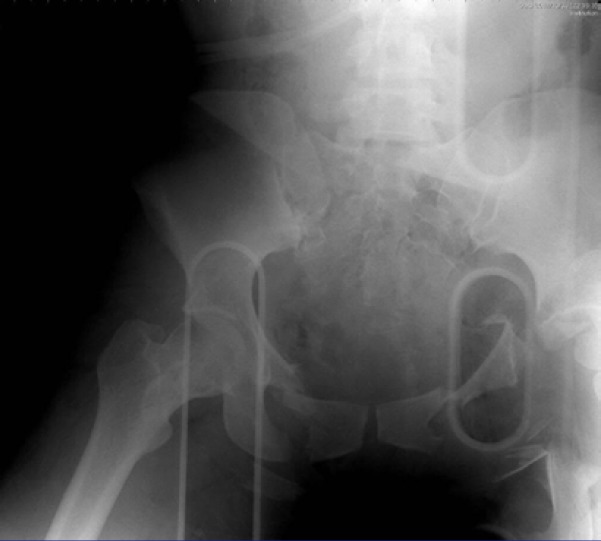
Portable chest and pelvic x-ray as an adjutant to primary survey were performed which showed type C pelvic fracture (Figure 1).
Figure 1.
Anterior-posterior X-ray of the patient’s pelvic
On her secondary survey, she had abrasion on her scalp, 1.5 cm laceration on her right tibia, deformity of her right thigh, and laceration in her genitalia with some vaginal bleeding. Direct pressure was applied and all lacerations were packed. According to negative e-FAST and pelvic fracture and shock, since the angiography was not available, it was decided to fix the pelvis with external fixator in the operation room. After the fixation, and because shock persisted, operative pelvic packing was undertaken. Unfortunately, she suffered cardiorespiratory arrest in the operating room and died.
EDUCATIONAL POINTs
Here are four important points which could improve patient management.
1. Airway is of highest priority in the management of severely injured patients. Tracheal intubation’s indications are: cardiac or respiratory arrest, failure to protect airway from aspiration (GCS less than 8), hypoxia and hypoventilation, and impending or existing airway obstruction. In refractory shock, rapid sequence intubation can be helpful since hypoperfusion can cause respiratory muscles fatigue, and intubation and paralysis can improve cardiac output up to 30% (1). As the patient was awake and the airway was intact, oxygenation with simple mask was started. The patient received 2 liters of normal saline and 3 units of packed cells but she still had hypotension, so rapid sequence intubation should have been considered at this stage.
2. On her circulation in primary survey after starting normal saline, 3 units of packed cells was transfused afterward while the patient was transferring to the operating room. Damage control resuscitation (DCR) is a primary approach to seriously injured patients and to preventing life-threatening events (hypothermia, coagulopathy and acidosis) (2). Massive transfusion protocol is activated in Assessment of Blood Consumption score of 2 or more (SBP less than 90 mmHg, HR>120/min, +FAST and penetrating torso injury), persistent hemodynamic instability and active bleeding (needs operation or angioembolization) (3).
So the sequence of life savers is:
1. Control bleeding (packing, wrapping, rolling)
2. Permissive hypotension strategy (if GCS=15)
3. Hemostatic resuscitation strategy
4. Early control surgery (less than 1 hour)
Traditional definition for massive transfusion is 10 units or more of packed cells within a 24-hour period but in modern massive transfusion protocols the aim is delivery of 1:1:1 ratio for packed cells, FFP, platelets, and cryoprecipitate. One of the major benefits is the avoidance of crystalloid volume during resuscitation (4). For massive transfusion, replacement of blood loss with warm blood is recommended as well as giving 10 cc of gluconate calcium 10% after 4 units of pack cells transfusion. So it is imperative to start massive transfusion for the patient after normal saline with plasma and platelet transfusion.
3. Unstable pelvic was wrapped with sheet. The goal of pelvic stabilization is to prevent additional vascular and tissue injuries and decrease pelvic volume. Circumferential sheet wrapping around the pelvis is a classic management of pelvic fractures (5). External pelvic compression devices like TPOD® and SAM® sling have been used for many years to provide fast and easy pelvic stabilization while the sheet wrap is readily available but is not easy to apply even by two people (6).
C-clamp® is another device for pelvic stabilization which can be used readily in the ED. Ileum fractures and communicated sacrum fractures are relative contraindicated factors in using c-clamp (7). It is preferable to use c-clamp instead of sheet in the Emergency Department and also external fixation for femur fracture in order to save time before the next step.
4. Damage control surgery (DCS) is a part of DCR. DCS tries to reduce early surgical intervention and focus on initial control of hemorrhage (2). In an unstable patient with pelvic fracture and negative e-FAST, therapeutic angiography with a probable diagnosis of life-threatening retroperitoneal bleeding should be proceeded (8). Angiography is better performed in a hybrid operating room and is indicated for arterial bleeding in case pelvic packing is not helpful. Nearly 85% of pelvic traumatic bleeding sites are venous in origin (9). The optimum outcomes require early access to interventional radiology with rapid readiness times after presentation to the ED (10). All of these factors put together and the lack of equipped and available settings provide the case for the patient to be transferred to the operating room earlier for pelvic packing in extra peritoneal space and bladder repair if needed, rather than spending time and waiting for angioembolization.
References
- 1.Puskarich M, Jones A. Shock. Rosen’s emergency medicine concepts and clinical practice. 9th ed 2018. [Google Scholar]
- 2.Giannoudi M, Harwood P. Damage control resuscitation: lessons learned. Eur J Trauma Emerg Surg. 2016;42:273–82. doi: 10.1007/s00068-015-0628-3. [DOI] [PMC free article] [PubMed] [Google Scholar]
- 3.Trauma ACoSCo. ACS TQIP massive transfusion in trauma guidelines. Chicago: American College of Surgeons; 2016. [Google Scholar]
- 4.Ball CG. Damage control resuscitation: history, theory and technique. Can J Surg. 2014;57(1):55–60. doi: 10.1503/cjs.020312. [DOI] [PMC free article] [PubMed] [Google Scholar]
- 5.Gerecht R, Larrimore A, Steuerwald M. Critical management of deadly pelvic injuries. JEMS. 2014;39(12):28–35. [PubMed] [Google Scholar]
- 6.Lustenberger T, Wutzler S, Störmann P, Marzi I. The role of pelvic packing for hemodynamically unstable pelvic ring injuries. Clin Med Insights Trauma Intens Med. 2015;6:CMTIM. [Google Scholar]
- 7.Lustenberger T, Meier C, Benninger E, Lenzlinger PM, Keel MJB. C-clamp and pelvic packing for control of hemorrhage in patients with pelvic ring disruption. J Emerg Trauma Shock. 2011;4(4):477–82. doi: 10.4103/0974-2700.86632. [DOI] [PMC free article] [PubMed] [Google Scholar]
- 8.Nichols J, Puskarich M. Abdominal Trauma. Rosen’s emergency medicine, concepts and clinical practice. 9th ed 2018. [Google Scholar]
- 9.Jeske HC, Larndorfer R, Krappinger D, Attal R, Klingensmith M, Lottersberger C, et al. Management of hemorrhage in severe pelvic injuries. J Trauma. 2010;68(2):415–20. doi: 10.1097/TA.0b013e3181b0d56e. [DOI] [PubMed] [Google Scholar]
- 10.Salcedo ES, Brown IE, Corwin MT, Galante JM. Pelvic angioembolization in trauma - Indications and outcomes. Int J Surg. 2016;33(Pt B):231–6. doi: 10.1016/j.ijsu.2016.02.057. [DOI] [PubMed] [Google Scholar]


