

Abstract
Fertility is one of the most economically important traits in both beef and dairy cattle production; however, only female fertility is typically subjected to selection. Male and female fertility have only a small positive genetic correlation which is likely due to the existence of a relatively small number of genetic variants within each breed that cause embryonic and developmental losses. Genomic tools have been developed that allow the identification of lethal recessive loci based upon marker haplotypes. Selection against haplotypes harbouring lethal alleles in conjunction with selection to improve female fertility will result in an improvement in male fertility. Genomic selection has resulted in a 2- to 4-fold increase in the rate of genetic improvement of most dairy traits in U.S. Holstein cattle, including female fertility. Considering the rapidly increasing rate of adoption of high-throughput single nucleotide polymorphism genotyping in both the U.S. dairy and beef industries, genomic selection should be the most effective of all currently available approaches to improve male fertility. However, male fertility phenotypes are not routinely recorded in natural service mating systems and when artificial insemination is used, semen doses may be titrated to lower post-thaw progressively motile sperm numbers for high-merit and high-demand bulls. Standardisation of sperm dosages across bull studs for semen distributed from young bulls would allow the capture of sire conception rate phenotypes for young bulls that could be used to generate predictions of genetic merit for male fertility in both males and females. These data would allow genomic selection to be implemented for male fertility in addition to female fertility within the U.S. dairy industry. While the rate of use of artificial insemination is much lower within the U.S. beef industry, the adoption of sexed semen in the dairy industry has allowed dairy herds to select cows from which heifer replacements are produced and cows that are used to produce terminal crossbred bull calves sired by beef breed bulls. Capture of sire conception rate phenotypes in dairy herds utilizing sexed semen will contribute data enabling genomic selection for male fertility in beef cattle breeds. As the commercial sector of the beef industry increasingly adopts fixed-time artificial insemination, sire conception rate phenotypes can be captured to facilitate the development of estimates of genetic merit for male fertility within U.S. beef breeds.
Keywords: Bull Fertility, Genomic Selection, Genome-Wide Association Study, Candidate Genes, Quantitative Trait Loci
Introduction
In cattle, fertility is commonly measured using a number of metrics including age at puberty, calving interval, non-return rate, number of services per conception and daughter pregnancy rate (Berry et al., 2014). Perhaps the most useful metric for evaluating fertility in both sexes is the probability of achieving a pregnancy as a result of a single mating to a randomly sampled, but fertile, member of the opposite sex from 4 to 16 hours following the onset of oestrus in the female. Because the majority of beef cattle are multi-sire mated and beef females are exposed to bulls for a period representing two to three oestrous cycles, this latter metric cannot be widely used in the beef industry. However, in the dairy industry, the majority of females are bred by artificial insemination (AI) allowing the outcomes of individual matings to be recorded. Despite this, genetic evaluations for fertility in the U.S. dairy industry are calculated only for females. The reason for this is that when service sire is included in the genetic evaluation model, the estimated additive genetic variance for male fertility is 0 (John B. Cole, USDA ARS, pers. comm.) and this appears to be due to the bull studs’ use of service sire conception rate (SCR) data to titrate the number of progressively motile pre-freeze spermatozoa until a uniform non-return rate is achieved, systematically eliminating naturally occurring variation in male fertility (Curt P. Van Tassell, USDA ARS, pers. comm.; DeJarnette et al., 2010). As a consequence, selection occurs only for female fertility in both the U.S. beef and dairy industries.
Although the heritabilities of male and female fertility are both generally low (Berry et al., 2014; Fortes et al., 2013a), response to selection is also governed by the extent of phenotypic variation and the length of the generation interval. The advent of genomic selection (Meuwissen et al., 2001) has dramatically reduced generation interval in the U.S. dairy industry (García-Ruiz et al., 2016) and is beginning to similarly impact the U.S. beef industry. This has enabled a remarkable increase in the rate of genetic improvement of production traits and also for female fertility within the U.S. Holstein population, which is shown in Figure 1 (García-Ruiz et al., 2016). While there do not appear to be a large number of studies in cattle, estimates of the genetic correlation between male and female fertility is positive but generally modest in most vertebrate species (from −0.30 to 0.20 for male and female non-return rate in Danish dairy cattle, Hansen, 1979; “slight” for male and female non-return rate in Norwegian dairy cattle, Syrstad, 1981; −.25, −.28 and −.41 between scrotal circumference and days to calving in Australian Hereford, Angus, and Zebu crosses, respectively, Meyer et al., 1991; 0.14 in Manech Tête Rousse sheep, David et al., 2007; 0.15 for male and female contributions to egg fertility in broiler chickens, Wolc et al., 2009; 0.34 for male and female contributions to conception in rabbits Piles and Tusell, 2012). Consequently, the majority of genes that create variation in male fertility have male specific functions and selection to improve female fertility will result in a positive, but less than optimal, increase in male fertility. Improvement of the overall efficiency of beef and dairy production will require the ability to identify and eliminate young bulls with sperm abnormalities and unacceptable semen quality (Taylor et al., 2018) and the development of predictors of genetic merit for male fertility that may be applied within and perhaps also across breeds. In this manuscript, we address the current state of knowledge concerning genetic variants responsible for variation in male fertility and the approaches that should be taken to enable the improvement of male fertility in beef and dairy cattle.
Figure 1.
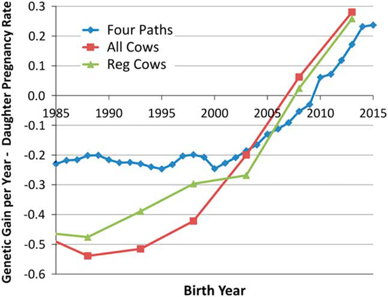
Average genetic gain per year for female fertility in U.S. Holsteins (García-Ruiz, 2016).
Mendelian variants causing variation in male and female fertility
Of the loci that create genetic variation in both male and female fertility, the most obvious are loss-of-function (LOF) mutations in genes that are essential for life. In the human genome, 7 168 (33.3%) of the 21 556 annotated genes are essential for life (Chen et al., 2017) meaning that the functionality of at least one copy of each of these genes is required for human life. The number and proportion of essential genes in cattle are probably very similar to those in humans. Mutations which disrupt the functionality of the proteins encoded by essential genes are LOF mutations and for genes located on the autosomes (non-sex chromosomes), homozygosity for a LOF mutation, or heterozygosity of two chromosomes each with a different LOF mutation in the same gene leads to lethality. Because these mutations are transmitted to progeny by both males and females, they are responsible for variation in genetic merit for both male and female fertility. The majority of LOF mutations produce early embryonic loss due to failure to implant or develop. These pregnancy losses are frequently not noticed, but calf losses may also occur in the second and third trimesters of pregnancy or postnatally, manifesting as a genetic defect. These loci are subject to purifying selection because homozygotes are removed every generation leading to relatively small decreases in allele frequency in each successive generation. However, the frequency of some of these LOF lethal alleles can be driven to high levels in a population by the extensive use of AI, which allows carrier bulls to transmit what might otherwise be a rare LOF mutation to a large number of progeny.
Because there is a large number of genes that are essential and these are all targets for mutation, there may be a very large number of lethal LOF mutations within a population. However, the frequency of the majority of these alleles is generally very low and the joint effect of these rare alleles on the mean fertility of the population is small. Because relatively few bulls have been whole genome sequenced (less than 4 000 world-wide), the majority of rare variants are yet to be found, either because they were not present in the sequenced animals or because they were detected only once in a sequenced animal and filtered as potentially being a sequence g. Of those that have been discovered, not all can confidently be predicted to be a LOF mutation. A mutation that produces a charged amino acid substitution that is predicted to not be tolerated may, or may not, result in the LOF of a protein. Several lethal LOF mutations have been found in cattle by applying the haplotypic insufficiency analytical technique first described by VanRaden et al. (2011). Using this technique, high density single nucleotide polymorphism (SNP) genotypes such as those produced by the 54 001 SNP BovineSNP50 assay (Matukumalli et al., 2009) are first phased so that for each genotyped individual, the two alleles present at each SNP genotype are assigned in a specific order to each of the chromosomes that are present in that individual. Each specific combination of SNP alleles present on a chromosome or chromosomal segment is called a haplotype and the specific pair of haplotypes present within each individual is called a diplotype. Next, the frequencies of haplotypes and diplotypes present in a sample of genotyped animals are tallied for small chromosome segments of, say 20 consecutive SNPs. The probability of observing no individuals that are homozygous for each haplotype is calculated based on the sample size and the assumption of random inheritance of haplotypes from each parent (Hardy-Weinberg equilibrium). To identify genomic regions that are likely to harbour an autosomal recessive lethal LOF allele, the method of VanRaden et al. (2011) identifies haplotypes that never occur in homozygous form when we would expect to see homozygotes in the sample based on the frequency of the haplotype. The logic behind this method is that if all of the chromosomes in the population that are identified by the same haplotype of 20 SNP alleles harbour a LOF lethal mutation, then every homozygote must be lethal and these individuals will never be seen in the population. Figure 2 provides a schematic representation of this process that shows that when 100 000 individuals have been genotyped and the chromosomes have been phased, 20 marker haplotypes that are at a frequency of 2% in the genotyped sample would be expected to be observed in homozygous form in 40 individuals. When in practice none are observed, the probability that this is due to chance alone is vanishingly small (PHWE = [1 – p2]N = 4 × 10⁻¹⁸) and we may conclude that the reason that we did not observe any homozygotes is that the haplotype harbours an autosomal recessive lethal LOF allele. Clearly the approach is limited by the need for many genotyped individuals, otherwise low frequency haplotypes will not appear in homozygous form simply due to chance. The approach also assumes that all chromosomes with the same haplotype identified based on alleles at 20 SNPs contain the lethal allele. This may not be the case when the mutation has recently occurred, in which case chromosomes in some individuals will carry the lethal mutation while chromosomes in other individuals will not carry the lethal allele despite the fact that the haplotypes all appear to be identical based upon the marker information. In this case, we will observe fewer homozygotes than expected based on the haplotype frequency, but certainly more than none. This reduces the power of the test and therefore much larger sample sizes are necessary to detect homozygote deficiency when the lethal allele is not perfectly associated with a single haplotype.
Figure 2.
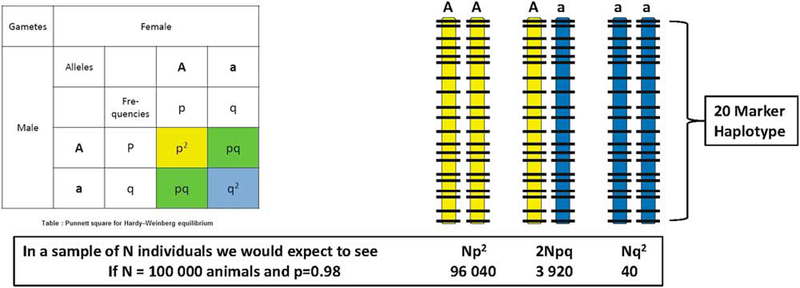
Two haplotypes (coloured yellow or blue) defined by alleles present at 20 consecutive single nucleotide polymorphism markers (locations shown as horizontal bars) are present at frequencies of p = 0.98 and q = 0.02 in a genotyped sample of size N = 100 000 cattle. On average, 40 animals would be expected to be homozygous for the rare (blue) haplotype.
A slightly different version of this test was discussed by VanRaden et al. (2011) and implemented by Hoff et al. (2017) that capitalizes on the available pedigree information in genotyped cattle populations. When trios of sire, dam and progeny or patrios of sire, maternal grandsire and progeny have all been genotyped and a particular haplotype is never observed in homozygous form, we can count the number of trios in which the sire and dam are both heterozygotes or the number of patrios where the sire and maternal grandsire are both heterozygotes and calculate the probability of not observing a progeny that is homozygous for the rare haplotype. This is demonstrated in Figure 3 for the case of a patrio where the dam has not been genotyped, the frequency of the rare haplotype in the population is q and the son has a probability of (2q + 1)/8 of being homozygous aa for the rare haplotype ‘a’ but is observed to be AA or Aa. If N such patrios in which the sire and maternal grandsire are both heterozygotes are counted as never producing a homozygous progeny, the probability of this being due to chance alone is PP = [0.75 – 0.25q]N and for haplotypes at a frequency of 2% in the population PP is 1.6 × 10⁻¹³ for as few as N = 100 patrios. This may represent many fewer than 300 individuals since sires and maternal grandsires may be common across many patrios. The probability calculation requires only independent assortment of parental alleles in each family and not independent families. The approach analyses either overlapping or non-overlapping 20 SNP marker windows sequentially along each chromosome in order to scan the entire genome for the presence of lethal mutations and so there is a multiple testing problem that requires adjustment of the probability values produced for each test to appropriately manage the false-discovery rate.
Figure 3.
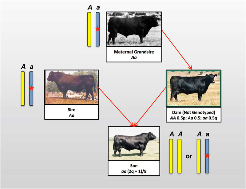
An example of a cattle patrio in which the sire and maternal grandsire are both heterozygous for a rare haplotype “a” at a frequency q = 1-p in the sample and for which no homozygotes are observed. The probability that the son is not homozygous for the a haplotype when it does not carry a lethal mutation is (2q + 1)/8.
Cole et al. (2017) reported 26 recessive haplotypes that are currently tracked in the U.S. dairy breed genomic evaluation system. Of these, two are fertility related in each of Jersey, Brown Swiss and Ayrshire and 8 are fertility related in Holsteins (Table 1). The frequencies of these 12 lethal haplotypes range from 0.37 to 13% and average 11.4%, 7.2%, 1.97% and 6.7% in Ayrshire, Brown Swiss, Holstein and Jersey, respectively. The causal mutations responsible for embryonic lethality have been discovered for only 9 of the 14 haplotypes. Fritz et al. (2013) performed a genome-wide scan in European dairy breeds for homozygous haplotype deficiency using 47 878 Holstein, 16 833 Montbeliarde and 11 466 Normande animals genotyped with the BovineSNP50 and found 18 haplotypes in Holstein, 11 in Montbeliarde and 6 in Normande with frequencies ranging from 1.7 to 9%. Nine of these haplotypes were found to be associated with reductions in fertility when directly tested against conception rate in both heifers and adult cows using heterozygous trio matings, validating the presence of lethal alleles. An additional 8 haplotypes were associated with conception rate in heifers or adult cows. Whole genome sequence data from 25 Holstein, 11 Montbeliarde and 9 Normande bulls with important individual contributions to their respective breeds (from 1.1 to 10.8%), sequenced to a depth of coverage from 8.9–39.2X were investigated in an attempt to identify the deleterious mutations associated with eight of the haplotypes associated with fertility in heifers and cows, leading to strong candidates for two lethal mutations in SHBG and SLC37A2 in the Montbeliarde. Six of the recessive lethal haplotypes detected in French Holsteins coincide with those segregating in the U.S. Holstein population (Cole et al., 2017) which is expected considering the broad international use of U.S. bulls. Sahana et al. (2013) identified 17 homozygote deficient haplotypes at frequencies of from 1.4 to 3.4% in 7 937 Nordic Holsteins genotyped with the BovineSNP50 BeadChip in an analysis in which haplotypes were based on 25 consecutive SNPs. These haplotypes appeared to define eight genomic regions likely to harbour lethal alleles, and of these, six regions were confirmed as having effects on fertility when tested for associations with either non-return rate or calving interval. Remarkably, of all the candidate lethal mutations found in Nordic Holsteins, only the locus on chromosome 21 fully overlaps the locus responsible for Brachyspina in U.S. Holsteins (Cole et al., 2017; Table 1).
Table 1.
Haplotypes or mutations responsible for embryonic lethality discovered by genome-scanning for haplotype or genotype homozygote insufficiency.
| Breed | Haplotype | OMIA1
9 913 ID |
Gene(s)2 | Freq (%) | BTA3 | Region (bp) |
|---|---|---|---|---|---|---|
| Ayrshire4 | AH1 | 1 934 | UBE3B | 13.00 | 17 | 65 921 497 |
| AH2 | 2 134 | RPAP2 | 9.80 | 3 | 51 267 548 | |
| Brown Swiss4 | BH1 | 1 825 | — | 6.67 | 7 | 42 811 272–47 002 161 |
| BH2 | 1 939 | TUBD1 | 7.78 | 19 | 11 063 520 | |
| Holstein-United States4 | HH0 | 151 | FANCI | 2.76 | 21 | 21 184 869–21 188 198 |
| HH1 | 1 | APAF1 | 1.92 | 5 | 63 150 400 | |
| HH2 | 1 823 | — | 1.66 | 1 | 94 860 836–96 553 339 | |
| HH3 | 1 824 | SMC2 | 2.95 | 8 | 95 410 507 | |
| HH4 | 1 826 | GART | 0.37 | 1 | 1 277 227 | |
| HH5 | 1 941 | TFB1M | 2.22 | 9 | 93 223 651–93 370 998 | |
| HHC | 1 340 | SLC35A3 | 1.37 | 3 | 43 412 427 | |
| HCD | 1 965 | APOB | 2.50 | 11 | 77 958 995 | |
| Holstein-France5 | BY | 151 | FANCI | 3.60 | 21 | 20 200 000–22 300 000 |
| HH1 | 1 | APAF1 | 2.60 | 5 | 61 400 000–66 200 000 | |
| HH2 | 1 823 | — | 1.70 | 1 | 93 000 000–97 400 000 | |
| HH3 | 1 824 | SMC2 | 2.50 | 8 | 94 000 000–96 500 000 | |
| HH4 | 1 826 | GART | 3.60 | 1 | 1 900 000–3 300 000 | |
| HH5/HH6 | 1 340 | SLC35A3 | 3.90–4.60 | 3 | 45 800 000–52 600 000 | |
| HH13 | 1 836 | — | 3.70 | 18 | 56 400 000–58 400 000 | |
| Holstein-Nordic6 | 05–1 351/05–1 476 | 1 907 | — | 1.60–2.02 | 5 | 106 713 645–114 405 063 |
| 07–501 | 1 909 | — | 1.92 | 7 | 34 633 456–36 127 497 | |
| 08–1 276/08–1 301/08–1 326/08–1 351 | — | — | 1.48–1.54 | 8 | 83 888 935–89 859 523 | |
| 11–926/11–976/11–1 001/11–1 026 | 1 910 | — | 1.35–1.37 | 11 | 55 345 639–63 759 322 | |
| 19–151 | 1 911 | — | 1.95 | 19 | 13 154 786–14 478 389 | |
| 21–276/21–30½1–326 | — | — | 1.94–2.05 | 21 | 20 477 690–24 844 501 | |
| Holstein-New Zealand7 | — | 2 036 | TTF1 | 3.52 | 11 | 102 485 897–102 515 271 |
| — | 2 037 | RABGGTB | 2.13 | 3 | 69 316 067–69 322 906 | |
| — | 2 038 | RNF20 | 1.82 | 8 | 92 911 255–92 935 750 | |
| Jersey-United States4 | JH1 | 1 697 | CWC15 | 12.10 | 15 | 15 707 169 |
| JH2 | 1 942 | — | 1.30 | 26 | 8 812 759–9 414 082 | |
| Jersey-New Zealand7 | — | 2 035 | OBFC1 | 6.59 | 26 | 24 700 354–24 737 868 |
| Montbeliarde5 | MH1 | 1 827 | SHBG | 9.00 | 19 | 27 600 000–29 400 000 |
| MH2 | 1 828 | SLC37A2 | 7.00 | 29 | 27 900 000–29 100 000 | |
| MH3 | 1 842 | — | 5.10 | 2 | 31 500 000–32 800 000 | |
| MH5 | 1 844 | — | 7.10 | 6 | 73 300 000–74 400 000 | |
| MH6 | 1 845 | — | 2.60 | 7 | 80 100 000–81 700 000 | |
| MH8 | 1 847 | — | 3.50 | 13 | 76 400 000–77 600 000 | |
| Normande5 | NH1 | 1 851 | — | 1.80 | 24 | 38 100 000–39 200 000 |
| NH2 | 1 852 | — | 3.80 | 1 | 145 700 000–146 800 000 | |
| NH5 | 1 829 | — | 1.90 | 7 | 3 600 000–4 600 000 | |
| NH6 | 1 855 | — | 1.90 | 15 | 59 800 000–61 100 000 | |
| Danish Swedish and Finnish Red Cattle8 | A27 | 1 901 | RNASEH2B | 6.50–16.00 | 12 | 20 101 696–20 755 193 |
| Nordic Red Cattle9 | — | BTBD9, GLO1, DNAH8 | 23 | 12 291 761–12 817 087 | ||
| Angus10 | ANH1 | — | — | 2.30 | 1 | 27 786 985–29 095 768 |
| ANH2 | — | — | 7.60 | 4 | 82 467 969–83 996 686 | |
| ANH3 | — | — | 2.30 | 8 | 62 040 920–63 000 189 | |
| ANH4 | — | — | 3.20 | 12 | 59 989 293–61 258 655 | |
| ANH5 | — | — | 3.80 | 15 | 82 317 986–83 144 172 | |
| ANH6 | — | — | 4.50 | 17 | 46 514 063–47 462 424 | |
| ANH7 | — | — | 4.40 | 29 | 43 043 207–44 243 444 | |
| Fleckvieh11 | FH1 | 1 957 | — | 2.90 | 1 | 1 668 494–6 187 555 |
| FH2 | 1 958 | SLC2A2 | 4.10 | 1 | 97 239 973 | |
| FH3 | 1 959 | — | 3.30 | 10 | 26 929 817–35 479 280 | |
| FH4 | 1 960 | SUGT1 | 3.30 | 12 | 11 131 497 | |
| Belgian Blue7 | — | 2 042 | EXOSC4 | 1.33 | 14 | 1 947 198–1 949 074 |
| — | 2 043 | MED22 | 1.15 | 11 | 104 305 076–104 311 650 | |
| — | 2 039 | MYH6 | 4.99 | 10 | 21 325 414–21 344 965 | |
| — | 2 041 | RPIA | 1.89 | 11 | 47 220 160–47 254 704 | |
| — | 2 040 | SNAPC4 | 5.13 | 11 | 103 884 749–103 905 548 | |
Online mendelian inheritance in animals. Taxon ID 9 913 represent cattle.
Multiple listed genes represent a deletion.
Bos taurus chromosome.
Hoff et al. (2017). Haplotypes not validated as fertility associated.
Pausch et al. (2015) performed a genome-wide scan for homozygous haplotype deficiency in 25 544 Fleckvieh cattle using the Illumina BovineSNP50 BeadChip and found four haplotypes (identified as FH1 through FH4) that were deficient in their observed numbers of homozygotes. Two haplotypes were never observed in homozygous form and the frequencies of all four haplotypes ranged from 2.9 to 4.1%. Insemination success was reduced by 6.64% and 5.99% in FH1 and FH4 carrier-to-carrier matings, respectively. A 4.06% decline in insemination success and a 4.3% reduced first-year survival rate of progeny was observed for FH3 carrier-to-carrier matings. Insemination success and stillbirth rate were not affected in FH2 carrier-to-carrier matings; however, juvenile mortality in progeny was increased by 6.6% compared to the survival of progeny from non-risk matings. Using whole genome sequence data from 263 animals from ten different cattle breeds (including 145 Fleckvieh and 15 Simmental) with an average of 10X sequence coverage, these authors identified strong functional candidate mutations underlying two of the haplotypes. A small indel producing a frameshift in SLC2A2 was shown to activate cryptic splice sites in the processed mRNA leading to aberrant splicing at exon 7, while a missense mutation in SUGT1 was predicted to be highly damaging to SGT1 protein function. With many fewer genotyped animals, Hoff et al. (2017) analysed BovineSNP50 data for 3 961 registered Angus animals and identified 7 haplotypes genome-wide that were predicted to harbour autosomal recessive lethal alleles. These were not validated to directly affect fertility or survival rates but ranged in frequency from 2.3 to 7.6%. Despite an analysis of sequence data from 109 bulls re-sequenced to an average 27X depth of coverage from which 1 to 27 bulls (average of 11.4 bulls were sequenced for each haplotype) were predicted to be carriers of each recessive lethal haplotype, no strong candidates for any of the lethal mutations were found despite 6 of the regions being detected as harbouring from 1 to 118 concordant (never homozygous) mutations. Because Hoff et al. (2017) restricted their attention to SNPs, many of the causal variants may be small or large insertions or deletions that are not easily or reliably detected from variant calling pipelines.
Advantages of the haplotype-based analysis approach are that it does not require any reproductive data and can be accomplished as a by-product of genotyping animals to enable genomic selection. As very large numbers of animals are genotyped within each breed (over 2 million Holsteins https://www.cdcb.us/Genotype/cur_density.html and over 300 000 registered Angus https://www.angus.org/pub/newsroom/releases/062717-single-step.html animals have now been genotyped in the U.S.) it becomes possible to detect haplotypes containing autosomal recessive lethal mutations that are at very low frequency within a population. For example, Holstein Haplotype 4 (HH4) is at a frequency of only 0.37% in the U.S. Holstein population (Cole et al., 2017). The disadvantage of the approach is that haplotype based selection is possible only when all chromosomes possessing the haplotype signature harbour the lethal allele leading to the complete absence of homozygotes for the haplotype. For recent mutations, where the lethal allele exists only on some of the chromosomes possessing the haplotype that has a homozygous deficiency, additional family analyses must be performed to identify which families transmit the lethal version of the haplotype and which transmit the viable haplotype. The problem is that the analysis does not actually identify which mutation within the genomic region spanned by the haplotype is the cause of the lethal phenotype. To accomplish this requires sequencing individuals that carry the lethal haplotype to identify candidate mutations that can then be directly tested by genotyping within the population for the absence of homozygotes. Fritz et al. (2013) warn that the existence of strong linkage disequilibrium may lead to the causative variants actually being located outside of the intervals defined by the haplotypes. This appears to be supported by the results in Table 1 which summarize studies performed in Holstein cattle world-wide and reveal that lethal haplotypes detected in different Holstein strains may not completely overlap, although we presume that the same causal variant was detected.
Mesbah-Uddin et al. (2018) utilized 10X average depth of coverage whole genome sequence data produced for 67 Holsteins, 27 Jerseys and 81 Nordic Red Cattle to identify 8 480 large deletions (199 bp – 773 kb; mean 4.5 kb; median 1 kb). The deletions were validated to have an overall false discovery rate of 8.8% using Illumina BovineHD genotype intensity data produced for 26 of the sequenced Holsteins, chromosome breakpoint assembly and alignment and the sequencing of PCR amplicons spanning breakpoints. By examining the deletion genotypes of the sequenced individuals, the authors found 5 000 deletions for which at least one sequenced individual was homozygous. Among these were 167 deleted genes that were demonstrated to be nonessential based on the occurrence of live homozygote individuals. This study is essentially the reciprocal of the haplotype-based approaches just discussed since it can unequivocally identify genes to be non-essential via the existence of living animals that are homozygous knockouts. While the resolution at which candidate lethal mutations are scanned is dramatically increased by considering whole genome sequence variation, the identification of putative lethal mutations is limited by the relatively small sample of sequenced individuals. However, a ~525 kb deletion on chromosome 23, which is known to cause stillbirth in Nordic Red Cattle (Sahana et al., 2016) was among the haplotypes that were not found in homozygous form in the sequenced animals.
Kadri et al. (2014) performed a genome-wide association study (GWAS) in 10 099 bulls from 5 European dairy breeds using estimated breeding values of the bulls for an index of cow fertility traits and found 14 quantitative trait loci (QTLs) at genome-wide significance, of which the most significant was located on chromosome 12. By repeating the analysis within each of the breeds, they identified that this QTL was primarily segregating in Finnish Ayrshire and Swedish Red, but was not detectable in Holstein-Friesian, Danish Red or Jerseys. A haplotype-based analysis identified a single haplotype (A27) that was found to contain a 660 kb deletion that contained four genes, that produced an increase in milk production in heterozygous animals but that was an embryonic lethal in homozygotes likely due to the loss of RNASEH2B. The frequency of this deletion haplotype was 6.5%, 11.5% and 16% in Danish, Swedish and Finnish Red Cattle, respectively.
Charlier et al. (2016) resequenced the genomes of 496 Holstein-Friesian x Jersey animals and 50 Belgian Blue Cattle to an average depth of 11X and resequenced the exomes of 78 animals from six Bos taurus breeds to an average depth of 40X. A total of 186 112 exonic variants, including 1 377 stop-gain, 112 stop-loss, 3 139 frame-shift, 1 341 splice-site, 85 338 missense, and 92 163 synonymous variants were discovered. Of the missense variants, 22 939 were predicted by SIFT and/or PolyPhen to be disruptive or damaging to protein function. From these, 3 779 candidate variants (frame-shift, splice-site, stop-gain and missense variants predicted to be damaging and/or deleterious) for embryonic loss were genotyped in ~35 000 New Zealand dairy cattle (296 LOF and 3 483 missense) and 1 050 were genotyped in ~6 300 Belgian Blue cattle (108 LOF, 942 missense). From the produced genotype data, the authors estimated that 15.5% of the tested LOF variants and 5.9% of the tested missense variants were embryonic lethal mutations. Nine common LOF variants were confirmed to be embryonic lethal mutations based upon the absence of homozygotes in carrier × carrier matings (Table 1).
Table 1 contains a list of haplotypes or mutations recently discovered via genome scans for homozygote insufficiency that have been demonstrated to affect fertility in several beef and dairy breeds. There are several remarkable observations from the data in this table. First, the average frequency of lethal mutations detected in cattle populations to date is about 3.8%, which is much larger than you would expect for loci subjected to strong purifying selection. This is clearly due to the extensive use of AI in these populations, which results in the strong enrichment of alleles present within the genomes of selected bulls that go on to have large numbers of progeny. However, the number of lethal loci detected to date in numerically large breeds such as Holstein (17) is far greater than for the numerically smaller breeds such as Jersey (3), Brown Swiss (2) and Ayrshire (2). Furthermore, the average frequency of lethal alleles in Holsteins (2.4%) is considerably lower than in Jersey (6.66%), Brown Swiss (7.23%) and Ayrshire (11.4%). This likely reflects a greater number of genotyped animals used to detect a larger number of rarer alleles, but it also may suggest that the larger number of bulls used in AI in numerically large breeds allows a greater enrichment of lethal loci in the breed, albeit at lower individual frequencies. While it is difficult to make conclusions about the identities of mutations based on haplotype data (Charlier et al., 2016 found three lethal mutations located within a 1.82 Mb region of chromosome 11 in two breeds) the data in Table 1 suggest that the majority of the common lethal alleles are breed specific. That is, these mutations occurred after breed formation and were driven to relatively high frequencies within the respective breeds by the use of selective breeding. On the other hand, there is also evidence for potentially different lethal loci segregating within subpopulations of Holsteins. Despite the widespread use of high merit U.S. Holstein bulls world-wide, the loci found by Sahana et al. (2013) in Nordic Holsteins are not completely consistent with those of Fritz et al. (2013) for French Holsteins which are largely consistent with those in the summary of Cole et al. (2017) for U.S. Holsteins. This would be expected if the majority of lethal mutations are quite recent in origin and if there is subdivision, possibly caused by different selection objectives in each of the subpopulations.
Cole et al. (2016) estimated that the economic losses due to reduced fertility and perinatal calf death in U.S. Holsteins was almost $11 million per year and Charlier et al. (2016) estimated the cost due to embryonic losses from 9 confirmed lethal loci to be NZ$13.8 million in New Zealand dairy cattle and €2.7 million in Belgian Blue cattle. The use of available genotype data to detect and avoid carrier x carrier matings is clearly an effective way to improve the fertility of cattle, but may become difficult to implement as the number of detected lethal mutations and genetic defects increases within individual breeds. Cole (2015) has suggested an alternative approach in which the estimated genetic merits of individuals for net merit are adjusted for the economic losses due to fetal losses and has shown the method to be effective at reducing the frequency of recessive lethal alleles, whilst maintaining current rates of genetic improvement. Lethal alleles are also excellent candidates for multiplex genome editing in which the heterozygous deleterious alleles are simply removed from the genomes of the bulls that are placed into AI each generation (Hickey et al., 2016).
Non-Mendelian effects on male fertility
Loss of function lethal mutations behave in a manner that is called Mendelian inheritance because the ability of an allele to impact the viability of a progeny is the same if the allele is transmitted through the male or female germ lines. However, loci that are transmitted through the mitochondrial genome (or genomes in cases of heteroplasmy; about 5% of individuals within mammalian species appear to have at least two mitochondrial genomes), on the Y chromosome, or on the X chromosome can cause patterns of inheritance that are non-Mendelian. There are also cases of variants that are located on the non-sex associated autosomes that lead to phenotypes that are inherited in a non-Mendelian fashion. Imprinted loci are transmitted through the germ-line with either alleles transmitted by females being silenced (female imprinting in which case the male inherited allele is expressed) usually by DNA methylation or alleles transmitted by males being silenced (male imprinting). Each generation, the methylation status of these alleles is reset according to the sex of individual. Flisikowski et al. (2010) reported the segregation of a lethal locus in which the mode of inheritance appeared to be autosomal dominant with incomplete penetrance (not all individuals possessing the lethal allele die). Semen from a Finnish Ayrshire bull was used to artificially inseminate 1 900 Finnish Ayrshire heifers and cows and field data collected by the AI Cooperative suggested that 42.6% of the late pregnancies attributed to the bull ended in stillbirths or abortions. This figure is close enough to a 50:50 segregation ratio to suggest that the bull was heterozygous for a mutation that was transmitted as a dominant lethal. The fact that lethality was slightly less than 50% suggested that there could have been some progeny that inherited the lethal allele that did not die, presumably because they were protected by variants at other loci and this might explain why the bull itself escaped death. However, the causal variant was shown to be a 110 kb deletion within the MIMT1 gene, which is part of the PEG3 (paternally expressed gene) domain. Since the alleles transmitted by males are expressed (the maternal alleles are silenced by methylation), both copies of MIMT1 were transmitted to progeny in activated form but progeny that inherited the deletion allele had no functional MIMT1 expression since all MIMT1 copies transmitted by their dams were silenced. The fact that the bull survived the inheritance of a lethal mutation was because he inherited the deletion MIMT1 allele in a silenced form from his dam and the functional MIMT1 allele was inherited from his sire. Thus, LOF mutations in MIMT1 have absolutely no effect on female fertility but are lethal when transmitted by the sire. MIMT1 encodes a non-coding RNA, which is not translated into a protein and has an unknown function. However, this naturally occurring mutation demonstrates that MIMT1 is essential to life. Magee et al. (2010) have also shown that SNPs within the PEG3 gene cluster are associated with calving, calf performance and fertility traits in Irish Holstein-Friesian cattle.
The bovine Y chromosome comprises a small pseudo-autosomal region with a homolog on the X chromosome and a much larger male-specific region which contains clusters of genes thought to be essential for male reproduction because they are primarily expressed during testicular development (Yang et al., 2011; Chang et al., 2013). The number of genes on the bovine Y chromosome appears to be surprisingly large relative to the number of genes found on the human Y chromosome suggesting a much greater potential role of the Y chromosome in phenotype determination, particularly male fertility, in cattle than in humans (Chang et al., 2013). However, there appears to have been surprisingly little work conducted to date to characterize the effects of mutations in bovine Y chromosome genes on male fertility. This is probably due to the fact that until recently, there was no reference sequence assembly for the bovine Y chromosome and due to the highly repetitive and palindromic nature of the Y chromosome sequence, the existing assembly is of much lower quality than the assembly for the autosomal genome. One study has shown that the number of copies of members of the HSFY and ZNF280BY gene families varies by almost an order of magnitude in cattle (from ~20 to over 300 copies) and that variation in copy number of both families was negatively correlated with testis size, but positively correlated with SCR (Yue et al., 2014). Because the number of copies of members of both families within individual bulls is positively correlated (Yue et al., 2014), it is not clear which family (if either) was causal for effects on male fertility. Copy number variation in members of the PRAMEY gene family encoding proteins found in the sperm head and tail has been shown to be negatively correlated with percentage of normal sperm and non-return rate, but not with SCR in Holstein bulls (Yue et al., 2013).
Mutations within genes or regulatory regions on the X chromosome have the potential to be severely deleterious to fertility in males who have only a single copy of the X chromosome and far less so in females who have two copies of the X chromosome, although one copy is presumably randomly inactivated in each cell within every tissue. Because of this, and like Y chromosome mutations, these variants are exposed to extremely strong purifying selection in males. Consequently, we might expect to find many fewer X and Y chromosome mutations affecting fertility than autosomal mutations. Despite this, using a GWAS in indicine and indicine x taurine bulls, Fortes et al. (2013b) found that the majority of genome-wide associations for scrotal circumference and percentage of normal spermatozoa at 24 months of age were located on the X chromosome. De Camargo et al. (2015) examined the effects of 7 SNPs responsible for amino acid substitutions in 7 genes located in regions of the X chromosome previously identified by the GWAS of Fortes et al. (2013b) in the same experimental population and detected significant associations for SNPs in LOC100138021, CENPI and TAF7L with percentage of normal spermatozoa (Table 2) and for SNPs in TEX11 and AR with scrotal circumference. None of the SNPs detected as being associated with male fertility were found to be associated with female fertility. A brief overview of effects of chromosomal aberrations and structural variation on male fertility is presented in Supplementary Material S1.
Table 2.
Genomic regions identified by genome-wide association analysis or candidate gene analysis as associated with sire conception rate.
| SNP | Location (Chr:bp) | Gene(s)1 |
|---|---|---|
| rs1361956182 | 7:41 208 950 | PROP1 |
| SNP116464,5 | 17:35 247 641–35 247 4906 | FGF2 |
| SNP121954,5 | 19:43 045 658–43 046 4776 | STAT5A |
| 1.5 Mb window7 | 5:105 357 507–106 813 133 | PARP11, AKAP3 |
| rs425593737,8 | 9:11 867 269 | RIMS1 |
| 1.5 Mb window7 | 13:58 456 868–59 951 247 | CTCFL, SPO11 |
| rs422961087 | 15:26 472 899 | CADM1 |
| 1.5 Mb window7 | 21:8 031 396–9 528 223 | IGF1R |
| 1.5 Mb window7 | 21:68 846 429–70 294 301 | TDRD9, CKB |
| rs1106973187 | 21:71 210 609 | BRF1 |
| 1.5 Mb window7,8 | 25:3 148 958–4 647 188 | MGRN1, SEPT12 |
| 1.5 Mb window7 | 25:26 736 589–28 233 820 | CCT6A |
| rs415675167 | 25:27 477 941 | KAT8 |
| rs4206518327,9 | 25:27 672 891 | ITGAM |
| rs416496817 | 25:28 711 626 | TYW1 |
| rs4164228310 | 2:24 837 034 | DYNC1I2 |
| rs10905943810 | 5:112 775 479 | LOC784935 |
| rs4157187810 | 18:54 965 977 | ZNF541 |
| rs1106296618,10 | 25:983 759 | CACNA1H |
| rs1097582488,10 | 25:2 270 518 | LOC617302 |
| rs1101298888,10 | 25:3 898 682 | ROGDI |
| rs1103832248,10 | 25:4 229 011 | LOC521021 |
| rs4248022310 | 29:14 271 174 | - |
| rs412571811,12 | 1:69 802 307 | ITGB5 |
| rs290248611,12 | 4:11 622 140 | COL1A2-AS1 |
| rs290155748,12 | 9:12 042 726 | RIMS1 |
| rs2901687512 | 10:6 003 240 | SFNX1, DRD1 |
SNP located within or near identified gene or window contains the listed genes.
Sire conception rate.
Estimated relative conception rate.
PCR amplicon contains identified SNP but exact reference genome position not provided.
QTL validated in two or more studies.
rs number corresponds to marker name but provided chromosomal coordinates are not correct.
Validated in follow-up analysis of expanded genotyped sample.
Quantitative trait loci responsible for variation in male fertility
The evolution of DNA methylation probably occurred as a method to silence the transcriptional activity of retrotransposons that were integrated into the germ-line via retroviruses (Nagamori et al., 2015). DNA methylation appears to be ubiquitous in mammalian genomes; however, variation in the extent of DNA methylation at specific loci is known to occur and this variation has been associated with the quantitative regulation of gene expression. By examining the methylation profiles of DNA extracted from the spermatozoa of bulls with high- and low-conception rates, Verma et al. (2014) found differentially methylated regions associated with 151 genes with functions in germ cell development, spermatogenesis, capacitation, and embryonic development in water buffalo. Using a similar approach in Holstein bulls identified as being extreme for SCR based upon at least 300 inseminations, Kropp et al. (2017) found 76 genomic regions to be differentially methylated in the DNA extracted from the spermatozoa of high and low-SCR bulls. What is not clear from these epigenetic studies is whether the detected differential methylation is the cause of differences in fertility or the effect of some other genomic mechanism that is responsible for the aberrant methylation of sequences regulating the expression of genes that are required for high fertility. Similarly, several studies have found differences in the spermatozoa mRNA transcript (Card et al., 2017), miRNA and piRNA (Capra et al., 2017) and protein (Peddinti et al., 2008) abundances. While these approaches have been pursued from the perspective of developing biomarkers of male fertility, the capability of the differentially abundant molecules to predict variation in male fertility has yet to be established.
There is an intrinsic relationship between GWAS studies and genomic selection. In GWAS, a large number of markers approximately evenly spread throughout the genome are assayed in a sample of phenotyped individuals and maker effects are individually tested to identify those that meet a pre-specified statistical threshold. This approach identifies the largest effect associations within the genome, based on the available sample size, and ignores the potential myriad of markers with small effects on the trait. These large effect markers can be used to generate molecular estimates of genetic merit, but typically these explain only small percentages of the overall additive genetic variance. In other words, there are relatively few markers of large effect and a much larger (but unknown) number of small effect markers. On the other hand, genomic selection attempts to use all of the markers (or a reasonably large subset of the markers when some Bayesian analyses are utilized) to predict genetic merit, capturing many more of the small effect variants, and can produce estimates of genetic merit that frequently explain at least 70% of the additive genetic variance in traits. In terms of application, it has historically been simpler and less expensive to genotype 10 or 20 markers in a large sample of individuals than many thousands of markers. However, with the recent deployment and very rapid adoption of high-density SNP arrays for genotyping in cattle and the impact that genomic selection has had on the improvement of female fertility in U.S. Holsteins (García-Ruiz et al., 2016), it is clear that the development of predictions of genetic merit for SCR should be a priority in cattle genomics. Two limitations to the approach are that all of the early industry genotyping was performed using the Illumina BovineSNP50 assay which did not include Y chromosome variants (although all of the newer assays do), and the currently utilized statistical analyses do not appropriately model the effects of X chromosome markers (Taylor, 2014). Imprinted loci are probably approximately correctly modelled in analyses of data from only a single sex, but are not correctly modelled in analyses of data from both sexes.
Feugang et al. (2009) estimated conception rates for 874 U.S. Holstein bulls with an average of 788 breedings (range 101 to 11 997) in a probit analysis after adjusting for herd-year-month, parity, cow, days in milk and sire proven status. The 10 highest and 10 lowest fertility bulls (mean difference 15.4% in SCR) were genotyped using the Affymetrix/ParAllele 9 919 SNP GeneChip and 8 207 polymorphic markers were analysed in a single marker regression of allele dosage on each bull’s SCR phenotype. The four most strongly associated SNPs (P<0.0001; Table 2) were then genotyped in a larger cohort of 100 low- and 101 high-fertility bulls and the SNPs on chromosomes 1 and 4 were validated as being associated with SCR (P<0.05). Peñagaricano et al. (2012) performed a GWAS in 1 755 Holstein bulls with SCR data using 38 650 SNPs with minor allele frequencies >5% using a linear model correcting for relatedness among bulls and testing the effects of SNPs individually either fit with genotypes as additive, or additive and dominance effects. After correcting for multiple testing, they found 8 SNPs defining 5 separate QTLs associated with SCR. Han and Peñagaricano (2016) performed a GWAS using 44 449 estimated SCR animal effects (additive genetic + non-additive genetic and permanent environment effects; Paul VanRaden, USDA ARS, pers. comm.) available on 10 884 U.S. Holstein bulls. Of these animals, 7 447 had high-density SNP genotype data and after filtering markers with minor allele frequencies <1% or that were sex-linked, 58 029 autosomal markers were analysed. A single-step BLUP analysis was used incorporating pedigree information for the animals that had not been genotyped. The percentage of additive genetic variance in SCR explained by all of the SNPs within 1.5 Mb genomic regions was estimated and significant regions were declared when they explained at least 0.5% of the SCR additive genetic variance. The authors also performed a single-SNP analysis in which the mixed model included the effects described above but only for the genotyped animals and found results that were consistent with those from the single-step BLUP. Han and Peñagaricano (2016) estimated that the SNPs explained 32% of the variance in SCR animal effects and, consequently, 68% of the unexplained variance was due to additive genetic variance not captured by the SNPs, and the service sire non-additive genetic and permanent environmental effects. Han and Peñagaricano (2016) also found six SCR QTLs located on chromosomes 5, 13, 21 and 25 that explained at least 0.5% of the SCR additive genetic variance (Table 2).
Since all three studies were conducted in U.S. Holstein bulls, we should expect considerable consistency among the results and also for candidate gene studies previously reporting significant associations with SCR (Khatib et al., 2010; Lan et al., 2013). However, Table 2 shows that only two loci were consistently detected in at least two different studies in U.S. Holsteins. Associations with RIMS1 on chromosome 9 between 11.8 and 12.1 Mb were detected by Feugang et al. (2009) and Han and Peñagaricano (2016). Associations were also detected on chromosome 25 in the region from 0.9 to 4.7 Mb by Peñagaricano et al. (2012) and Han and Peñagaricano (2016). This is somewhat typical of GWAS studies particularly when they are underpowered and there are few variants of large effect responsible for trait variation. Druet et al. (2009) performed a GWAS with 148 microsatellite markers in 10 families containing 515 French Holstein bulls for semen production phenotypes of ejaculated volume and sperm concentration, number of spermatozoa, motility, velocity, percentage of motile spermatozoa after thawing and abnormal spermatozoa. Of the 11 detected QTL, only two affecting ejaculated volume (chromosome 15 at 22 cM) and sperm motility (chromosome 7 at 34 cM) appear to overlap with the SCR QTL reported in Table 2.
Abdollahi-Arpanahi et al. (2017) used the SCR data for 7 447 bulls that was employed by Han and Peñagaricano (2016) for GWAS to evaluate the utility of genomic selection to predict the genetic merit of AI bulls for SCR using a 5-fold cross-validation scheme and SNP feature selection. Using all 54 706 fitted SNP, the average correlation between predicted genetic merit and SCR phenotype was 0.34 corresponding to a prediction accuracy of approximately 0.63. Selecting SNPs within genes with Gene Ontology and Medical Subject Heading terms including reproduction, fertilization, sperm motility or sperm capacitation did not improve prediction accuracy. However, restricting the analysis to the 18 659 SNPs detected as being associated (Pnominal<0.05) with SCR increased the correlation slightly to 0.35. Non-linear models uniformly outperformed linear models for accuracy of prediction, but the improvement was generally fairly small.
Conclusions
Male and female fertility are positively correlated but the correlation is low and genetic predictions for fertility are currently only produced for females. Genomic selection has produced dramatic increases in female fertility in a relatively short period of time in U.S. Holsteins demonstrating that a low heritability is not the sole determinant of selection response. While this improvement should also have produced a small correlated response in male fertility, this is an unsatisfactory solution considering the economic importance of fertility to cattle production and the need to increase the efficiency and quantity of animal-based food proteins world-wide.
In dairy cattle, there is an opportunity to rapidly develop genomic predictions for male fertility (in both sexes) considering the large number of genotyped animals and the availability of SCR phenotypes. However, these phenotypes should be based on inseminations made by yearling bulls in which sperm dosages have been standardised and this will require collaboration between AI organizations. In the U.S. beef industry, the majority of genetic improvement in all traits is created by selection within the registered sector. Despite the reduced use of AI relative to the dairy industry, it should similarly be possible to capture the benefits of increased rates of genotyping to develop genetic predictions for SCR. The increasing use of sexed semen to produce heifer replacements within the U.S. dairy industry also presents an opportunity for the generation of SCR data for beef bulls, since sexed male semen from beef bulls is increasingly being used to breed dairy cows that were not selected to produce heifer replacements. Scoring conception rates in dairy cows is agnostic to the breed of bulls used in AI. Finally, increasing the rate of use of AI in commercial beef herds via the use of synchronization of oestrus and ovulation to facilitate fixed-time AI of beef cows has an enormous opportunity for the collection of SCR phenotypes in beef bulls. If 10% of the commercial beef cows in the U.S. were bred by AI, the industry could collect more SCR phenotypes than are currently produced within the entire dairy industry. If genomic predictions of merit for male fertility are to be produced for both males and females, efforts should be invested to develop and evaluate models that appropriately model the effects of sex chromosome and imprinted variants.
Supplementary Material
Implications.
Male and female fertility are lowly heritable in cattle, but the reduction in generation interval achieved through the deployment of genomic selection has allowed a rapid improvement of female fertility in U.S. dairy cattle. For several logistical reasons, genetic evaluations are not widely produced for male fertility in either beef or dairy cattle. Because the genetic correlation between male and female fertility is low, little improvement in male fertility is expected as a correlated response to selection for female fertility. To maximise food production from cattle, new approaches must be developed to improve male fertility independently of female fertility.
Acknowledgements
The authors appreciate the support of NIH-USDA Dual Purpose with Dual Benefit Program grant number NIH 1R01HD084353. PS is also supported by USDA-NIFA grant 2015–67015-23231 and by seed funding from the Food for the 21st Century Program of the University of Missouri. JT and RS are supported by USDA-NIFA grants 2013–68004-20364, 2016–67015-24923 and 2017–67015-26760. The authors also appreciate the constructive criticisms of two anonymous reviewers who appreciably improved the manuscript.
Footnotes
Ethics Statement
No animals were used in this review study.
Software and data repository resources
No software or unpublished data were used in this review.
Declaration of interest
The authors have no conflicts of interest.
References
- Abdollahi-Arpanahi R, Morota G and Peñagaricano F 2017. Predicting bull fertility using genomic data and biological information. Journal of Dairy Science 100, 9656–9666. [DOI] [PubMed] [Google Scholar]
- Berry DP, Wall E and Pryce JE 2014. Genetics and genomics of reproductive performance in dairy and beef cattle. Animal 8 (suppl. 1), 105–121. [DOI] [PubMed] [Google Scholar]
- Capra E, Turri F, Lazzari B, Cremonesi P, Gliozzi TM, Fojadelli I, Stella A and Pizzi F 2017. Small RNA sequencing of cryopreserved semen from single bull revealed altered miRNAs and piRNAs expression between High- and Low-motile sperm populations. BMC Genomics 18, 14. [DOI] [PMC free article] [PubMed] [Google Scholar]
- Card CJ, Krieger KE, Kaproth M and Sartini BL 2017. Oligo-dT selected spermatozoal transcript profiles differ among higher and lower fertility dairy sires. Animal Reproduction Science 177, 105–123. [DOI] [PubMed] [Google Scholar]
- Chang TC, Yang Y, Retzel EF and Liu WS 2013. Male-specific region of the bovine Y chromosome is gene rich with a high transcriptomic activity in testis development. Proceedings of the National Academy of Science U S A 110, 12373–12378. [DOI] [PMC free article] [PubMed] [Google Scholar]
- Charlier C, Li W, Harland C, Littlejohn M, Coppieters W, Creagh F, Davis S, Druet T, Faux P, Guillaume F, Karim L, Keehan M, Kadri NK, Tamma N, Spelman R and Georges M 2016. NGS-based reverse genetic screen for common embryonic lethal mutations compromising fertility in livestock. Genome Research 26, 1333–1341. [DOI] [PMC free article] [PubMed] [Google Scholar]
- Chen W-H, Lu G, Chen X, Zhao X-M and Bork P 2017. OGEE v2: an update of the online gene essentiality database with special focus on differentially essential genes in human cancer cell lines. Nucleic Acids Research 45, D940–D944. [DOI] [PMC free article] [PubMed] [Google Scholar]
- Cole JB 2015. A simple strategy for managing many recessive disorders in a dairy cattle breeding program. Genetics Selection Evolution 47, 94. [DOI] [PMC free article] [PubMed] [Google Scholar]
- Cole JB, Null DJ and VanRaden PM 2016. Phenotypic and genetic effects of recessive haplotypes on yield, longevity, and fertility. Journal of Dairy Science 99, 7274–7288. [DOI] [PubMed] [Google Scholar]
- Cole JB, VanRaden PM, Null DJ, Hutchison JL, Cooper TA and Hubbard SM 2017. Haplotype tests for recessive disorders that affect fertility and other traits. Available at: https://aipl.arsusda.gov/reference/recessive_haplotypes_ARR-G3.html Accessed 11/30/17. [Google Scholar]
- David I, Bodin L, Lagriffoul G, Leymarie C, Manfredi E and Robert-Granié C 2007. Genetic analysis of male and female fertility after artificial insemination in sheep: comparison of single-trait and joint models. Journal of Dairy Science 90, 3917–3923. [DOI] [PubMed] [Google Scholar]
- De Camargo GM, Porto-Neto LR, Kelly MJ, Bunch RJ, McWilliam SM, Tonhati H, Lehnert SA, Fortes MR and Moore SS 2015. Non-synonymous mutations mapped to chromosome X associated with andrological and growth traits in beef cattle. BMC Genomics 16, 384. [DOI] [PMC free article] [PubMed] [Google Scholar]
- DeJarnette JM, Nebel RL and Marshall CE 2010. Understanding estimates of AI sire fertility. Available at http://www.selectsires.com/programs/docs/UnderstandingAISireFertility.pdf?version=20170404 Accessed 12/18/2017.
- Druet T, Fritz S, Sellem E, Basso B, Gérard O, Salas-Cortes L, Humblot P, Druart X and Eggen A 2009. Estimation of genetic parameters and genome scan for 15 semen characteristics traits of Holstein bulls. Journal of Animal Breeding and Genetics 126, 269–277. [DOI] [PubMed] [Google Scholar]
- Feugang JM, Kaya A, Page GP, Chen L, Mehta T, Hirani K, Nazareth L, Topper E, Gibbs R and Memili E 2009. Two-stage genome-wide association study identifies integrin beta 5 as having potential role in bull fertility. BMC Genomics 10, 176. [DOI] [PMC free article] [PubMed] [Google Scholar]
- Flisikowski K, Venhoranta H, Nowacka-Woszuk J, McKay SD, Flyckt A, Taponen J, Schnabel R, Schwarzenbacher H, Szczerbal I, Lohi H, Fries R, Taylor JF, Switonski M and Andersson M 2010. A novel mutation in the maternally imprinted PEG3 domain results in a loss of MIMT1 expression and causes abortions and stillbirths in cattle (Bos taurus). PLoS One 5, e15116. [DOI] [PMC free article] [PubMed] [Google Scholar]
- Fortes MRS, DeAtley KL, Lehnert SA, Burns BM, Reverter A, Hawken RJ, Boe-Hansen G, Moore SS and Thomas MG 2013a. Genomic regions associated with fertility traits in male and female cattle: Advances from microsatellites to high-density chips and beyond. Animal Reproduction Science 141, 1–19. [DOI] [PubMed] [Google Scholar]
- Fortes MR, Reverter A, Kelly M, McCulloch R and Lehnert SA 2013b. Genome-wide association study for inhibin, luteinizing hormone, insulin-like growth factor 1, testicular size and semen traits in bovine species. Andrology 1, 644–650. [DOI] [PubMed] [Google Scholar]
- Fritz S, Capitan A, Djari A, Rodriguez SC, Barbat A, Baur A, Grohs C, Weiss B, Boussaha M, Esquerre D, Klopp C, Rocha D and Boichard D 2013. Detection of haplotypes associated with prenatal death in dairy cattle and identification of deleterious mutations in GART, SHBG and SLC37A2. PLoS One 8, e65550. [DOI] [PMC free article] [PubMed] [Google Scholar]
- García-Ruiz A, Cole JB, VanRaden PM, Wiggans GR, Ruiz-López FJ and Van Tassell CP 2016. Changes in genetic selection differentials and generation intervals in US Holstein dairy cattle as a result of genomic selection. Proceedings of the National Academy of Sciences U S A 113, E3995–E4004. [DOI] [PMC free article] [PubMed] [Google Scholar]
- Han Y and Peñagaricano F 2016. Unravelling the genomic architecture of bull fertility in Holstein cattle. BMC Genetics 17, 143. [DOI] [PMC free article] [PubMed] [Google Scholar]
- Hansen M 1979. Genetic investigations on male and female fertility in cattle. Livestock Science 6, 325–334. [Google Scholar]
- Hickey JM, Bruce C, Whitelaw A and Gorjanc G 2016. Promotion of alleles by genome editing in livestock breeding programmes. Journal of Animal Breeding and Genetics 133, 83–84. [DOI] [PubMed] [Google Scholar]
- Hoff JL, Decker JE, Schnabel RD and Taylor JF 2017. Candidate lethal haplotypes and causal mutations in Angus cattle. BMC Genomics 18, 799. [DOI] [PMC free article] [PubMed] [Google Scholar]
- Kadri NK, Sahana G, Charlier C, Iso-Touru T, Guldbrandtsen B, Karim L, Nielsen US, Panitz F, Aamand GP, Schulman N, Georges M, Vilkki J, Lund MS and Druet T 2014. A 660-Kb deletion with antagonistic effects on fertility and milk production segregates at high frequency in Nordic Red cattle: additional evidence for the common occurrence of balancing selection in livestock. PLoS Genetics 10, e1004049. [DOI] [PMC free article] [PubMed] [Google Scholar]
- Khatib H, Monson RL, Huang W, Khatib R, Schutzkus V, Khateeb H and Parrish JJ 2010. Short communication: Validation of in vitro fertility genes in a Holstein bull population. Journal of Dairy Science 93, 2244–2249. [DOI] [PubMed] [Google Scholar]
- Kropp J, Carrillo JA, Namous H, Daniels A, Salih SM, Song J and Khatib H 2017. Male fertility status is associated with DNA methylation signatures in sperm and transcriptomic profiles of bovine preimplantation embryos. BMC Genomics 18, 280. [DOI] [PMC free article] [PubMed] [Google Scholar]
- Lan XY, Peñagaricano F, DeJung L, Weigel KA and Khatib H 2013. Short communication: A missense mutation in the PROP1 (prophet of Pit 1) gene affects male fertility and milk production traits in the US Holstein population. Journal of Dairy Science 96, 1255–1257. [DOI] [PubMed] [Google Scholar]
- Magee DA, Sikora KM, Berkowicz EW, Berry DP, Howard DJ, Mullen MP, Evans RD, Spillane C and MacHugh DE 2010. DNA sequence polymorphisms in a panel of eight candidate bovine imprinted genes and their association with performance traits in Irish Holstein-Friesian cattle. BMC Genetics 11, 93. [DOI] [PMC free article] [PubMed] [Google Scholar]
- Matukumalli LK, Lawley CT, Schnabel RD, Taylor JF, Allan MF, Heaton MP, O’Connell J, Moore SS, Smith TPL, Sonstegard TS and Van Tassell CP 2009. Development and characterization of a high density SNP genotyping assay for cattle. PLoS One 4, e5350. [DOI] [PMC free article] [PubMed] [Google Scholar]
- Mesbah-Uddin M, Guldbrandtsen B, Iso-Touru T, Vilkki J, De Koning DJ, Boichard D, Lund MS and Sahana G 2018. Genome-wide mapping of large deletions and their population-genetic properties in dairy cattle. DNA Research 25, 49–59. [DOI] [PMC free article] [PubMed] [Google Scholar]
- Meuwissen TH, Hayes BJ and Goddard ME 2001. Prediction of total genetic value using genome-wide dense marker maps. Genetics 157, 1819–1829. [DOI] [PMC free article] [PubMed] [Google Scholar]
- Meyer K, Hammond K, Mackinnon MJ and Parnell PF 1991. Estimates of covariances between reproduction and growth in Australian beef cattle. Journal of Animal Science 69, 3533–3543. [DOI] [PubMed] [Google Scholar]
- Nagamori I, Kobayashi H, Shiromoto Y, Nishimura T, Kuramochi-Miyagawa S, Kono T and Nakano T 2015. Comprehensive DNA methylation analysis of retrotransposons in male germ cells. Cell Reports 12, 1541–1547. [DOI] [PubMed] [Google Scholar]
- Pausch H, Schwarzenbacher H Burgstaller J Flisikowski K Wurmser C Jansen S Jung S Schnieke A Wittek T and Fries R 2015. Homozygous haplotype deficiency reveals deleterious mutations compromising reproductive and rearing success in cattle. BMC Genomics 16, 312–325. [DOI] [PMC free article] [PubMed] [Google Scholar]
- Peddinti D, Nanduri B, Kaya A, Feugang JM, Burgess SC and Memili E 2008. Comprehensive proteomic analysis of bovine spermatozoa of varying fertility rates and identification of biomarkers associated with fertility. BMC Systems Biology 2, 19. [DOI] [PMC free article] [PubMed] [Google Scholar]
- Peñagaricano F, Weigel KA and Khatib H 2012. Genome-wide association study identifies candidate markers for bull fertility in Holstein dairy cattle. Animal Genetics 43 (suppl. 1), 65–71. [DOI] [PubMed] [Google Scholar]
- Piles M and Tusell L 2012. Genetic correlation between growth and female and male contributions to fertility in rabbit. Journal of Animal Breeding and Genetics 129, 298–305. [DOI] [PubMed] [Google Scholar]
- Sahana G, Nielsen US, Aamand GP, Lund MS and Guldbrandtsen B 2013. Novel harmful recessive haplotypes identified for fertility traits in Nordic Holstein cattle. PLoS ONE 8, e82909. [DOI] [PMC free article] [PubMed] [Google Scholar]
- Sahana G, Iso-Touru T, Wu X, Nielsen US, de Koning DJ, Lund MS, Vilkki J and Guldbrandtsen B 2016. A 0.5-Mbp deletion on bovine chromosome 23 is a strong candidate for stillbirth in Nordic Red cattle. Genetics Selection Evolution 48, 35. [DOI] [PMC free article] [PubMed] [Google Scholar]
- Syrstad O 1981. Selection for fertility on the basis of AI data. Livestock Production Science 8, 247–252. [Google Scholar]
- Taylor JF 2014. Implementation and accuracy of genomic selection. Aquaculture 420–421, S8–S14. [Google Scholar]
- Taylor JF, Schnabel RD and Sutovsky P 2018. Identification of genomic variants causing sperm abnormalities and reduced male fertility. Animal Reproduction Science pii: S0378–4320(17)31072–2. [DOI] [PMC free article] [PubMed] [Google Scholar]
- VanRaden PM, Olson KM, Null DJ and Hutchison JL 2011. Harmful recessive effects on fertility detected by absence of homozygous haplotypes. Journal of Dairy Science 94, 6153–6161. [DOI] [PubMed] [Google Scholar]
- Verma A, Rajput S, De S, Kumar R, Chakravarty AK and Datta TK 2014. Genome-wide profiling of sperm DNA methylation in relation to buffalo (Bubalus bubalis) bull fertility. Theriogenology 82, 750–759. [DOI] [PubMed] [Google Scholar]
- Wolc A, White IM, Olori VE and Hill WG 2009. Inheritance of fertility in broiler chickens. Genetics Selection Evolution 41, 47. [DOI] [PMC free article] [PubMed] [Google Scholar]
- Yang Y, Chang TC, Yasue H, Bharti AK, Retzel EF and Liu WS 2011. ZNF280BY and ZNF280AY: autosome derived Y-chromosome gene families in Bovidae. BMC Genomics 12, 13. [DOI] [PMC free article] [PubMed] [Google Scholar]
- Yue XP, Chang TC, DeJarnette JM, Marshall CE, Lei CZ and Liu WS 2013. Copy number variation of PRAMEY across breeds and its association with male fertility in Holstein sires. Journal of Dairy Science 96, 8024–8034. [DOI] [PubMed] [Google Scholar]
- Yue XP, Dechow C, Chang TC, DeJarnette JM, Marshall CE, Lei CZ and Liu WS 2014. Copy number variations of the extensively amplified Y-linked genes, HSFY and ZNF280BY, in cattle and their association with male reproductive traits in Holstein bulls. BMC Genomics 15, 113. [DOI] [PMC free article] [PubMed] [Google Scholar]
Associated Data
This section collects any data citations, data availability statements, or supplementary materials included in this article.