. 2019 May 3;8:e44760. doi: 10.7554/eLife.44760

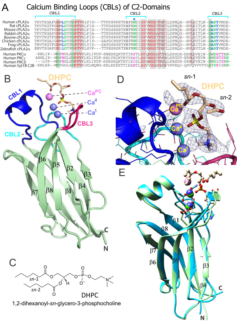
Figure 1. Structure of cPLA2α C2‒domain containing bound DHPC and calcium.

(A) Sequence alignment of C2-domain calcium-binding loop (CBL) regions in cPLA2α from different eukaryotes compared to human PKCs and Syt1. Residues that bind Ca2+ are green. Residues interacting directly with PC in our structural complex (blue or blue asterisk) are absolutely conserved among eukaryotic cPLA2α proteins but not in PKCs and Syt1. Conversely, residues that interact with PS in the PKCα-PS structure (magenta) are highly conserved in PKCs and Syt1, but not in cPLA2α. Shaded residues are identical. The human and chicken cPLA2α CBL sequences are 92% identical and 94.5% highly conserved (see Figure 1—figure supplement 1 for full-length sequence alignment). (B) Ribbon structure representation of the cPLA2α C2-domain bound to 1,2-dihexanoyl-sn-glycero-3-phosphocholine (DHPC). The DHPC molecule (beige stick) straddles the β1–β2 loop (CBL1, blue), β3–β4 loop (CBL2, cyan) and β5–β6 loop (CBL3, red). Ca1 and Ca4 (blue spheres) are in a similar position in the apo-form structure; whereas CaPC (magenta sphere) is unique to the DHPC-bound form. (C) DHPC structural formula. (D) Fo-Fc omit electron density map for the bound DHPC molecule at the 2.5σ contour level. (E) Superimposition of the chicken cPLA2α C2-domain with bound DHPC (colored as in Figure 1B) on the human lipid-free structure (PDB: 1RLW, cyan). Root mean square deviation = 0.7 Å after superimposition of Cα atoms.

Figure 1.

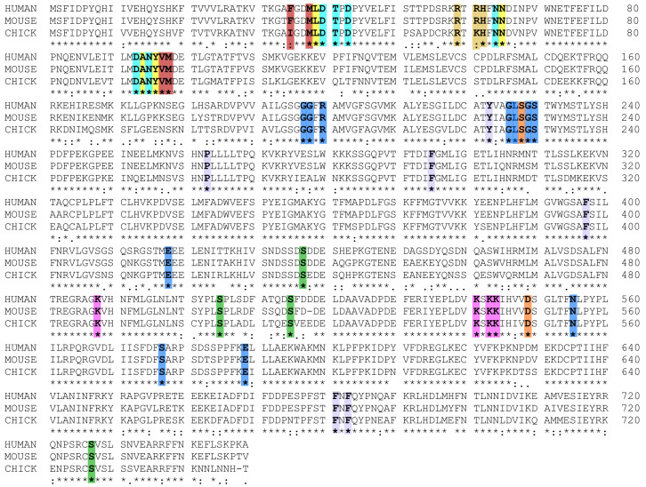
Figure 1—figure supplement 1. Sequence alignment of cPLA2α for human, mouse, and chicken proteins.

Figure 1—figure supplement 1.

In the C2-Domains, Ca2+ binding residues are cyan, DHPC binding residues are yellow; C1P binding residues are beige, and membrane interaction residues are burgundy. In the Catalytic Domains, active site residues are orange, PAPC polar headgroup interacting residues are blue, PAPC acyl chain interacting residues are lavender, PIP2 interacting residues are magenta, and phosphorylation sites are green. Sequence alignment was generated using Clustal W (Larkin et al., 2007).
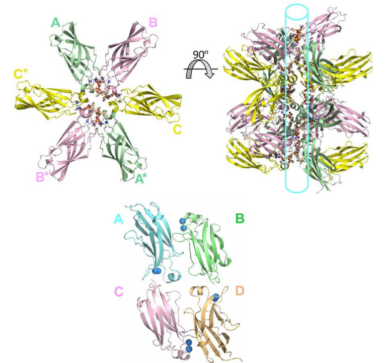
Figure 1—figure supplement 2. Tubular topology formed in the crystal lattice of the cPLA2α C2-domain–DHPC structural complex.

Figure 1—figure supplement 2.

(Upper left panel) Cross-sectional view of three cPLA2α C2-PC complexes in the asymmetric unit (labeled A, B, C) and three complexes in the neighboring asymmetric unit (A*, B* and C*) that together form a ring-like structure. The hexanoyl acyl chains associated in one complex are separated from hexanoyl acyl chains in adjacent complexes by 4.9 to 7.2 Å. (Upper right panel) Rotation by 90° reveals PC lipids packed into a ring-like topology that forms a tubular-like scaffold. (Lower panel) The crystal packing lattice of lipid-free human cPLA2α C2-domain (PDB: 1RLW). The incorporated Ca2+ is shown as blue spheres. Protomer A packs with protomers B and C, but not with protomer D.