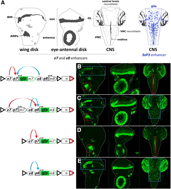
Figure 1.
e7p7 and e8p8 interact in cis. (A) Schematics of a wing disk, an eye-antennal disk, and two central nervous systems (CNSs), with the areas of m7 and m8 expression marked in shades of black. WM, wing margin; AMPs, adult muscle precursors; OL, optic lobe; VNC, ventral nerve cord. In the second CNS, a set of glial cells is indicated in shades of blue, corresponding to the expression pattern of the artificial 3xP3 enhancer (see later in Figure 4). (B and D) GFPm7 and (C and E) GFPm8 expression in wing disk, eye-antennal disk, and CNS (in left, middle, and right columns, respectively) from the EGFP-tagged constructs shown in the diagrams on the left panel; red dotted rectangles highlight e7-specific expression in the AMPs and in the midline of the VNC; blue dotted rectangles indicate e8-specific expression in the WM and in neuroblasts of the central brain and VNC. Also note that both enhancers drive expression in some common areas, e.g., the eye morphogenetic furrow. In the constructs’ schematics enhancers are shown as ovals (e7 and e8), promoters as bent arrows (p7 and p8), and insulators as triangles: black triangle: gypsy insulator (GI); red triangle: Wari insulator (WI), included in the 3′ of the mini-white marker gene. Blue and red curved arrows in the diagrams depict, respectively, e7 and e8 activities, which are shared between p7 (B) and p8 (C) in the wing disk.

