



Abstract
Lichen planus is a chronic inflammatory mucocutaneous disorder that is seen in skin and oral mucosa. Definitive etiology for oral lichen planus remains unknown. It may or may not be associated with skin lesions. Different clinical patterns such as reticular, plaque, erosive, bullous, and atrophic are seen in oral mucosa of which bullous lichen planus is a rare entity. We present a unique case of bullous lichen planus in a 20-year-old male without skin manifestations along with the review of literature comprising various case reports of bullous lichen planus.
KEYWORDS: Biopsy, bullous lichen planus, histopathology, review
INTRODUCTION
Oral lichen planus is a chronic inflammatory mucocutaneous disorder that is T-cell mediated. Prevalence of oral lichen planus in Indian population is 2.6%.[1] Women have higher predisposition to oral lichen planus and the condition is seen at the peak age of 30–60 years.[2] Commonly seen variant of oral lichen planus is reticular type whereas erosive type of lichen planus is the second most common type.[3]
Bullous lichen planus (BLP) is relatively a rare variant of lichen planus. Most patients with BLP are associated with multifocal involvement and skin lesions.[4] BLP is commonly associated with burning sensation and pain. Presence of bullae is seen in association with the white striae. Clinically, at times BLP can be presented as erosive lichen planus as the fragile bullae formed can easily get ruptured, which makes it difficult to diagnose. If the erosive component is severe, epithelial separation from the underlying mucosa may occur, which leads to bullae formation.[5] An unambiguous aspect of the etiology and pathogenesis of BLP is not well established, owing to a relatively low number of cases reported.
CASE REPORT
A 21-year-old male patient reported to the Dental outpatient department of Chettinad Dental College and Research Institute with a chief complaint of reddish white patches on his right inner cheek region and palate for past 2 months with associated burning sensation. Patient gave a clinical history of a small swelling that was seen in the palate initially. He felt fluid-filled swelling that ruptured subsequently. The reddish-white lesion in the palate enlarged to the present size. A similar lesion was noted around the same period in the right buccal mucosa. The patient was previously under topical antifungal therapy that temporarily reduced the burning sensation.
Patient had no relevant medical, dental, or familial history and maintained good oral hygiene. He had no chewing or smoking habit and consumes spicy foods.
On inspection of right buccal mucosa [Figure 1], a single erythematous lesion with radiating white striae was seen of size 4 × 2 cm at the level of occlusal plane extending anteriorly from 45 to distal aspect of 47 posteriorly. It was soft in consistency with elevated white irregular margins. Over the palate [Figure 2], a single elevated erythematous lesion interspersed with white striae was seen measuring 4 × 4 cm; extending anteriorly 1 cm behind the incisive papilla; posteriorly up to fovea palatina; laterally 5 mm below the free gingival margins of 16, 17, 26, and 27; and posteriorly extending to 1 cm above the soft palate. The lesion did not affect the palatal rugae and incisive papilla but had covered the mid-palatine raphae. The lesion contained multiple minute round erythematous projections of varying sizes surrounded by white striae. The surface was rough and non-tender on palpation. The patient presented with no other skin abnormalities.
Figure 1.

Lesion in buccal mucosa
Figure 2.
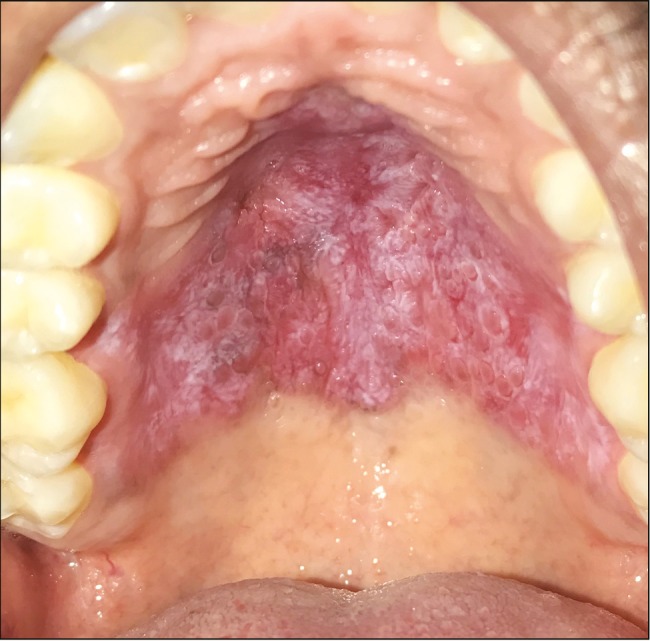
Lesion in hard palate
Differential diagnosis
Correlating with the clinical history and presentation, the lesion was provisionally diagnosed as “bullous lichen planus,” along with lichen planus pemphigoides, pemphigus, pemphigoid, allergic stomatitis, erythroleukoplakia, and erythematous candidiasis.
Investigations
Patient’s basic hematological parameters were evaluated and found to be within normal limits. Patient was referred for incisional biopsy of the lesion.
Histopathological findings
Macroscopic findings: Two bits of soft tissue specimen were placed in 10% formalin. Specimens were taken from right buccal mucosa and palate. Both the tissues processed for histopathological evaluation.
Microscopic findings: Orthokeratinized stratified squamous epithelium of varying thickness with flat to elongated rete pegs. Areas of basal cell degeneration were evident. Juxta-epithelial connective tissue shows dense infiltration of chronic inflammatory cells of lymphocytes interspersed with increased capillaries. Presence of intraepithelial cleft [Figures 3 and 4].
Figure 3.

Acanthosis, juxta epithelial connective tissue shows dense infiltration of inflammatory cells interspersed with increased capillaries
Figure 4.

The rate process are flat to elongated areas of basal cell degeneration; presence of intra-epithelial cleft is evident
Final diagnosis
On the basis of the histopathological features, the lesion in right buccal mucosa and palate was diagnosed as BLP.
Treatment
Patient was advised for medical and stress management. He was advised to take prednisolone 20 mg and was tapered over period of 6 weeks. Topical application of Kenacort was also prescribed to the patient. Regression of lesion was seen completely over the end of 6 weeks.
DISCUSSION
Lichen planus is a chronic inflammatory autoimmune disease in which autoantibodies are generated against the basal keratinocytes, leading to its degeneration. The etiology of the lesion is not well defined although stress is a commonly associated factor in these patients. Cell-mediated immunity is mostly responsible for the pathogenesis of oral lichen planus (OLP). It could be either triggered by endogenous or exogenous factors. Exogenous factors include drugs, restoration, infections, and food allergies. Endogenous factors include genetic factors and autoimmunity.[23]
The characteristics of lichen planus are flattened, polygonal, pruritic, and violaceous papules with grayish-white scaly surfaces. The sites affected usually include flexor surfaces of wrists, forearms, dorsal surfaces of hands, shins, and genital areas.[6]
Andreasen[22] has classified oral lichen planus into reticular, papular, plaque, atrophic, erosive, and bullous. The commonly presented variant is reticular, which presents the characteristic clinical feature called Wickham’s striae that is seen as interlacing white lines. Most frequently affected site is buccal mucosa. Other commonly affected sites are tongue, gingiva, and lower lip.[5]
Other rare variants of lichen planus that are seen are annular lichen planus, lichen planus pigmentosus, and lichen planus planopilaris. Erosive lichen planus in which the genital areas and gingiva are affected together is referred as vulvovaginal-gingival syndrome (female equivalent)/penogingival syndrome (male equivalent) and collectively called as “genito-gingival syndrome.”[25] Lichen planus lesions replacing hair follicles with scarring tissue have also been reported. This type is called lichen planus planopilaris, which is more seen in women. This type has also been reported under BLP cases. There are three types: classic lichen planus planopilaris, with frontal fibrosing alopecia, which mostly affects postmenopausal women[21] and Graham Little syndrome in which scarring alopecia is seen in scalp only, involving loss of hair in pubis or axilla or other areas.[11]
Bullous lichen planus
There are two variants of BLP: familial and nonfamilial. The familial variant is more common than the nonfamilial variant. Familial BLP occurs at an earlier age with longer duration of disease and more extensive eruptions, and increased tendency to involve nails is seen in familial BLP when compared to nonfamilial variants. Pathogenesis of familial variants maybe attributable to genetic factors.[7]
In BLP, blisters develop adjacent to or on preexisting lichen plants lesions in oral mucosa as well as lower extremities. It might be associated with alopecia. BLP that affects nails appear as hemorrhagic crusty lesions resulting in exposure of nail bed and finally nail atrophy.[8]
Oral BLP is associated with cutaneous lesions.[4] The lesion can be seen with an intact bullae or eroded surface superimposed or juxtaposed with white striae.
Demographic data and history
On the review of the aforementioned articles [Table 1], oral BLP shows no clear gender predilection with peak of incidence occurring at third to sixth decade of life. The medical and dental history does not reveal a clear establishment between systemic conditions and oral BLP. However, a special condition of oral BLP occurs due to the usage of a drug—labetalol. It shows the importance of ruling out the drug history in diagnosing oral BLP. Two case reports show patients with dermal disease developing oral BLP. No habitual or familial history is seen in these patients. The patients mostly have a complaint of burning sensation or pain or both in their oral cavity. Most commonly associated factor in these case reports is the consumption of spicy food. The presented case of 21-year-old male patient has spicy dietary habits and was subjected to a stressful lifestyle.
Table 1.
Review of case reports (demographic data and history)
| S.No | Author, Year | Age | Sex | Place reported | Medical & Dental History | Habits | F.H | Duration of Lesion | Burning (B)/ Pain (P) | Associative Factors |
|---|---|---|---|---|---|---|---|---|---|---|
| 1 | Rashmi et al- 2011[10] | 40 | M | Chandigarh, India | Dental Filling 3 months before lesion | N | N | 15 days | B | Spicy food |
| 2 | Kotya et al- 2014[11] | 35 | F | Telangana, India | N | N | N | 4 mon | B | Spicy Food |
| 3 | Preethi et al- 2015[12] | 44 | F | Karnataka, India | N | N | N | 2 y | P | N.A |
| 4 | Biljana et al- 2011[13] | 5 | M | Serbia | Atopic Dermatitis since 3 y | N | N | 1.5 mon | N | N.A |
| 5 | Archana et al- 2011[14] | 34 | F | Karnataka, India | Gen. Pruritis for 10 yrs and is under medication. Anal hemorrhoids was treated. extraction of molar and gingival flap | N | N | 4 days | B | Spicy food. Stress |
| 6 | Gange et al- 1978[15] | 67 | M | England | Under labetalol 400 mg, Clonidine 0.1 mg/day | N | N | 10 weeks | P | N.A |
| 7 | Ashok et al- 2016[16] | 60 | F | Odisha, India | N | N | N | 1 y | B & P | Spicy Food |
| 8 | Verma et al- 2014[17] | 45 | M | Maharastra, India | N | N | N | 3 mon | N | N.A |
| 9 | Guru et al 2016[18] | 40 | F | Andhra, India | N | N | N | 4 months | P | N.A |
| 10 | Dhanraj et al 2017[19] | 23 | M | Puducherry, India | N | N | N | 2 weeks | N | N.A |
| 11 | Kumari et al 2017[20] | 48 | M | Bihar, India | Asthmatic- use steroidal inhaler | N | N | 8 months | B | Aggravated by Spicy foods Stress |
Y = present; N = not present; N.A = not applicable
Clinical features
On the review of the aforementioned articles [Table 2], oral BLP shows skin manifestations in almost all cases except one. Oral BLP shows an earlier onset of lesions over the extremities especially the lower and later spreads to the body. Pruritic violaceous papular appearance is seen in most cases, which is accompanied by bullae formation later associated with pigmentations. The bullae contain clear fluid. Most commonly affected intraoral site is buccal mucosa, seldom arising in tongue, palate, and labial mucosa. Oral BLP shows bilateral occurrence in almost all cases. The lesion appears to be seen as an erythematous area/intact bullae in the center surrounded by white striae at the periphery. The patient presented with only oral manifestations without the involvement of any skin lesions. The unilateral buccal lesion in this case shows erythematous center surrounded by interspersed white striae and light pigmentation. It was soft in consistency. The palatal lesion was elevated with multiple minute round erythematous projections of varying sizes interspersed with white striae.
Table 2.
Review of case reports (clinical features)
| S.No | Author | Extra-oral Site | I-O Lesion | Intra oral site | U / B | Centre of lesion | Border of lesion |
|---|---|---|---|---|---|---|---|
| 1 | Rashmi et al[10] | Violaceous annular plaque over glans penis | Y | Bu Mu | U | Single round tense vesicle | Reticular whitish streaks |
| 2 | Kotya et al[11] | Multiple papule on face and scalp black healed pigmented lesions on face. Alopecia | Y | Bu Mu Lat. Border of Tongue | B B | Areas of erosion & pigmentation Area of erosion and intact bullae | Diffuse white striae Fine linear striae |
| 3 | Preethi et al[12] | Vesicles and bullae were seen on hands | Y | BuMu | B | Erythmatous area | White lacy pattern |
| 4 | Biljana et al[13] | All over skin except face and oral mucosa Bilateral dorsal part of hands and lower legs | N | N | N | N | N |
| 5 | Archana et al[14] | Isolateral ulcerative lesion on scalp associated with severe alopecia. Black healed pigmented lesions on legs, hands and back | Y | Bu Mu Hard Palate | B | Bullae with clear filled fluid in L, area of erythema in R. White papules intersperse with striae | Reticular whitish streaks Normal |
| 6 | Gange et al[15] | Penis-white striae, Trunk-red papules and macules, Legs-bullous, Limbs-typical lichen planus. | N | N | N | N | N |
| 7 | Ashok et al[16] | Typical fine lacy white streaks at junction of cheeks and lips. Koebners phenomenon- (+) | Y | BuMu | B | N.A | N.A |
| 8 | Verma et al[17] | multiple, itchy, red raised papules over both forearms spread over trunk and all extremities. A few vesicles developed over the pre-existing papules containing clear fluid. Multiple, discrete to confluent, erythematous to violaceous papules coalescing to form plaques on the extremities, trunk and buttocks | N | N | N | N | N |
| 9 | Guru et al[18] | Intial lesion-multiple itchy hyper pigmented raised lesion over lower extremities then to trunk. Developed with few bullae on pre-existing lesion leaving erythematous areas after rupture. Then multiple, discrete to confluent, erythematous to violaceous papules coalescing to form plaques on the extremities and back of the trunk. Tense bullae filled with clear and haemorrhagic fluid present | Y | Bu Mu | N.A | Oral cavity showed reticulate lesions over buccal mucosa | |
| 10 | Dhanaraj et al[19] | N | Y | Bu Mu | B | solitary vesicle of was evident on the right buccal mucosa presented with bullae filled with clear fluid. White striae in mndibular vestibule | white colored striae. |
| 11 | Kumari et al[20] | N | Y | Bu Mu Labial Mu | B | Irregular Erythematous area with pseudomembranous slough | White striae |
Y = present; N = not present; N.A = not applicable; U = unilatereal; B = bilateral; Bu = buccal mucosa; Mu = mucosa; Lat = Lateral
Differential diagnosis
Diagnosis of oral lichen planus can be clinically verified with the presence of pathognomic appearance of Wickham’s striae in reticular lichen planus bilaterally. The definitive diagnosis for oral lichen planus can be rendered only with histological evaluation of the lesion.
The differential diagnosis of oral BLP can be lichen planus pemphigoides, pemphigus, pemphigoid, allergic stomatitis, erythroleukoplakia, and erythematous candidiasis.
BLP is commonly misdiagnosed as lichen planus pemphigoid. In lichen planus lichenoids (LPP), the lesions that developed were mostly de novo are on previously existing lichen planus (LP) lesions. Rarely in LPP, bullae would develop only in lesion without affecting the surrounding skin. Hence this makes the differentiation even more difficult. LPP has the presence of circulating autoantibodies in the blood that are absent in BLP.[9] Possibility of drug/restorative material induced lichenoid reactions should also be ruled out.
Investigations
Commonly performed investigations in BLP are hemogram, renal and liver function tests, anti-hepatitis B and C tests, and urine analysis [Table 3]. Blood picture is commonly taken to rule out any nutritional deficiencies such as iron deficiency or immunodeficiency, which could be the underlying cause for lichen planus. There have been several surveys conducted, which established a strong relationship between hepatitis B/hepatitis C infected patients to occurrence of oral lichen planus in them.[23] Liver function tests are performed to rule out chronic liver disease, which shows strong relation to OLP. The most common ones are chronic active hepatitis and primary biliary cirrhosis.[23] LP patients are prone to urolithiasis. Renal function tests/urine analysis can also be performed in precaution to see the presence of any renal problem.[24] Immunofluorescence is carried out to rule out lichen planus pemphigoides.[23] Cytosmear is taken to rule out any candida super-infection.[23] Biopsy is the confirmatory test to diagnose BLP. The set criteria of histopathological features can only confirm the presence of BLP. In the case presented, blood picture and biopsy were performed to confirm the diagnosis of BLP.
Table 3.
Review of case reports (investigations and treatment)
| S.No | Author | Keratosis | Acanthosis | Basal Cell Degeneration | Melanin incontinence | Rete Ridges | lymphocyte band | Epithelial Clefts | Others |
|---|---|---|---|---|---|---|---|---|---|
| 1 | Rashmi et al[10] | Parakeratosis | N.A | Y | N.A | N.A | moderate sub-epithelial band | N.A | Mild Basal Cell hyperplasia, Hypergranulosis |
| 2 | Kotya et al[11] | N.A | N.A | N.A | N.A | N.A | N.A | N.A | N.A |
| 3 | Preethi et al[12] | Atrophic | Y | Y-dense | Y | N.A | |||
| 4 | Biljana et al[13] | Orthokeratosis | Y | Y | Y | Saw- tooth | Y-dense | Max-joseph spaces progressing to sub- epidermal bullae | Civatte bodies Hypergranulosis |
| 5 | Archana et al[14] | N.A | N.A | N.A | N.A | N.A | N.A | N.A | N.A |
| 6 | Gange et al[15] | N.A | N.A | Y | N.A | N.A | Y (upper dermis) | Bulla formation | Necrotic keratinocytes in epidermis |
| 7 | Ashok et al[16] | N.A | N.A | N.A | N.A | N.A | N.A | N.A | N.A |
| 8 | Verma et al[17] | N.A | N.A | Y | Y | N.A | Dense- moderate (sub- epithelial) | Derm- epidermal clefts | Foci of Colloid bodies, Spongiotic epidermis |
| 9 | Guru et al[18] | Parakeratotic | N.A | Y | N | N.A | moderate Dermo- epidermal | Sub- epidermal clefts and bullae | Colloid bodies |
| 10 | Dhanaraj et al[19] | Hyperkeratotic, irregular hyperplasia | Y | N.A | N | N.A | Y- Dense (Papillary dermis) | N.A | Civatte bodies, focal hypergranulosis |
| 11 | Kumari et al[20] | N.A | N.A | N.A | N.A | Saw- tooth | N.A | Large Max- joseph spaces | Hypergranulosis |
Y = present; N = not present; N.A = not applicable
Histopathology
In most cases, the lesions depict hyperkeratosis with hypergranulosis or acanthosis or both [Table 4]. Moderate-to-dense lymphocytic band is seen mostly in papillary dermis. Basal cell degeneration is seen in most of the cases reported. Presence of saw-tooth rete ridges is seen rarely. Clear epithelial or subepithelial clefts are present in most of the cases with the presence of civatte or colloid bodies. The histopathology of our patient was also similar to that of BLP in the literature with absence of civatte or colloidal bodies.
Table 4.
Review of case reports (histopathology)
| S.No | Author | Investigations | Treatment | Prognosis |
|---|---|---|---|---|
| 1 | Rashmi et al[10] | Biopsy, Hemogram, Anti-Hepatitis B&C Tests, Renal & Liver Function tests and lipid profile | 25 mg Acitretin q.d, 16 mg met. Prednisolone q.d, 0.1% triamcinolone t.i.d | Good |
| 2 | Kotya et al[11] | Hemogram,Renal & Liver Function tests, Biopsy | Oral minipulse therapy bethamethasone 5 mg q.d (tapered later) | Excellent |
| 3 | Preethi et al[12] | Biopsy, Hemogram | N | N |
| 4 | Biljana et al[13] | Hemogram, Anti-Hepatitis B&C Tests, Biopsy | 1 mg/kg/day predinsolone, BB-UVB radiation,Topical Fluocinonide, Pimecrolimus | Excellent |
| 5 | Archana et al[14] | Biopsy Hemogram, OPG, Cytosmear, Blood Glucose | Levamisole 150mg, prednisolone 10 mg, Benzydamine oral rinse, Ranitidine. Triamcinolone after intial treatment | Excellent |
| 6 | Gange et al[15] | Biopsy | Labetalol & clonidine replaced by atenlol & moduretic. Topical Steroids | Excellent |
| 7 | Ashok et al[16] | N | Prednisolone 20 mg qd. Topical Triamcinalone | Good |
| 8 | Verma et al[17] | Biopsy, Direct Immunofluorescence, Hemogram, Urine Analysis | Oral Prednisolone 40 mg/day. Tapered after 6 weeks | Excellent |
| 9 | Guru et al[18] | Routine blood tests.urinalysis. Skin bioposy. Biopsy of a tense blister. Immunofluorescence. | oral prednisolone 40 mg/day, which was tapered off in 6 weeks. | N |
| 10 | Dhanaraj et al[19] | Incisional Biopsy.direct immunofluorescence. Anti-hepatitis C virus (HCV) test | Clobetasol mouthwash Topical 0.1% Tacrolimus | Poor Excellent |
| 11 | Kumari et al[20] | complete hemogram, blood sugar, cytosmear, and incisional biopsy, Immunofluorescence | Initial-Tab prednisolone 20mg topical clotrimazole (1%), benzydamine oral rinse 0.15% ranitidine 150 mg for two weeks. The dosage was tapered for the next 6 weeks. Later topical triamicnalone 1% was given | Good |
Y = present; N = not present; N.A = not applicable
Treatment
Systemic drugs such as prednisolone or topical application such as triamcinolone when used separately or together give good prognosis [Table 3]. Phototherapy such as broadband ultraviolet-B therapy has proven to help in the eradication of BLP. Usage of retinoids such as tretinoin can also help in controlling the lesions. Usage of immunomodulators such as tacrolimus/pimecrolimus has helped in the control of autoimmune activity, which helps in the regression of BLP. Oral minipulse therapy in which betamethasone 5 mg is given once daily over two consecutive days in a week. This has proven to be very effective in eradicating the lesion. Clobetasol has poor prognosis as the lesion persisted even after using it. Adjuvant drugs such as benzydamine oral rinse and topical clotrimazole can also be given. The patient was under the medication of prednisolone 20 mg and topical application of triamcinolone 0.1% for 6 weeks. The dosage of prednisolone was tapered over 6 weeks. He was also advised for stress management. The lesion regressed completely after 6 weeks.
Declaration of patient consent
The authors certify that they have obtained all appropriate patient consent forms. In the form the patient(s) has/have given his/her/their consent for his/her/their images and other clinical information to be reported in the journal. The patients understand that their names and initials will not be published and due efforts will be made to conceal their identity, but anonymity cannot be guaranteed.
Financial support and sponsorship
Nil.
Conflicts of interest
There are no conflicts of interest.
REFERENCES
- 1.Murti PR, Daftary DK, Bhonsle RB, Gupta PC, Mehta FS, Pindborg JJ. Malignant potential of oral lichen planus: Observations in 722 patients from India. J Oral Pathol. 1986;15:71–7. doi: 10.1111/j.1600-0714.1986.tb00580.x. [DOI] [PubMed] [Google Scholar]
- 2.Gupta S, Jawanda MK. Oral lichen planus: An update on etiology, pathogenesis, clinical presentation, diagnosis and management. Indian J Dermatol. 2015;60:222–9. doi: 10.4103/0019-5154.156315. [DOI] [PMC free article] [PubMed] [Google Scholar]
- 3.Edwards PC, Kelsch R. Oral lichen planus: Clinical presentation and management. J Can Dent Assoc. 2002;68:494–9. [PubMed] [Google Scholar]
- 4.Varghese SS, George GB, Sarojini SB, Vinod S, Mathew P, Mathew DG, et al. Epidemiology of oral lichen planus in a cohort of south Indian population: A retrospective study. J Cancer Prev. 2016;21:55–9. doi: 10.15430/JCP.2016.21.1.55. doi:10.15430/JCP.2016.21.1.55. [DOI] [PMC free article] [PubMed] [Google Scholar]
- 5.Shirasuna K. Oral lichen planus: Malignant potential and diagnosis. Oral Sci Int. 2014;11:1–7. doi:10.1016/S1348-8643(13)00030-X. [Google Scholar]
- 6.Weston G, Payette M. Update on lichen planus and its clinical variants. Int J Womens Dermatol. 2015;1:140–9. doi: 10.1016/j.ijwd.2015.04.001. doi:10.1016/j.ijwd.2015.04.001. [DOI] [PMC free article] [PubMed] [Google Scholar]
- 7.Huang C, Yan X, Yang L, Jing Z, Jin T, Jiawen L, et al. A retrospective and comparative study of familial and non-familial bullous lichen planus. J Huazhong Univ Sci Technol. 2007;27:336. doi: 10.1007/s11596-007-0331-7. doi:10.1007/s11596-007-0331-7. [DOI] [PubMed] [Google Scholar]
- 8.Liakopoulou A, Rallis E. Bullous lichen planus—A review. J Dermatol Case Rep. 2017;11:1–4. doi: 10.3315/jdcr.2017.1239. doi:10.3315/jdcr.2017.1239. [DOI] [PMC free article] [PubMed] [Google Scholar]
- 9.Gawkrodger DJ, Stavropoulos PG, McLaren KM, Buxton PK. Bullous lichen planus and lichen planus pemphigoides—Clinico-pathological comparisons. Clin Exp Dermatol. 1989;14:150–3. doi: 10.1111/j.1365-2230.1989.tb00914.x. [DOI] [PubMed] [Google Scholar]
- 10.Jindal R, De D, Kanwar AJ. Bullous oral lichen planus: An unusual variant. Indian Dermatol Online J. 2011;2:39–40. doi: 10.4103/2229-5178.79853. doi:10.4103/2229–5178.79853. [DOI] [PMC free article] [PubMed] [Google Scholar]
- 11.Maloth KN, Sunitha K, Boyapati R, Shravan Kumar DR. Bullous lichen planus treated with Oral Minipulse Therapy: A rare case report. J Clin Diagn Res. 2014;8:ZD17–9. doi: 10.7860/JCDR/2014/11465.5338. doi:10.7860/JCDR/2014/11465.5338. [DOI] [PMC free article] [PubMed] [Google Scholar]
- 12.Nayak PB, Desai D, Shetty SJ, Alva ML, Rebello A. A rare variant of oral lichen planus-bullous form. IJSS Case Rep Rev. 2015;2:35–7. [Google Scholar]
- 13.Arsov B, Popadić S, Nikolić M. Bullous lichen planus in childhood—A case report/Bulozni lichen planus u dečijem uzrastu. Serbian Journal of Dermatology and Venereology. 2011;3:28–32. doi:10.2478/v10249-011-0035-7. [Google Scholar]
- 14.Patil A, Prasad S, Ashok L, Sujatha GP. Oral bullous lichen planus: Case report and review of management. Contemp Clin Dent. 2012;3:344–8. doi: 10.4103/0976-237X.103634. doi:10.4103/0976-237X. 103634. [DOI] [PMC free article] [PubMed] [Google Scholar]
- 15.Gange RW, Jones EW. Bullous lichen planus caused by labetalol. Br Med J. 1978;1:816–7. doi: 10.1136/bmj.1.6116.816-a. [DOI] [PMC free article] [PubMed] [Google Scholar]
- 16.Jena AK, Sirka CS, Mohapatra M. Atypical bullous lichen planus in palate: A case study. Oral Health Case Rep. 2016;2:126. doi:10.4172/2471–8726.1000126. [Google Scholar]
- 17.Verma R, Vasudevan B, Kinra P, Vijendran P, Badad A, Singh V. Bullous lichen planus. Indian J Dermatol Venereol Leprol. 2014;80:279. doi: 10.4103/0378-6323.132275. [DOI] [PubMed] [Google Scholar]
- 18.Prasad PG, Kumar R, Ramani JR, Jhanvi Bullous lichen planus: A case report. J Evolution Res Dermatol Venereol. 2016;2:4–5. [Google Scholar]
- 19.Dhanraj T, Paulose S, Kavya SY. Oral lichen planus bullous variety: A case report. J Med Radiol Pathol Surg. 2017;4:21–3. [Google Scholar]
- 20.Kumari A, Singh PK. Bullous lichen planus: A case report with review of literature. J Case Rep Stud. 2017;5:403. doi:10.15744/2348-9820.5.403. [Google Scholar]
- 21.Kossard S, Lee MS, Wilkinson B. Postmenopausal frontal fibrosing alopecia: A frontal variant of lichen planopilaris. J Am Acad Dermatol. 1997;36:59–66. doi: 10.1016/s0190-9622(97)70326-8. [DOI] [PubMed] [Google Scholar]
- 22.Andreasen JO. Oral lichen planus. 1. A clinical evaluation of 115 cases. Oral Surg Oral Med Oral Pathol. 1968;25:31–42. doi: 10.1016/0030-4220(68)90194-1. [DOI] [PubMed] [Google Scholar]
- 23.Scully C, Beyli M, Ferreiro MC, Ficarra G, Gill Y, Griffiths M, et al. Update on oral lichen planus: Etiopathogenesis and management. Crit Rev Oral Biol Med. 1998;9:86–122. doi: 10.1177/10454411980090010501. [DOI] [PubMed] [Google Scholar]
- 24.Oguz U, Takci Z, Oguz ID, Resorlu B, Balta I, Unsal A. Are patients with lichen planus really prone to urolithiasis? Lichen planus and urolithiasis. Int Braz J Urol. 2016;42:571–7. doi: 10.1590/S1677-5538.IBJU.2015.0364. doi:10.1590/S1677-5538.IBJU.2015.0364. [DOI] [PMC free article] [PubMed] [Google Scholar]
- 25.Cribier B, Ndiaye I, Grosshans E. [Peno-gingival syndrome. A male equivalent of vulvo-vagino-gingival syndrome?] Rev Stomatol Chir Maxillofac. 1993;94:148–51. [PubMed] [Google Scholar]