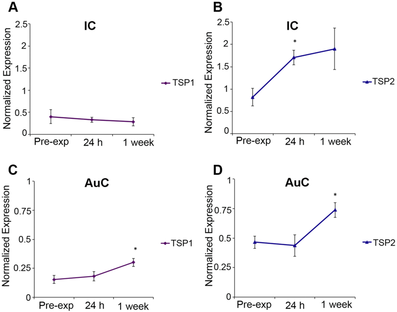
Figure 7. Noise-induced changes in TSP gene expression in auditory brain regions.
Quantitative qPCR was used to determine expression of TSP1 (A, C) and TSP2 (B, D) genes in mouse inferior colliculus (IC) and auditory cortex (AuC) before (pre-exposure) and after noise exposure (24 hours and 1 week). In the IC significant upregulation of TSP2 (B), but not TSP1 (A), gene expression was observed 24 hours after noise exposure. TSP2 gene expression remains upregulated one week after noise. In the AuC, both TSP1 and TSP2 mRNA levels were significantly higher at 1 week after noise exposure compared to pre-exposure levels (C and D respectively). Data shown is the expression of the gene of interest relative to the internal standard, GAPDH. Results are expressed as mean ± standard error of the mean (SEM). Significant differences are indicated by * = p<0.05.

