Correction to: BMC Res Notes (2019) 12:242 10.1186/s13104-019-4281-5
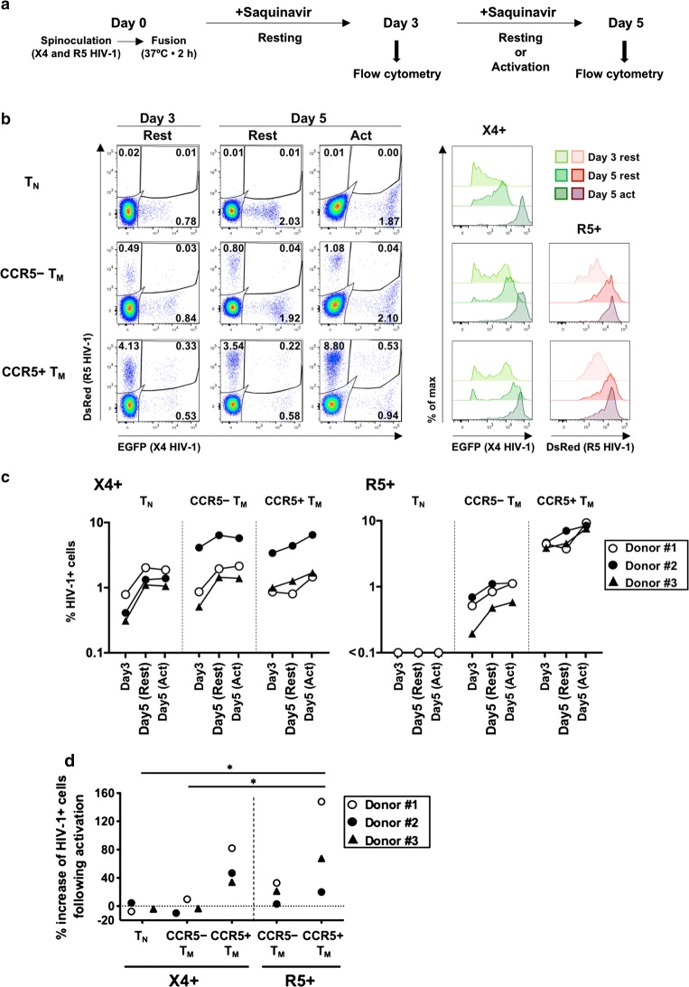
After publication of the original article [1], the authors became aware of a miscalculation in the original Fig. 2d.
should be calculated as:
The corrected Fig. 2d is shown in this erratum.
Fig. 2.
HIV-1 infection and culture of resting CD4+ T-cell subsets isolated by cell sorting. Subsets of naïve T cells (TN), or CCR5+ or CCR5− memory T cells (TM), were separately infected and cultured. a Schematic of the protocol of HIV-1 infection and culture. b Representative flow-cytometry profiles of cells from Donor #1 at day 3 and day 5 post-infection (resting or activated), separated according to reporter expression indicating the presence of X4 or R5 HIV-1, with the percentage of each subset indicated (left panels). The intensity of fluorescence for each viral reporter in each cell subset [except for the very low percentage of DsRed+ cells (R5+) in TN cells] is shown in the right-hand panels. c Percentages of HIV-1+ cells in each CD4+ T-cell subset in three donors. d Percentage increases in frequencies of HIV-1+ cells following activation were estimated by comparing percentages of HIV-1+ cells in the activation condition with those in the resting condition at day 5 post-infection. Significant differences (*P < 0.05, **P < 0.01) were determined by repeated-measures one-way ANOVA followed by Tukey’s multiple comparison test. In c and d, HIV-1+ cells include the corresponding reporter (either EGFP or DsRed) single-positive cells and double-positive cells
Although the statistical significances have been altered, the hierarchical mode between cell-subset groups remains the same. It is still shown that numbers of X4 HIV-1+ cells increased consistently in the CCR5+ TM subset of all three donors tested. Therefore, the correction does not change the scientific conclusion.
Footnotes
Publisher's Note
Springer Nature remains neutral with regard to jurisdictional claims in published maps and institutional affiliations.
Contributor Information
Kazutaka Terahara, Email: tera@nih.go.jp.
Ryutaro Iwabuchi, Email: ryutaroi@niid.go.jp.
Masahito Hosokawa, Email: m.hosokawa@aoni.waseda.jp.
Yohei Nishikawa, Email: nishikawa_yohei@fuji.jp.
Haruko Takeyama, Email: haruko-takeyama@waseda.jp.
Yoshimasa Takahashi, Email: ytakahas@nih.go.jp.
Yasuko Tsunetsugu-Yokota, Email: yyokota@nih.go.jp.
Reference
- 1.Terahara K, Iwabuchi R, Hosokawa M, Nishikawa Y, Takeyama H, Takahashi Y, Tsunetsugu-Yokota Y. A CCR5+ memory subset within HIV-1-infected primary resting CD4+ T cells is permissive for replication-competent, latently infected viruses in vitro. BMC Res Notes. 2019;12:242. doi: 10.1186/s13104-019-4281-5. [DOI] [PMC free article] [PubMed] [Google Scholar]


