Abstract
Male genital tract angiomyofibroblastoma (AMF) is a rare benign tumour, with a total of 34 cases reported in literature. We are presenting a case of AMF of the glans penis in a 68-year-old man who presented with a progressively increasing in size large lesion located on the tip of his penis. Following routine investigations, the lesion was surgically excised with no adjuvant treatment, the patient was followed-up for 5 years with no evidence of local, nodal or distant recurrence. As AMF of the glans penis is extremely rare, there is not enough literature to support management guide lines, but it appears that AMF responds very well to complete surgical excision; occasional cases of recurrence have been previously reported, so a long-term follow-up is advised.
Keywords: urology, sexual health, genital ulcers
Background
We are presenting a rare case of penile angiomyofibroblastoma (AMF) which was managed with surgical resection, with no adjuvant treatment, followed-up for 5 years with neither local or nodal recurrence nor distant metastasis.
AMF was first described in 1992 by Fletcher et al as a benign neoplasm, distinct from aggressive angiomyxoma, which histologically consist of prominent blood vessels and stromal cells.1
Genital tract AMF is predominantly common in females rather than males. Male genital tract AMF is considered a rare benign tumour, with a total of 34 cases only reported in the literature.2
Case presentation
A 68-year-old man presented to our outpatient clinic with penile lesion of about 2 years duration with a slowly progressive course. His medical history included paraplegia and incontinence (managed with a long term urinary catheter) following a road traffic accident 5 years prior to presentation which lead to spinal injury. He smoked cigarettes for 20 years but stopped approximately 10 years ago.
General examination was unremarkable and abdominal examination revealed no palpable masses or organomegaly. Inguinal examination revealed a large right sided uncomplicated (reducible) indirect inguinal hernia, with no palpable inguinal or iliac lymphadenopathy.
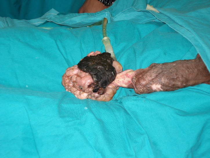
Genital examination revealed a lobulated swelling at the tip of the glans penis not involving the urethral meatus measuring 10 x8 x7 cm with the surrounding penile skin, proximal to the glans, showing thick irregular areas of hyper and hypopigmentation (figure 1).
Figure 1.
Preoperative photo showing the penile lesion.
Investigations
Incisional biopsy was taken form the glans penis lesion and surrounding skin. The results came back showing AMF, with surrounding penile skin mild non-specific chronic inflammatory changes.
Routine blood tests were within normal limits and preoperative staging CT scan did not show any nodal or distant metastasis.
Differential diagnosis
Differential diagnosis of AMF include aggressive angiomyxoma, myxoid leiomyoma, peripheral nerve sheath tumours as well as superficial angiomyxoma.
Treatment
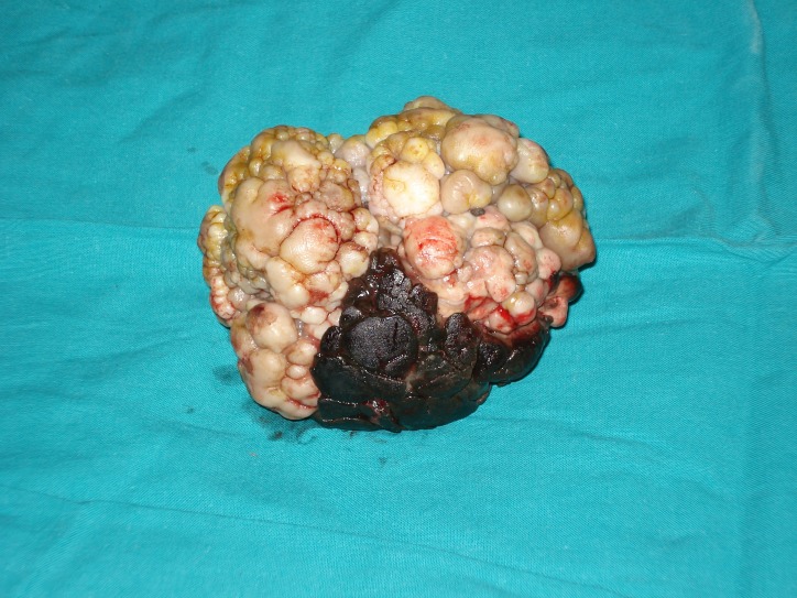
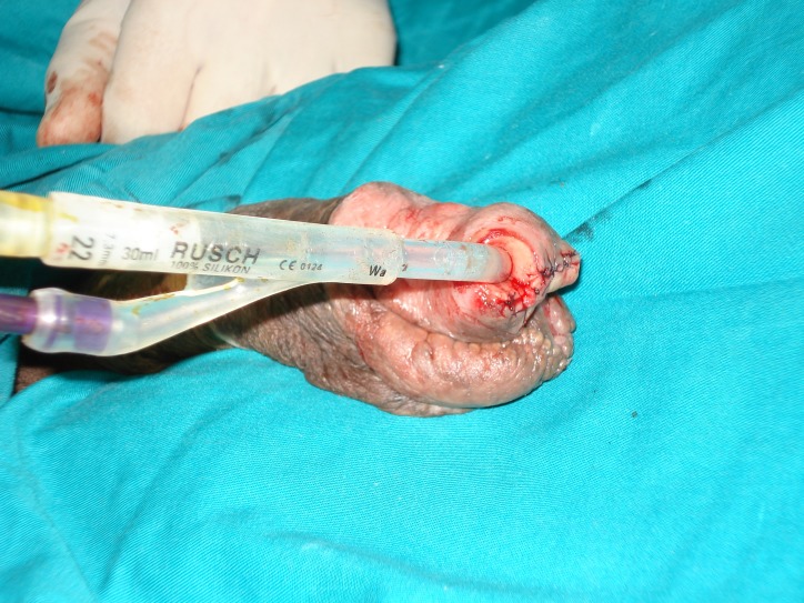
The lesion was resected under general anaesthesia with adequate safety margin at the base of its pedicle (figure 2), tumour bed was closed with interrupted 3/0 Vicryl (figure 3). Postoperative histopathology revealed the same pathology with clear resection margins.
Figure 2.
The penile lesion specimen after resection.
Figure 3.
Intraoperative photo showing primary closure of resection bed.
Outcome and follow-up
The patient was seen in the outpatient clinic at 1, 6 and 12 months postoperatively then followed-up for 5 years with no signs of local or nodal recurrence or distant metastasis.
Discussion
AMF is defined histologically as a benign, well-circumscribed myofibroblastic mesenchymal tumour, which is more common in females than males, normally present around middle age, follow a slow progressive, non-invasive course, usually arise from lower genital tract; vulva in females, scrotum, perineum or spermatic cord in males.3
Clinically it normally present with a painless, slow growing mass, usually females of reproductive age.
Eleven cases of males genital tract AMF were reported by Laskin et al in 1998.4 In 2011 Flucke et al reported that only 14 studies of males genital tract AMF have been reported in the literature,5 Recently in 2017 Msakni et al reported that a total of 34 cases have been reported in the literature.2
Male genital tract AMF normally follow a benign clinical course with the exception of one invasive case reported by Garcia Mediero et al 6 and one locally recurrent case reported by Laskin et al which suggested sarcomatous degeneration.3
Histologically AMF can be distinguished from aggressive angiomyxoma by its circumscribed borders, much higher cellularity, more numerous blood vessels, frequent presence of plump stromal cells, minimal stromal mucin and rarity of erythrocyte extravasation.1
To ensure patient’s favourable outcome; AMF should be differentiated from other lesions as aggressive angiomyxoma, myxoid leiomyoma, peripheral nerve sheath tumours as well as superficial angiomyxoma.7
Treatment of AMF include wide excision with adequate clear margins, followed by long term follow-up due to the fact that occasional cases of recurrence have been previously reported6
Learning points.
In spite of the fact that angiomyofibroblastoma of the glans penis is a very rare entity, yet it should be included in the differential diagnosis of any penile lesions.
Surgical resection with adequate safety margin is the corner stone of management.
Long follow-up is required following resection but the duration and frequency of follow-up is not yet clear.
Footnotes
Contributors: MI was the author responsible for the surgery, ongoing care of the patient in hospital. SM conceived the case report, researched and drafted the manuscript. Both authors read and approved the final manuscript.
Funding: The authors have not declared a specific grant for this research from any funding agency in the public, commercial or not-for-profit sectors.
Competing interests: None declared.
Provenance and peer review: Not commissioned; externally peer reviewed.
Patient consent for publication: Obtained.
References
- 1. Fletcher CD, Tsang WY, Fisher C, et al. Angiomyofibroblastoma of the vulva. A benign neoplasm distinct from aggressive angiomyxoma. Am J Surg Pathol 1992;16:373–82. [DOI] [PubMed] [Google Scholar]
- 2. Msakni I, Ghachem D, Bani MA, et al. Paratesticular Angiomyofibroblastoma-Like Tumor: Unusual Case of a Solidocystic Form. Case Rep Med 2017;2017:1273531 10.1155/2017/1273531 [DOI] [PMC free article] [PubMed] [Google Scholar]
- 3. Bouhajja L, Rammeh SA, Sayari S, et al. Angiomyofibroblastoma of the spermatic cord: a case report. Pathologica 2017;109:368–70. [PubMed] [Google Scholar]
- 4. Laskin WB, Fetsch JF, Mostofi FK. Angiomyofibroblastomalike tumor of the male genital tract: analysis of 11 cases with comparison to female angiomyofibroblastoma and spindle cell lipoma. Am J Surg Pathol 1998;22:6–16. [DOI] [PubMed] [Google Scholar]
- 5. Flucke U, van Krieken JH, Mentzel T. Cellular angiofibroma: analysis of 25 cases emphasizing its relationship to spindle cell lipoma and mammary-type myofibroblastoma. Mod Pathol 2011;24:82–9. 10.1038/modpathol.2010.170 [DOI] [PubMed] [Google Scholar]
- 6. García Mediero JM, Alonso Dorrego JM, Núñez Mora C, et al. [Scrotal invasive angiomyofibroblastoma. First reported case]. Arch Esp Urol 2000;53:827–9. [PubMed] [Google Scholar]
- 7. Cubilla A, Chaux A. Angiomyofibroblastoma. PathologyOutlines.com website http://www.pathologyoutlines.com/topic/penscrotumangiomyofibroblastoma.html [Accessed 25 May 2019].


