

Introduction
Autologous fat transfer is an increasingly popular surgical procedure with many applications in the cosmetic realm and beyond. It is found to be an efficacious procedure, but there is growing concern over adverse effects.1, 2 Potential risks include common complications such as fat necrosis, seroma formation, and cellulitis.1 More serious complications have also been reported, including life-threatening infections and potentially fatal fat emboli, especially in gluteal fat transfer.1, 3 The American Society of Plastic Surgeons recently issued a warning identifying gluteal fat grafting as the aesthetic procedure with the highest mortality rate (1 in 3000 procedures).4 Historically, use of silicone for gluteal enhancement has resulted in numerous cases of granulomatous foreign body–like reactions.5, 6 Theoretically, the use of autologous tissue would decrease the incidence of these reactions. Here we report the first case, to our knowledge, of a granulomatous reaction to autologous fat transfer in the gluteal region.
Case report
A 46-year-old white woman underwent gluteal augmentation with autologous fat transfer in July 2017 for the first time at an outside facility. Lipoinjection methods including a closed system and sterile technique were used to transfer approximately 500 mL of adipose tissue from the bilateral thighs to each buttock. In September 2017 erythematous, tender nodules developed on the buttocks with significant drainage (Fig 1). Superficial skin bacterial cultures of the draining lesions, performed at the outside facility, were negative. At that time she was treated with 2 series of 2-week courses of oral ciprofloxacin with minimal improvement.
Fig 1.

This image was obtained by the patient on her cell phone 2 months postoperatively. The photo shows multiple erythematous to violaceous crusted nodules in the linear array across the mid buttock.
She presented to the dermatology department in November 2017 with depressed, geometric, brown-purple plaques on the bilateral mid buttock region (Figs 2 and 3). Examination also found an area extruding white keratotic material in the left mid buttock that was mildly tender. The patient stated her condition had been slowly improving over the last 2 months, but she continued to experience significant discomfort. A complete review of systems found she had been experiencing fatigue and leg swelling over this period. Her medical history was significant for a positive antinuclear antibody (titer, 1:640); however, additional autoimmune workup and evaluation by the rheumatology department did not result in a diagnosis of systemic lupus erythematosus or other autoimmune condition. This laboratory abnormality was not felt to be contributory to her presenting cutaneous condition.
Fig 2.

The patient's left buttock, 4 months postoperative, shows multiple purple-brown depressed firm, tender, macules. Three in the mid buttock show a linear pattern suggesting external etiology. The central lesion, extruding white hyperkeratotic material, was used to obtain the skin biopsy.
Fig 3.
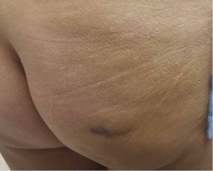
The patient's right buttock, 4 months postoperative, shows one purple-brown depressed macular scar with some central white hyperkeratotic material.
Two punch biopsies were performed on the day of initial evaluation: a 6-mm telescoping punch of skin followed by a 4-mm punch biopsy on the left buttock in the area of extruding material, into the subcutaneous fat. Results showed scarring and chronic granulomatous inflammation with histiocytic giant cells, lymphocytes, plasma cells, neutrophils, and eosinophils in the dermis and subcutis (Fig 4). There was no evidence of fat necrosis on histologic examination. Periodic acid–Schiff, acid-fast bacilli, and Gram stains were negative, making fungal, mycobacterial, and bacterial causes unlikely. An additional 4-mm punch biopsy of skin was sent for tissue culture, which showed suspected skin flora of rare coagulase-negative staphylococcus and was negative for fungi and acid-fast bacilli.
Fig 4.

Dermis shows pockets of inflammation; there are histiocytic giant cells, lymphocytes, plasma cells, neutrophils and eosinophils. (Hematoxylin-eosin stain; original magnification: ×40.)
At a subsequent visit in May 2018, 10 months after the initial procedure, she had a tender erythematous nodule on the lateral right buttock. This lesion was treated with 0.4 mL of intralesional triamcinolone acetonide, 5 mg/mL injection. The patient reported dramatic improvement in the days after injection.
In late summer 2018, the patient subsequently underwent an additional autologous fat transfer procedure, at the same outside facility, attempting to rectify the depressions created from the initial procedure. This procedure did not produce the patient's desired cosmetic outcome. She has not had any new inflammatory lesions thus far.
Discussion
Gluteal augmentation via liposuction and autologous fat transfer is becoming increasingly widely performed. Approximately 20,301 of these procedures were performed in the United States in 2017, a 10% increase from 2016.7 Using this technique, surgeons are able to produce dramatic body contouring results by reducing the mass of areas adjacent to the gluteal region and increasing the mass of the gluteal region itself.2 When compared with gluteal implantation, this procedure improves cosmetic appearance of scars, reduces complications, and has led to increased patient satisfaction with outcomes.1, 2 Common complications with implants and artificial fillers include seromas, capsular contractures, implant migration, wound healing complications, thinning of native tissues, infections, and foreign body responses.3, 5, 6 Lipogranulomas may be an example of foreign body reactions to lipid or oil-like substances, which result from the inability of the body to metabolize exogenous lipids in the tissue interstitium. Another proposed mechanism is endogenous degeneration of lipids secondary to allergic reactions or trauma.8
A literature review of lipogranuloma after autologous fat transfer returned no cases in the gluteal region. However, several case reports and case series of granulomatous reactions to fat transfer in the face have been reported. Two of the largest retrospective case series, describing lipogranulomas in the eyelid, cheek, and forehead, were published by Seo and Sa9 and Lee et al.10
Seo and Sa9 reviewed the outcomes of 27 patients with oral prednisone, intralesional steroid injection, surgery, observation, or combination therapy with the end point of full resolution. Steroids were found to be 70% effective (21 patients), surgery was 100% effective (2 patients), and observation was 50% effective (4 patients). Lee et al10 found oral steroids to be 75% effective (41 patients), but 2 patients went on to require surgery. The authors of this article noted that 78% of their patients had lipogranulomas upon second injection with cryopreserved fat, indicating cryopreserved fat is a risk factor for granuloma formation.10
Currently, more research needs to be conducted, as definitive treatment for this condition remains unclear, although steroids and surgical excision appear to be promising options. Unfortunately, surgical excision is not always practical depending on the location and extent of the lesion. Other sources conjecture that complications such as these might be prevented by proper technique in the operating room to ensure viability of the fat cells.3 However, it seems that these complications may be an inherent risk of the procedure. We present our case to serve as a warning about one of the potential complications of autologous fat transfer.
Footnotes
Funding sources: None.
Conflicts of interest: None disclosed.
References
- 1.Sinno S., Chang J.B., Brownstone N.D., Saadeh P.B., Wall S., Jr. Determining the safety and efficacy of gluteal augmentation: a systematic review of outcomes and complications. Plast Reconstr Surg. 2016;137(4):1151–1156. doi: 10.1097/PRS.0000000000002005. [DOI] [PubMed] [Google Scholar]
- 2.Ghavami A., Villanueva N.L. Gluteal augmentation and contouring with autologous fat transfer: part I. Clin Plast Surg. 2018;45(2):249–259. doi: 10.1016/j.cps.2017.12.009. [DOI] [PubMed] [Google Scholar]
- 3.Rapkiewicz A.V., Kenerson K., Hutchins K.D., Garavan F., Lew E.O., Shuman M.J. Fatal complications of aesthetic techniques: the gluteal region. J Forensic Sci. 2018;63(5):1406–1412. doi: 10.1111/1556-4029.13761. [DOI] [PubMed] [Google Scholar]
- 4.American Society of Plastic Surgeons Gluteal fat grafting advisory. https://www.plasticsurgery.org/for-medical-professionals/advocacy/key-issues/gluteal-fat-grafting-advisory
- 5.Singh M., Solomon I.H., Calderwood M.S., Talbot S.G. Silicone-induced granuloma after buttock augmentation. Plast Reconstr Surg Glob Open. 2016;4(2):e624. doi: 10.1097/GOX.0000000000000618. [DOI] [PMC free article] [PubMed] [Google Scholar]
- 6.Camuzard O., Dumas P., Foissac R. Severe granulomatous reaction associated with hypercalcemia occurring after silicone soft tissue augmentation of the buttocks: a case report. Aesthetic Plast Surg. 2014;38(1):95–99. doi: 10.1007/s00266-013-0167-4. [DOI] [PubMed] [Google Scholar]
- 7.American Society of Plastic Surgeons Plastic surgery statistics report. 2017. https://www.plasticsurgery.org/documents/News/Statistics/2017/plastic-surgery-statistics-full-report-2017.pdf
- 8.Park H.E., Kim H.T., Lee C.H., Bae J.H. Delayed lipogranuloma of the cheek following autologous fat injection: report of 2 cases. Int J Clin Exp Pathol. 2014;7(9):6391–6394. [PMC free article] [PubMed] [Google Scholar]
- 9.Seo J.W., Sa H.S. Periorbital lipogranuloma following facial autologous fat injections: non-surgical treatment. Aesthetic Plast Surg. 2015;39(6):946–952. doi: 10.1007/s00266-015-0554-0. [DOI] [PubMed] [Google Scholar]
- 10.Lee S., Lee J.H., Choi H.S. Clinical characteristics of patients with a periorbital mass after autologous fat injection for facial augmentation and short-term outcomes of steroid treatment. J Plast Reconstr Aesthet Surg. 2015;68(11):1498–1503. doi: 10.1016/j.bjps.2015.07.002. [DOI] [PubMed] [Google Scholar]