Abstract
Fetal growth restriction (FGR) is caused by placental insufficiency and can lead to short and long-term neurodevelopmental delays. Taurine, one of the most abundant amino acids in the brain, is critical for the normal growth and development of the nervous system; however, the mechanistic role of taurine in neural growth and development remains unknown. The present study investigated the role of taurine in FGR. Specifically, we explored the proteomic profiles of fetal rats at 6 h postpartum by two-dimensional difference gel electrophoresis combined with matrix assisted laser desorption ionization-time-of-flight (TOF)/TOF tandem mass spectrometry; the findings were verified via reverse transcription-quantitative polymerase chain reaction. A total of 31 differentially expressed protein spots were selected. Among these, 31 were matched, including dihydropyrimidinase-related protein 2 and, CRK and peroxiredoxin 2. Functional analysis using the Gene Ontology database and Ingenuity Pathway Analysis demonstrated that the differentially expressed proteins were mainly associated with neuronal differentiation, 'metabolic process', 'biological regulation' and developmental processes. The present study identified several proteins that were differentially expressed in rats with FGR in the presence or absence of taurine administration. The results of the present study suggest a potential role for taurine in the treatment and prevention of FGR.
Keywords: fetal growth restriction, taurine, 2D DIGE, MALDI-TOF/TOF MS
Introduction
Fetal growth restriction (FGR) refers to a condition in which the birth weight of a fetus is significantly lower than normal due to the failure to attain the ideal growth potential in the womb; FGR is affected by a variety of factors, including, the health of a pregnant female, the placenta and the fetus itself (1). An epidemiological survey revealed that the incidence of FGR in China was as high as 8.8% (1,2). FGR may have serious short-term adverse consequences on fetal and neonatal health, including high perinatal mortality, asphyxia of surviving fetuses, premature delivery and hypoglycemia (1). In addition, FGR may be associated with significantly high incidences of disease in various systems, such as the respiratory system (meconium aspiration, respiratory distress syndrome and bronchopulmonary dysplasia), and the circulatory and nervous systems (intracranial hemorrhage, periventricular white matter damage and hypoxic-ischemic encephalopathy), leading to an increased risk of hospitalization with extended hospital stay (1,3). Additionally, FGR may have serious long-term effects, including coronary heart disease, hypertension, insulin resistance and type II diabetes, kidney disease, obesity, dyslipidemia, metabolic syndrome and adrenal dysfunction (3,4). The fetal period, particularly during the first 10-18 weeks of pregnancy, is the most active period of neuron proliferation. Providing a fetus is exposed to unfavorable conditions during this window of development, neuronal proliferation and axonal extension will be directly affected (5). This may lead to decreases in the number and volume of fetal brain cells, and severe effects on interneuronal connections (5). Samuelsen et al (6) used an optical fractionator to analyze cells in various developmental regions of the brain affected by FGR and reported that the growth rate of brain cells in the cerebral cortex peripheral region of the FGR group was significantly lower than that of the control group, and the total number of cerebral cortical cells was significantly reduced. Tatli et al (7) studied the brain volume of premature infants with FGR and infants with normal weight using quantitative three-dimensional magnetic resonance imaging. The total brain volume and cerebral cortical volume of premature infants were significantly reduced compared with the normal control group, and remained smaller than the latter at 2 months of age (7).
Our previous study with transmission electron microscopy demonstrated that the brains of fetal rats with FGR were characterized by a notably disordered cerebral cortex structure, increased apoptosis of brain cells, decreased number of neurons and synapses with a sparse structure and glial cell proliferation (8). Clinical studies have reported that FGR can affect fetal brain function and development; infants with FGR have considerably hypofunctional brains at birth, as manifested by a lack of a sleep cycle, poor continuity of brain waves, extended burst intervals, and significantly reduced minimum and maximum voltages (9,10). It has been identified via long-term follow-up that FGR not only leads to various adverse outcomes for infant patients, including long-term retardation of neurological development, movement and behavioral disorders, language barriers, and short attention focusing, but has also been considered as the most important independent risk factor for severe neurological impairments in children with cerebral palsy (3,11). As the incidence of FGR is increasing, brain development disorders have become an important determinant of survival and long-term quality of life of fetuses and newborn children (12). Therefore, research promoting brain development in fetuses with FGR is important not only for its theoretical and clinical significance, but also for its impact on social and personal well-being. As brain developmental disorders of fetuses with FGR begin in the womb, postnatal intervention is often insufficient (13). Therefore, enhancing the effectiveness of prenatal (intrauterine) intervention is likely the best strategy to improve the poor prognosis of the nervous system in fetuses with FGR.
Taurine is an important amino acid in the human body and the most abundant free amino acid in the central nervous system. It is an essential amino acid in the fetal-neonatal-infant growth process, particularly for the development of the nervous system (14). Taurine has a wide range of beneficial effects on the brain, including protection against antioxidative stress and free radicals, stabilization of the cell membrane, reducing apoptosis of brain cells and relieving brain edema (15). A previous study has demonstrated that maternal gestational protein malnutrition is the main cause of FGR and metabolic disorders (16), and fetuses can only obtain taurine from their mother via the placenta, as fetuses cannot synthesize taurine. Previous research has confirmed that a low level of taurine in the brain structure of fetal rats with FGR leads to the increased apoptosis of brain cells, decreased proliferation of neurons and disordered brain ultrastructure, which is an important cause of brain development disorder and nervous system disability of fetal rats with FGR (8,17). Taurine content in the brain structures of fetal rats with FGR can be significantly increased by providing supplemental taurine to pregnant rats, thereby decreasing the apoptosis of brain cells, promoting neuron proliferation and improving brain ultrastructure; thus, the brain and body weights of fetal rats with FGR at birth can increase (17,18). The proliferation and differentiation of neural stem cells (NSCs) are the basis of the growth and development of the central nervous system (18); however, whether taurine supplementation in pregnant rats promotes the proliferation and differentiation of NSC of fetal rats with FGR, facilitating neuronal proliferation and brain development, and the underlying mechanism remain unknown. In the present study, comparative proteomics analysis of rat brain tissue of FGR and taurine-treated FGR models were used to identify the proteins and regulatory pathways that may be involved in mediating the beneficial effects of taurine on neuronal proliferation and brain development.
Materials and methods
Establishment of FGR model
As previously reported by Jansson et al (19), the whole-course low-protein diet method was used to establish an FGR model of Sprague Dawley rats. Taurine was administered according to our previous studies (17,18). Briefly, a total of 15 9-week-rat weighing 250-300 g (purchased from the Animal Science Department of Capital Medical University, Beijing, China) were selected, and then randomly divided into three groups: Control, FGR and FGR + taurine supplement (300 mg/kg weight), with each containing 5 pregnant rats. The rats were housed under a 12 h light/dark cycle (22±2°C, humidity: 70%, ventilation rate: 8 times/h) with full access to water. The control group was provided a standard diet (composition ratio: 20% protein, 67% carbohydrate and 13% fat), while the FGR and taurine groups were fed low protein diets (composition ratio: 8% protein, 79% carbohydrate and 13% fat; taurine was added starting from the 12th day of pregnancy) until spontaneous delivery. Newborn rats were weighed with an electronic precision balance within 6 h of birth, and those with a birth weight <2 standard deviations below the mean weight (5.5 g, data not shown) of the control group were identified as FGR rats. Mother rats were then housed in clean environment. The animal experiments were approved by the Ethics Committee of The First People's Hospital of Chenzhou (Chenzhou, China; approval no. 2017019). Mother rats were euthanized with pentobarbital sodium (135 mg/kg) following spontaneous delivery; lack of heart beat and breathing were considered to indicate animal death.
Protein extract preparation
A total of 24 fetal rats were sacrificed within 6 h after spontaneous delivery to obtain brain tissues. Fetal rats were then (~8-12 fetal rats from each mother rat) were sacrificed by cervical dislocation, and then positioned on an operating table in a supine position; an incision was made on the chest to expose the heart. Following normal saline perfusion (200-250 ml) via the left ventricle, the fetal rats were turned into a prone position to conduct an incision on the scalp. The skull was gently clamped into position at the posterior fontanelle using tweezers; the skull was cut and removed to expose the brain, and the cerebellum and brain stem were cut to allow removal of brain tissue. Specimens (n=8 per group) were weighed, and 50 mg of the tissue was cut and placed into a homogenate tube, with 400 μl of lysate and protease inhibitor phenylmethylsulfonyl fluoride at 1:100. Then, the mixture was placed in a pre-cooled homogenizer for stirring at 5,500 r/min for 5 min at 0-4°C. Following homogenization, the liquid was left at 20°C for 2 h and then aspirated into an EP tube for centrifugation at 22,000 × g for 60 min at 0-4°C. The protein supernatant was collected and transferred to an EP tube following centrifugation. The protein concentration was measured with Bicinchoninic Acid reagent (Thermo Fisher Scientific, Inc., Waltham, MA, USA).
2D difference gel electrophoresis (DIGE) analysis
This procedure were performed as described by Zhou et al (20). A total of 500 μg of protein samples from each group was dissolved in rehydration buffer (GE Healthcare, Little Chalfont, UK) and allowed to rehydrate with 24 cm immobilized pH gradient (IPG) strips (pH 3.0-10.0; GE Healthcare) overnight at 0-4°C. The IPGphor apparatus (GE Healthcare) was used to perform isoelectric focusing. Then, the strips were equilibrated and proteins were separated by 12.5% SDS-PAGE as the second dimension of separation. Electrophoresis was then performed using the Ettan Dalt six electrophoresis systems (GE Healthcare). Subsequently, gel plates were obtained and stained with Coomassie brilliant blue following fixation with ethyl alchol and glacial acetic acid for 30 min at room temperature. Then, stained gel plates were scanned using an image scanner (UMAX image scanner, PowerLook 2100XL, UMAX Technologies, New Taipei City, Taiwan). The gel images were eventually analyzed with DeCyder™ 2D Differential Analysis Software (DeCyder 2D V8.0, GE Healthcare). Protein spots were marked and selected for identification providing alterations in the abundance ratio were observed as >1.5-fold and P<0.05.
Matrix assisted laser desorption ionization (MALDI)/time-of-flight (TOF)/TOF tandem mass spectrometry (MS) analysis
This Spots of interest were excised and destained with 15 mM potassium ferricyanide and 50 mM sodium thiosulphate at room temperature for 20 min. The spots were then reduced in 10 mM DTT at 56°C for 1 h followed by alkylation in 55 mM iodoacetamide at room temperature for 45 min. Following washing with 50% acetonitrile (ACN) and dehydration with 100% ACN, the gel fragments were dried in a speed vacuum at room temperature. The dried gel fragment was then digested with 7 ng/μl trypsin (Promega Corporation, Madison, WI, USA) overnight at 25°C. The digested proteins were extracted twice with 50% ACN followed by 100% ACN. The digested peptides were spotted on a MALDI plate. MALDI-TOF-MS and TOF/TOF tandem MS were performed on an ABI 4800 mass spectrometer (Applied Biosystems; Thermo Fisher Scientific, Inc.). TOF/TOF tandem MS fragmentation spectra were acquired for each sample, averaging 4,000 laser shots per fragmentation spectrum on each of the 5-10 most abundant ions present in each sample excluding trypsin autolytic peptides and other known background ions. The resulting peptide mass and the associated fragmentation spectra were submitted to the Mascot search engine (Matrix Science Ltd., London, UK, http://www.matrixscience.com/) in order to search the non-redundant database of SwissProt (International Protein Index human 3.62 database, https://www.uniprot.org/). Searches were performed without restricting the protein molecular weight or isoelectric point, with variable carbamidomethylation of cysteine and oxidation of methionine residues and with one missed cleavage allowed in the search parameters. Candidates with either a protein score confidence interval >95% or a Mascot score >61 were considered significant as positive identifications. The maximum errors permitted in the search were MS 0.2 Da and MS/MS 0.3 Da.
Bioinformatics analysis
For an overview of the cellular localization, the Gene Ontology (GO) database was used to analyze the molecular function and biological processes of the identified proteins. Pathway analysis was performed for proteins with differential abundance among samples from the control, FGR and taurine-treated FGR rats. International Protein Index accession numbers were imported into the Ingenuity Pathway Analysis (IPA) program (Ingenuity Systems; Qiagen, Inc., Valencia, CA, USA). The identified proteins were mapped to the most significant networks generated from previous publications (21) and public protein interaction databases (http://www.ingenuity.com). A P-value calculated with the right-tailed Fisher's exact test was used to determine a network's score. Networks were ranked according to their degree of association with our data set.
Validation experiments by reverse transcription-quantitative polymerase chain reaction (RT-qPCR)
Total RNA in brain tissue from three groups of rats was extracted using TRIzol reagent (Thermo Fisher Scientific, Inc.). cDNA was synthesized from 1 μg of total RNA using PrimeScript™ RT Master Mix (Takara Bio, Inc., Otsu, Japan) according to manufacturer's protocols. qPCR was performed in a CFX96™ real-time detection system (ABI PRISM 7500, Applied Biosystems; Thermo Fisher Scientific, Inc.) with a total volume of 20 μl, containing 10 μl 2X TransStart Tip Green qPCR Super Mix (Trans) (Transgene Biotech, Beijing, China), 1 μl of each primer, 1 μl cDNA templates and 7 μl ddH2O. The specific primer sequences are listed in Table I. The amplification conditions were as follows: Initial denaturation at 94°C for 30 sec, 40 cycles of denaturation at 94°C for 5 sec, annealing at 56°C for 15 sec and final extension at 72°C for 10 sec. The relative expression levels were determined using the 2−ΔΔCq method (22). Each treatment was repeated in triplicate.
Table I.
Primer sequences used in reverse transcription-quantitative polymerase chain reaction.
| Gene | Forward primers (5′-3′) | Reverse primer (5′-3′) |
|---|---|---|
| Adprh | GGAGTCTCAGTTGCCCCATAA | GGTGTATCTTCTCCCCATCCC |
| Cct2 | GGCTTGTACCATTGTACTTCGTG | ATCTCAGAACAACCTCCTCCATAA |
| Ckb | AGACTGACCTCAACCCAGACAAC | AGGCTGGACAGGGCTTCTACT |
| Commd3 | GCAGTTACAGGATTTGGTGGG | TCCTCTAGGCTCCTGGGTCA |
| Crk | TCAGTGGGAAGGGGAGTGTAA | TTGTTCTCATCTGTCAGCAAAGTG |
| Dctn2 | GGAACGAGCCAGATGTTTATGA | TCAAGCCCCTTTGTCCCTAC |
| Dpysl2 | AGGCTTTTATGAATCCAATCTGC | AACAACAAACCCAAAATACCGA |
| Dpysl3 | GGGCCATTGCTCAAGTTCAT | AGACCACATTTCCTTTCTTCCTG |
| Hn1 | GCCCACAGGGACTCCTAATG | ACAGGGATGCGACTTTCTTCA |
| Hnrnpk | GGACCAGATACAGAACGCACAG | CAGCAAAGATTTCACTACACCTCA |
| Hspd1 (Hsp60) | GGTTTGGGGACAACAGGAAG | AGTTTAGCAAGTCGCTCGTTCA |
| Ina | TCTACAACCTCCAAAGTCTCATCC | GCATTCCCTCTTAGGCATCG |
| Kng1 | GAGTTAGAAAGCGGCAACCAG | GCGTCCTTGAAGTCACAGTCC |
| Ldhb | GTTGGACAAGTTGGAATGGCT | TTAGAATTGGCTGTCACGGAGTA |
| Pdia3 | ACGTGCTGATCGAGTTTTATGC | TGGCTGGTGAGAAGTAGATGGTA |
| Pgls | TCATCACGCCTGGCTTATTG | GGACCGATTTCTCGCATTTTA |
| Prdx2 | GACCTACCTGTGGGACGCTC | TGGAGAAGTATTCCTTGCTGTCA |
| Psmc3 | TCCAAGCCATGAAAGACAAAAT | GGGGTCAACATCCAGCAACT |
| Sae1 (Uble1a) | CTTGACTCATCGGAGACCACC | TGCGGAACTTCAGCAACACT |
| Snca | TGTCGTTGTACCCACTGTCCTAA | AGAGCCTGCTACCATGTATTCACT |
| Stub1 | TGAGAATGGCTGGGTAGAGGA | AGAAGGGAGACAAGGGAGGC |
| Tmod2 | ACATCGACGAGGACGAGCTT | AATCCAGCCGGTAGTGTTGC |
| Tubb3 | GGCCTTCCTCCACTGGTACA | CTCCTCGTCGTCATCTTCATACA |
| Uchl1 | CGGAGAAGTTGTCCCCTGAA | TGGCCGTCCACATTATTGAA |
Statistical analysis
The data were presented as the mean ± standard deviation. One-way analysis of variance followed by a Tukey's post hoc test was used to compare the expression levels between different groups by RT-qPCR. All statistical analyses were conducted using SPSS 17.0 (SPSS, Inc., Chicago, IL, USA) and were performed in triplicate. P<0.05 was considered to indicate a statistically significant difference.
Results
Determination of the protein expression profile of fetal rat brain tissues by 2D DIGE analysis and identification of differentially expressed protein by MALDI-TOF/TOF MS
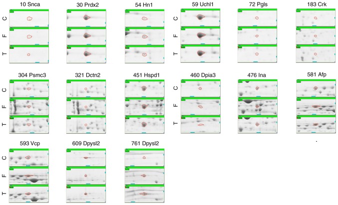
To identify the differentially expressed proteins in the FGR, FGR + taurine, and control groups, a 2D DIGE analysis was performed. A total of 269 spots were detected and matched successfully. Following a Mascot database search using the acquired MS data, 31 spots were identified by MALDI-TOF/TOF MS as differentially expressed in the three groups. The corresponding identity and sequence coverage was determined (Table SI). Of the detected spots, 30 exhibited statistically significant differences in expression (P<0.05) using the standard of ≥1.5-fold variation in expression (Fig. 1, Table SII). The abundance of 15 identified spots, including α-synuclein (Snca), peroxiredoxin 2 (Prdx2) and hematological and neurological expressed 1 (Hn1), was relatively lower in tissues from the FGR group, but was elevated following administration with taurine (Fig. 2). On the contrary, the abundance of 7 spots, including lactate dehydrogenase B chain (Ldhb), SUMO-activating enzyme subunit 1 (Sae1) and creatinine kinase B-type (Ckb), was higher in the FGR group and then markedly suppressed by taurine (Fig. 3). Other proteins (9 spots in total) did not meet these standards and are presented in Fig. 4.
Figure 1.
Overview of two-dimensional electrophoresis PAGEs of control (normal), FGR rats and FGR rats with taurine prenatally. Spots in green circles represented identified proteins. FGR, fetal growth restriction. Turquoise squares represented the order of identified proteins.
Figure 2.
Expression of identified proteins with significant difference in C (control, normal rats), F and T. Typical examples of identified differentially expression protein spots which presented lower expression in FGR rats compared with control group and reversed by taurine. C, control, normal rats; F, fetal growth restriction; T, fetal growth restriction with prenatal taurine administration.
Figure 3.
Expression of identified proteins with significantly different expression in the C, F and T groups. Examples of the identified proteins with differential expression that presented higher expression in the F group compared with the C group, and suppressed by taurine. C, control, normal rats; F, fetal growth restriction; T, fetal growth restriction with prenatal taurine administration.
Figure 4.
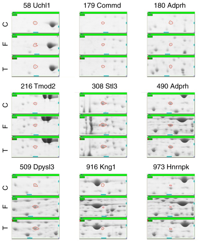
Typical examples of identified differentially expression protein spots without notable alterations across the treatment groups. C, control; F, fetal growth restriction; T, fetal growth restriction with taurine prenatally.
Analysis of networks via GO and IPA
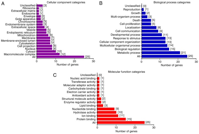
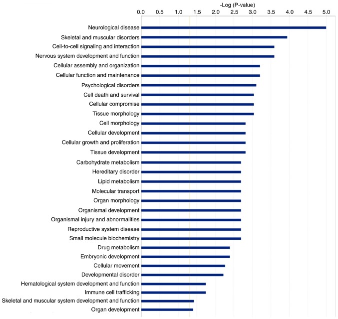
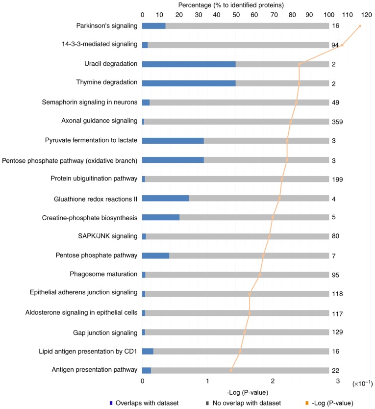
GO data was used to reveal the different functions of the proteins and processes in which they are involved. GO analysis based on 'cellular component' categories demonstrated that the identified proteins were mainly associated with macromolecular complexes in the cytosol and nucleus (Fig. 5A). Conversely, GO analysis based on 'biological process' categories indicated that other identified proteins were involved in 'metabolic process', 'biological regulation' and 'multicellular organismal processes' (Fig. 5B). The largest proportion of the identified proteins was involved in binding and hydrolysis based on major 'molecular function' categories (Fig. 5C). IPA of functional associations within our set of proteins revealed that the proteins were highly associated with neurological diseases, and nervous system development and function, such as Parkinson's signaling and glutathione redox reactions (Figs. 6 and 7). The majority of these proteins were localized to and functioned in the cytoplasm (Fig. 8).
Figure 5.
Gene ontology categories of the identified proteins. Proteins were classified into (A) 'cellular component', (B) 'biological process' and (C) 'molecular function' depending on the number of involved genes.
Figure 6.
Enriched pathway identified using IPA for differentially expressed genes. Identified proteins with significant differences were used to process IPA analysis for pathway enrichment. IPA, Ingenuity Pathway Analysis.
Figure 7.
Ingenuity Pathway Analysis of differentially expressed genes to identify the canonical pathways.
Figure 8.
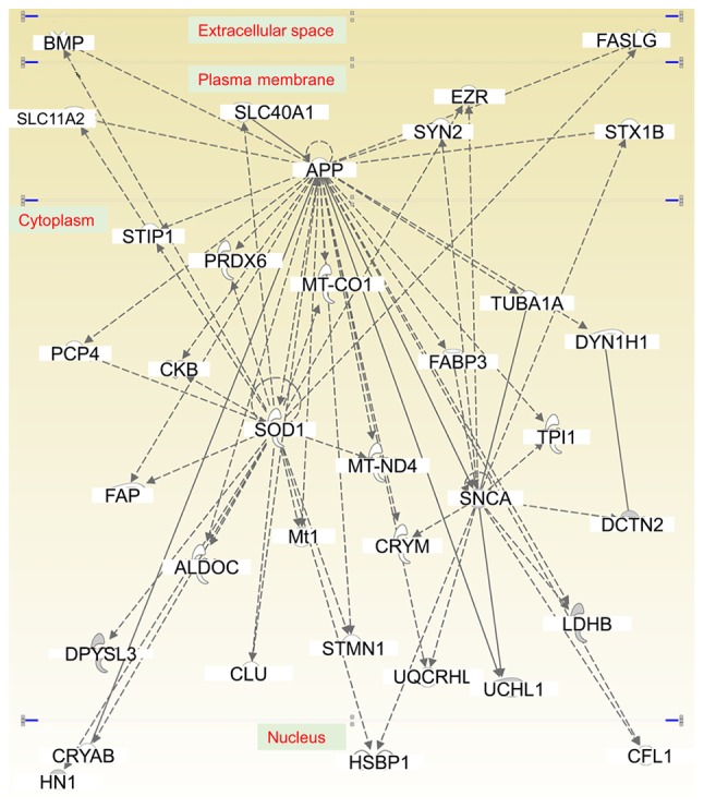
Functional gene networks identified using Ingenuity Pathway Analysis for differentially expressed genes.
Validation of identified proteins by RT-qPCR
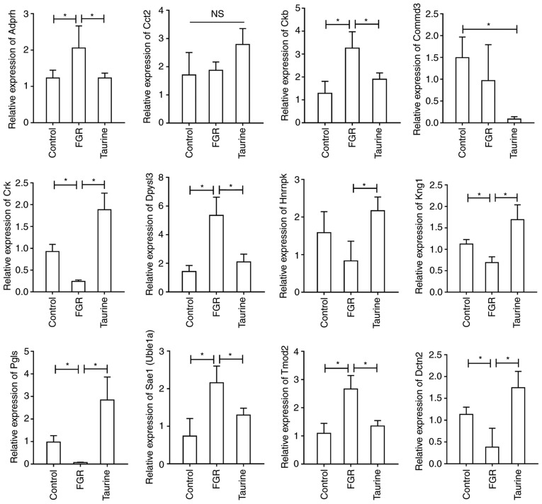
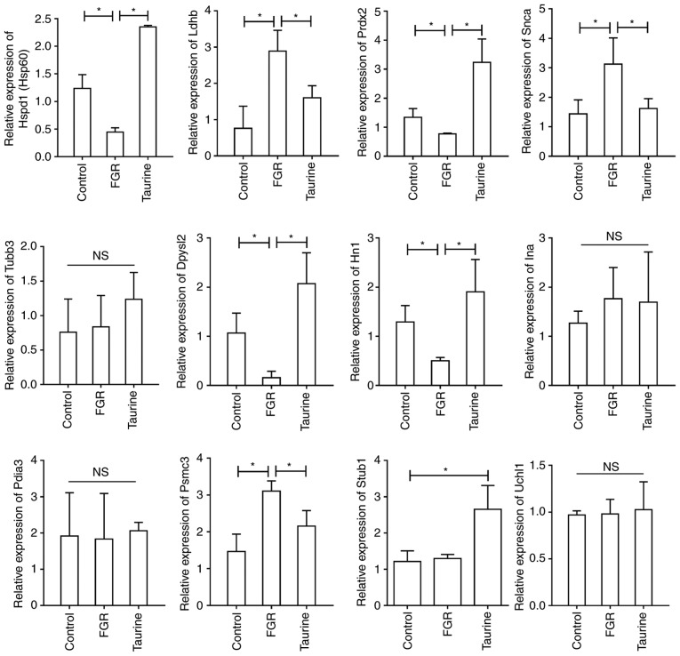
The gene expression levels of the corresponding proteins identified in the control, FGR and FGR + Taurine groups were determined by RT-qPCR. As presented in Figs. 9 and 10, the mRNA expression levels of certain genes with potential significance in these three groups were determined. Significantly reduced expression of Crk, dynactin subunit 2 (Dctn2), dihydropyridinase like (Dpysl)2, Hn1, heat shock protein family D (Hsp60) member 1, kininogen 1, 6-phosphogluconolactonase and Prdx2 was observed in the FGR group compared with the control, but was reversed by taurine administration. On the contrary, the expression of Ckb, Sae1, tropomodulin 2, Dpysl3, Ldhb, Snca and proteasome 26S subunit ATPase 3 was significantly increased in FGR rats compared with the control, but was suppressed in taurine-treated FGR rats. In addition, the expression of internexin neuronal intermediate filament protein a, protein disulfide isomerase family A member 3, ubiquitin C-terminal hydrolase L1, chaperonin containing TCP1 subunit 2 and tubulin β3 class III was not significantly different among the three groups. Interestingly, the expression of COMM domain containing 3 was significantly lower in taurine-treated FGR rats compared with the control and FGR groups. We detected significantly higher expression of Stub1 in taurine-treated FGR rats compared with the control and FGR rats. Additionally, the expression of heterogenous nuclear ribonucleoprotein K was significantly lower in the FGR group compared with the control; however, no significant difference was observed. Of note, its expression was significantly promoted in taurine-treated FGR rats compared with FGR alone.
Figure 9.
Relative expression of Adprh, Cct2, Ckb, Commd3, Crk, Dpysl3, Hnrnpk, Kng1, Pgls, Sae1, Tmod2 and Dctn2 in control, FGR, and taurine (FGR + Taurine). Expression of identified proteins was detected using qPCR. *P<0.05. NS, not significant. Adprh, ADP-ribosylarginine hydrolase; Cct2, chaperonin containing TCP1 subunit 2; Ckb, creatinine kinase B-type; Commd3, COMM domain containing 3; Dctn2, dynactin subunit 2; Dpysl3, dihydropyridinase like 3; FGR, fetal growth restriction; Hnrnpk, heterogenous nuclear ribonucleoprotein K; Kng1, kininogen 1; Pgls, 6-phosphogluconolactonase; Sae1, SUMO-activating enzyme subunit 1; Tmod2, tropomodulin 2.
Figure 10.
Relative expression of Hspd1, Ldhb, Prdx2, Snca, Tubb3, Dpysl2, Hn1, Ina, Pdia3, Psmc3, Stub1 and Uchl1 in control, FGR, and taurine (FGR + Taurine). Expression of identified proteins was detected using reverse transcription-quantitative polymerase chain reaction. *P<0.05. NS, not significant. Dpysl2, dihydropyridinase like 2; Hn1, hematological and neurological expressed 1; Hspd1, heat shock protein family D (Hsp60) member 1; Ina, n neuronal intermediate filament protein a; Ldhb, lactate dehydrogenase B chain; Pdia3, protein disulfide isomerase family A member 3; Psmc3, proteasome 26S subunit ATPase; Prdx2, peroxiredoxin 2; Snca, α-synuclein; Stub1, STIP1 homology and U-box containing protein 1; Tubb3, tubulin β3 class III; Uchl1, ubiquitin C-terminal hydrolase L1.
Discussion
FGR is a clinical problem in perinatology. The incidence of intrauterine growth restriction in newborns ranges between 3-7% in the total population, and is a contributor to perinatal morbidity and mortality (23). In the past decade the incidence has increased, resulting partly from poor dietary habits, particularly protein malnutrition during pregnancy (24). Clinical research has revealed that FGR can induce a multitude of adverse effects, including short and long-term neurodevelopmental delays, and cognitive dysfunction (25,26). A previous study reported that antenatal taurine supplementation enhances the brain ultrastructure of FGR rats (8). In the present study, rat model of FGR was established and taurine was administered antenatally. A combination of 2D DIGE and MALDI-TOF/TOF MS analysis was conducted to determine differential protein expression. The results revealed that taurine could significantly alter the expression of certain proteins associated with neuronal development and function, which are important in ameliorating the adverse effects of FGR.
The relationship between taurine and the growth of infants and young children has received considerable attention; however, the findings of numerous studies are controversial. Animal and cell-based studies have demonstrated that taurine could promote body weight gain, cognitive development and neural cell differentiation, while control experiments evaluating physical growth, fat absorption and neural development in infants and young children have mostly negative results. Whether taurine promotes growth and development requires further investigation into the differences and associations between animal and cell-based experiments; studies for the analysis of various populations should also be conducted. In the present study, FGR rats were established using a low-protein diet. Samples obtained from fetal rats were used for 2D DIGE analysis, and differentially expressed proteins spots were identified by MALDI-TOF/TOF MS. Finally, these proteins were further analyzed by RT-qPCR. These proteins were determined to be associated with neuronal differentiation, nerve injury and regeneration, the PKA-CREB signaling pathway and the Rho-ROCK signaling pathway (27,28).
Taurine is the most abundant amino acid in the brain, and is indispensable to normal growth and development of the nervous system (17). Among the differentially expressed proteins identified by MALDI-TOF/TOF MS in the present study, several of these have a potential role in nerve growth and differentiation. DPYSL2 belongs to the DPYSL family of proteins (29), which includes five members; these proteins serve important roles in the maturation of the nervous system in humans and in rats (30). DPYSL2, also reported as collapsin response mediator protein 2, and isocitrate dehydrogenase (NADP) have been proposed to be plasma membrane-associated proteins involved in neuronal differentiation, axon growth and guidance, neuronal growth cone collapse and cell migration (31). Tan et al (30) demonstrated elevated DPYSL3 expression in neuroblastoma via negative regulation mediated by MYCN. When co-expressed in COS-7 cells, DPYSLs are physically associated with DPYSL3 to form a large complex in vivo. The expression of DPYSL is brain-specific, high in fetal and neonatal rat brains, and is reduced to very low levels in the adult brain. In addition, DPYSL expression is upregulated during the neuronal differentiation of embryonic carcinoma P19 and PC12 cells (32). Finally, immunoprecipitation analysis of rat brain extracts revealed the co-precipitation of DPYSL with proteins possessing tyrosine kinase activity (32). In the present study, the expression of DPYSL2 was lower in FGR rats and increased following taurine administration; however, this was not observed for DPYSL3.
The literature suggests that DCTN2 is involved in synapse stability and is an essential factor in maintaining normal neuronal function (33). Puls et al (34) indicated that mutation of dynactin will lead to dysfunction of dynactin-mediated transport, inducing human motor neuron disease. Evidence has indicated that dynactin, encoded by Dctn2, participates in promoting axonal growth and multiorganelle transport via a phosphatidylinositol 3-kinase catalytic subunit type 3/ankyrin-B/dynactin pathway (35). In the present study, Dctn2 expression was decreased in the FGR group compared with the control group, but increased following antenatal taurine administration. The effects of taurine on Dctn2 expression suggests that it may have a positive role in FGR rats.
The role of CRK in neuronal differentiation remains unclear. Researchers suggest that overexpression of CRK is not sufficient to promote the neuronal differentiation of PC12 cells, as a growth factor stimulatory signal is required (36); however, Matsuda et al (37) demonstrated that microinjection of the CRK has a positive role in neuronal differentiation. This may be due to the differing binding potential of CRK to phosphorylated tyrosine-containing proteins (38); however, whether CRK can be mediated by taurine remains unknown.
Taurine can reduce neuronal apoptosis in the brain during FGR (17); however, the mechanisms are not fully understood. Researchers have demonstrated that DPYSL3 is involved in oxidative damage in primary cortical neurons (39). Specifically, oxidative stress caused by H2O2 can induce a surge of reactive oxygen species (ROS), which is characterized by the degradation of DPYSL3 via nitric oxide synthase (NOS) mediation. Furthermore, the administration of a NOS inhibitor can prevent N-methyl-D-aspartate-induced ROS formation and an increase in the intracellular levels of Ca2+ (40). Finally, it has been reported that increased DPYSL3 expression results in rescue from cell death (40). Homeostatic alterations caused by ROS-dependent activation of L-voltage-gated calcium channels are sufficient to induce possible calpain-mediated DPYSL3 truncation; this finding was the first demonstration of a mechanistic role of ROS in glutamate induction, calpain activation, and DPYSL3 protein degradation (40). In fact, a large body of research has demonstrated that taurine can reduce ROS in vitro (41). In the present study, suppressed DPYSL3 expression was observed in FGR rats, which was partially reversed by taurine. Therefore, taurine may exert a neuroprotective role via the suppression of ROS levels induced by oxidative stress.
In conclusion, the present study identified proteins that are differentially expressed in normal, FGR rats and taurine-treated FGR rats using 2D DIGE combined with MALDI-TOF/TOF MS/MS. Of the 37 protein spots that were identified, 31 were matched. GO category analysis indicated that these proteins were involved in metabolic processes, biological regulation, developmental processes and cell proliferation. Furthermore, proteins such as DPYSLs, CRK and DCTN2 may be associated with neuronal differentiation and neuroprotection; however, further investigation is required. To the best of our knowledge, the present study is the first to explore role of taurine in the treatment of FGR based on proteomics analysis.
Supplementary Materials
Acknowledgments
Not applicable.
Funding
This study was supported by National Natural Science Foundation of China (grant no. 81170577).
Availability of data and materials
All data generated or analyzed during this study are included in this published article.
Authors' contributions
HL and WF designed and performed the experiments; YW provided assistance with 2D-DIGE; HFL and JL analyzed the 2D-DIGE data and prepared the MALDI-TOF/TOF MS sample; HL drafted the manuscript; WF revised the manuscript and provided administrative support. All authors reviewed the manuscript. All authors read and approved the final manuscript.
Ethics approval and consent to participate
The animal experiments were approved by the Ethics Committee of The First People's Hospital of Chenzhou (Chenzhou, China; approval no. 2017019).
Patient consent for publication
Not applicable.
Competing interests
Not applicable.
References
- 1.Giabicani E, Pham A, Brioude F, Mitanchez D, Netchine I. Diagnosis and management of postnatal fetal growth restriction. Best Pract Res Clin Endocrinol Metab. 2018;32:523–534. doi: 10.1016/j.beem.2018.03.013. [DOI] [PubMed] [Google Scholar]
- 2.Liu J, Wang XF, Wang Y, Wang HW, Liu Y. The incidence rate, high-risk factors, and short- and long-term adverse outcomes of fetal growth restriction: A report from Mainland China. Medicine (Baltimore) 2014;93:e210. doi: 10.1097/MD.0000000000000210. [DOI] [PMC free article] [PubMed] [Google Scholar]
- 3.Longo S, Bollani L, Decembrino L, Di Comite A, Angelini M, Stronati M. Short-term and long-term sequelae in intrauterine growth retardation (IUGR) J Matern Fetal Neonatal Med. 2013;26:222–225. doi: 10.3109/14767058.2012.715006. [DOI] [PubMed] [Google Scholar]
- 4.Verschuren MT, Morton JS, Abdalvand A, Mansour Y, Rueda-Clausen CF, Compston CA, Luyckx V, Davidge ST. The effect of hypoxia-induced intrauterine growth restriction on renal artery function. J Dev Orig Health Dis. 2012;3:333–341. doi: 10.1017/S2040174412000268. [DOI] [PubMed] [Google Scholar]
- 5.Iruretagoyena JI, Davis W, Bird C, Olsen J, Radue R, Teo Broman A, Kendziorski C, Splinter BonDurant S, Golos T, Bird I, Shah D. Differential changes in gene expression in human brain during late first trimester and early second trimester of pregnancy. Prenat Diagn. 2014;34:431–437. doi: 10.1002/pd.4322. [DOI] [PubMed] [Google Scholar]
- 6.Samuelsen GB, Pakkenberg B, Bogdanović N, Gundersen HJ, Larsen JF, Graem N, Laursen H. Severe cell reduction in the future brain cortex in human growth-restricted fetuses and infants. Am J Obstet Gynecol. 2007;197:56.e1–e7. doi: 10.1016/j.ajog.2007.02.011. [DOI] [PubMed] [Google Scholar]
- 7.Tatli M, Guzel A, Kizil G, Kavak V, Yavuz M, Kizil M. Comparison of the effects of maternal protein malnutrition and intrauterine growth restriction on redox state of central nervous system in offspring rats. Brain Res. 2007;1156:21–30. doi: 10.1016/j.brainres.2007.04.036. [DOI] [PubMed] [Google Scholar]
- 8.Liu J, Liu L, Chen H. Antenatal taurine supplementation for improving brain ultrastructure in fetal rats with intrauterine growth restriction. Neuroscience. 2011;181:265–270. doi: 10.1016/j.neuroscience.2011.02.056. [DOI] [PubMed] [Google Scholar]
- 9.Cohen E, Wong FY, Wallace EM, Mockler JC, Odoi A, Hollis S, Horne RSC, Yiallourou SR. EEG power spectrum maturation in preterm fetal growth restricted infants. Brain Res. 2018;1678:180–186. doi: 10.1016/j.brainres.2017.10.010. [DOI] [PubMed] [Google Scholar]
- 10.Fu LC, Lv Y, Zhong Y, He Q, Liu X, Du LZ. Tyrosine phosphorylation of Kv1.5 is upregulated in intrauterine growth retardation rats with exaggerated pulmonary hypertension. Braz J Med Biol Res. 2017;50:e6237. doi: 10.1590/1414-431x20176237. [DOI] [PMC free article] [PubMed] [Google Scholar]
- 11.Baschat AA. Neurodevelopment following fetal growth restriction and its relationship with antepartum parameters of placental dysfunction. Ultrasound Obstet Gynecol. 2011;37:501–514. doi: 10.1002/uog.9008. [DOI] [PubMed] [Google Scholar]
- 12.Nardozza LM, Caetano AC, Zamarian AC, Mazzola JB, Silva CP, Marçal VM, Lobo TF, Peixoto AB, Araujo Júnior E. Fetal growth restriction: Current knowledge. Arch Gynecol Obstet. 2017;295:1061–1077. doi: 10.1007/s00404-017-4341-9. [DOI] [PubMed] [Google Scholar]
- 13.Albu AR, Horhoianu IA, Dumitrascu MC, Horhoianu V. Growth assessment in diagnosis of fetal growth restriction. Review. J Med Life. 2014;7:150–154. [PMC free article] [PubMed] [Google Scholar]
- 14.Metges CC, Görs S, Lang IS, Hammon HM, Brüssow KP, Weitzel JM, Nürnberg G, Rehfeldt C, Otten W. Low and high dietary protein: Carbohydrate ratios during pregnancy affect materno-fetal glucose metabolism in pigs. J Nutr. 2014;144:155–163. doi: 10.3945/jn.113.182691. [DOI] [PubMed] [Google Scholar]
- 15.Li S, Guan H, Qian Z, Sun Y, Gao C, Li G, Yang Y, Piao F, Hu S. Taurine inhibits 2,5-hexanedione-induced oxidative stress and mitochondria-dependent apoptosis in PC12 cells. Ind Health. 2017;55:108–118. doi: 10.2486/indhealth.2016-0044. [DOI] [PMC free article] [PubMed] [Google Scholar]
- 16.Ripps H, Shen W. Review: Taurine: A 'very essential' amino acid. Mol Vis. 2012;18:2673–2686. [PMC free article] [PubMed] [Google Scholar]
- 17.Liu J, Liu L, Wang XF, Teng HY, Yang N. Antenatal supplementation of taurine for protection of fetal rat brain with intrauterine growth restriction from injury by reducing neuronal apoptosis. Neuropediatrics. 2012;43:258–263. doi: 10.1055/s-0032-1324730. [DOI] [PubMed] [Google Scholar]
- 18.Liu J, Wang X, Liu Y, Yang N, Xu J, Ren X. Antenatal taurine reduces cerebral cell apoptosis in fetal rats with intrauterine growth restriction. Neural Regen Res. 2013;8:2190–2197. doi: 10.3969/j.issn.1673-5374.2013.23.009. [DOI] [PMC free article] [PubMed] [Google Scholar]
- 19.Jansson N, Pettersson J, Haafiz A, Ericsson A, Palmberg I, Tranberg M, Ganapathy V, Powell TL, Jansson T. Down-regulation of placental transport of amino acids precedes the development of intrauterine growth restriction in rats fed a low protein diet. J Physiol. 2006;576:935–946. doi: 10.1113/jphysiol.2006.116509. [DOI] [PMC free article] [PubMed] [Google Scholar]
- 20.Zhou X, Wang P, Zhang YJ, Xu JJ, Zhang LM, Zhu L, Xu LP, Liu XM, Su PP. Comparative proteomic analysis of melanosis coli with colon cancer. Oncol Rep. 2016;36:3700–3706. doi: 10.3892/or.2016.5178. [DOI] [PubMed] [Google Scholar]
- 21.Yan-Fang T, Zhi-Heng L, Li-Xiao X, Fang F, Jun L, Gang L, Lan C, Na-Na W, Xiao-Juan D, Li-Chao S, et al. Molecular mechanism of the cell death induced by the histone deacetylase pan inhibitor LBH589 (Panobinostat) in Wilms tumor cells. PLoS One. 2015;10:e0126566. doi: 10.1371/journal.pone.0126566. [DOI] [PMC free article] [PubMed] [Google Scholar]
- 22.Livak KJ, Schmittgen TD. Analysis of relative gene expression data using real-time quantitative PCR and the 2-(Delta Delta C(T)) method. Methods. 2001;25:402–408. doi: 10.1006/meth.2001.1262. [DOI] [PubMed] [Google Scholar]
- 23.Cruz-Lemini M, Crispi F, Van Mieghem T, Pedraza D, Cruz-Martínez R, Acosta-Rojas R, Figueras F, Parra-Cordero M, Deprest J, Gratacós E. Risk of perinatal death in early-onset intrauterine growth restriction according to gestational age and cardiovascular doppler indices: A multicenter study. Fetal Diagn Ther. 2012;32:116–122. doi: 10.1159/000333001. [DOI] [PubMed] [Google Scholar]
- 24.Romo A, Carceller R, Tobajas J. Intrauterine growth retardation (IUGR): Epidemiology and etiology. Pediatr Endocrinol Rev. 2009;6(Suppl 3):S332–S336. [PubMed] [Google Scholar]
- 25.Figueras F, Cruz-Martinez R, Sanz-Cortes M, Arranz A, Illa M, Botet F, Costas-Moragas C, Gratacos E. Neurobehavioral outcomes in preterm, growth-restricted infants with and without prenatal advanced signs of brain-sparing. Ultrasound Obstet Gynecol. 2011;38:288–294. doi: 10.1002/uog.9041. [DOI] [PubMed] [Google Scholar]
- 26.Bassan H, Stolar O, Geva R, Eshel R, Fattal-Valevski A, Leitner Y, Waron M, Jaffa A, Harel S. Intrauterine growth-restricted neonates born at term or preterm: How different? Pediatr Neurol. 2011;44:122–130. doi: 10.1016/j.pediatrneurol.2010.09.012. [DOI] [PubMed] [Google Scholar]
- 27.Park SY, Jung WJ, Kang JS, Kim CM, Park G, Choi YW. Neuroprotective effects of α-iso-cubebene against glutamate-induced damage in the HT22 hippocampal neuronal cell line. Int J Mol Med. 2015;35:525–532. doi: 10.3892/ijmm.2014.2031. [DOI] [PubMed] [Google Scholar]
- 28.Kubo T, Hata K, Yamaguchi A, Yamashita T. Rho-ROCK inhibitors as emerging strategies to promote nerve regeneration. Curr Pharm Des. 2007;13:2493–2499. doi: 10.2174/138161207781368657. [DOI] [PubMed] [Google Scholar]
- 29.Turck CW, Webhofer C, Nussbaumer M, Teplytska L, Chen A, Maccarrone G, Filiou MD. Stable isotope metabolic labeling suggests differential turnover of the DPYSL protein family. Proteomics Clin Appl. 2016;10:1269–1272. doi: 10.1002/prca.201600078. [DOI] [PubMed] [Google Scholar]
- 30.Tan F, Wahdan-Alaswad R, Yan S, Thiele CJ, Li Z. Dihydropyrimidinase-like protein 3 expression is negatively regulated by MYCN and associated with clinical outcome in neuroblastoma. Cancer Sci. 2013;104:1586–1592. doi: 10.1111/cas.12278. [DOI] [PMC free article] [PubMed] [Google Scholar]
- 31.Arimura N, Kaibuchi K. Neuronal polarity: From extracellular signals to intracellular mechanisms. Nat Rev Neurosci. 2007;8:194–205. doi: 10.1038/nrn2056. [DOI] [PubMed] [Google Scholar]
- 32.Inatome R, Tsujimura T, Hitomi T, Mitsui N, Hermann P, Kuroda S, Yamamura H, Yanagi S. Identification of CRAM, a novel unc-33 gene family protein that associates with CRMP3 and protein-tyrosine kinase(s) in the developing rat brain. J Biol Chem. 2000;275:27291–27302. doi: 10.1074/jbc.M910126199. [DOI] [PubMed] [Google Scholar]
- 33.Eaton BA, Fetter RD, Davis GW. Dynactin is necessary for synapse stabilization. Neuron. 2002;34:729–741. doi: 10.1016/S0896-6273(02)00721-3. [DOI] [PubMed] [Google Scholar]
- 34.Puls I, Jonnakuty C, LaMonte BH, Holzbaur EL, Tokito M, Mann E, Floeter MK, Bidus K, Drayna D, Oh SJ, et al. Mutant dynactin in motor neuron disease. Nat Genet. 2003;33:455–456. doi: 10.1038/ng1123. [DOI] [PubMed] [Google Scholar]
- 35.Lorenzo DN, Badea A, Davis J, Hostettler J, He J, Zhong G, Zhuang X, Bennett V. A PIK3C3-Ankyrin-B-Dynactin pathway promotes axonal growth and multiorganelle transport. J Cell Biol. 2014;207:735–752. doi: 10.1083/jcb.201407063. [DOI] [PMC free article] [PubMed] [Google Scholar]
- 36.Hempstead BL, Birge RB, Fajardo JE, Glassman R, Mahadeo D, Kraemer R, Hanafusa H. Expression of the v-crk oncogene product in PC12 cells results in rapid differentiation by both nerve growth factor- and epidermal growth factor-dependent pathways. Mol Cell Biol. 1994;14:1964–1971. doi: 10.1128/MCB.14.3.1964. [DOI] [PMC free article] [PubMed] [Google Scholar]
- 37.Matsuda M, Hashimoto Y, Muroya K, Hasegawa H, Kurata T, Tanaka S, Nakamura S, Hattori S. CRK protein binds to two guanine nucleotide-releasing proteins for the Ras family and modulates nerve growth factor-induced activation of Ras in PC12 cells. Mol Cell Biol. 1994;14:5495–5500. doi: 10.1128/MCB.14.8.5495. [DOI] [PMC free article] [PubMed] [Google Scholar]
- 38.Birge RB, Fajardo JE, Mayer BJ, Hanafusa H. Tyrosine-phosphorylated epidermal growth factor receptor and cellular p130 provide high affinity binding substrates to analyze Crk-phosphotyrosine-dependent interactions in vitro. J Biol Chem. 1992;267:10588–10595. [PubMed] [Google Scholar]
- 39.Kowara R, Moraleja KL, Chakravarthy B. Involvement of nitric oxide synthase and ROS-mediated activation of L-type voltage-gated Ca2+ channels in NMDA-induced DPYSL3 degradation. Brain Res. 2006;1119:40–49. doi: 10.1016/j.brainres.2006.08.047. [DOI] [PubMed] [Google Scholar]
- 40.Kowara R, Kristoffer L, Chakravarthy B. Evidence that NMDA-induced DPYSL3 truncation involves ROS-mediated activation of nitric oxide synthase and L-type voltage-gated Ca2+ channels. Brain Res. 2006;1119:40–49. doi: 10.1016/j.brainres.2006.08.047. [DOI] [PubMed] [Google Scholar]
- 41.Kim YS, Kim EK, Jeon NJ, Ryu BI, Hwang JW, Choi EJ, Moon SH, Jeon BT, Park PJ. Antioxidant effect of taurine-rich paroctopus dofleini extracts through inhibiting ROS production against LPS-induced oxidative stress in vitro and in vivo model. Adv Exp Med Biol. 2017;975(Pt 2):1165–1177. doi: 10.1007/978-94-024-1079-2_93. [DOI] [PubMed] [Google Scholar]
Associated Data
This section collects any data citations, data availability statements, or supplementary materials included in this article.
Supplementary Materials
Data Availability Statement
All data generated or analyzed during this study are included in this published article.


