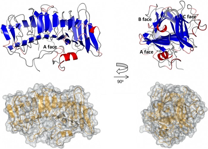
FIG 3.
Detail of the β-solenoid domain from ComZ. (Upper) Two orthogonal ribbon plot views of the β-solenoid domain from ComZ, colored by secondary structure: β-strand (blue), α-helix (red), turn (pink), and coil (gray). Faces of the solenoid are labeled on the right. (Lower) Surface models, in the same orientations as that of the upper panel, superimposed on ribbon plot.

