Anopheles gambiae mosquitos are the main vectors of malaria, threatening around half of the world’s population. The bacterial symbiont Wolbachia can interfere with disease transmission by other important insect vectors, but until recently, it was thought to be absent from natural A. gambiae populations. Here, we critically analyze the genomic, metagenomic, PCR, imaging, and phenotypic data presented in support of the presence of natural Wolbachia infections in A. gambiae. We find that they are insufficient to diagnose Wolbachia infections and argue for the need of obtaining robust data confirming basic Wolbachia characteristics in this system. Determining the Wolbachia infection status of Anopheles is critical due to its potential to influence Anopheles population structure and Plasmodium transmission.
KEYWORDS: endosymbionts, malaria, metagenomics, vector biology
ABSTRACT
Wolbachia (Alphaproteobacteria, Rickettsiales) is an intraovarially transmitted symbiont of insects able to exert striking phenotypes, including reproductive manipulations and pathogen blocking. These phenotypes make Wolbachia a promising tool to combat mosquito-borne diseases. Although Wolbachia is present in the majority of terrestrial arthropods, including many disease vectors, it was considered absent from Anopheles gambiae mosquitos, the main vectors of malaria in sub-Saharan Africa. In 2014, Wolbachia sequences were detected in A. gambiae samples collected in Burkina Faso. Subsequently, similar evidence came from collections all over Africa, revealing a high Wolbachia 16S rRNA sequence diversity, low abundance, and a lack of congruence between host and symbiont phylogenies. Here, we reanalyze and discuss recent evidence on the presence of Wolbachia sequences in A. gambiae. We find that although detected at increasing frequencies, the unusual properties of these Wolbachia sequences render them insufficient to diagnose natural infections in A. gambiae. Future studies should focus on uncovering the origin of Wolbachia sequence variants in Anopheles and seeking sequence-independent evidence for this new symbiosis. Understanding the ecology of Anopheles mosquitos and their interactions with Wolbachia will be key in designing successful, integrative approaches to limit malaria spread. Although the prospect of using Wolbachia to fight malaria is intriguing, the newly discovered strains do not bring it closer to realization.
OBSERVATION
Wolbachia is an obligate intracellular, intraovarially transmitted bacterium living in symbiosis with many invertebrates (1). Depending on host and symbiont genotypes and environmental conditions, Wolbachia has been shown either to affect the biology of its hosts in striking ways or to exert only mild phenotypes. Some of the conspicuous Wolbachia phenotypes include reproductive manipulations, where maternally inherited symbionts favor the survival and reproduction of transmitting females over those of noninfected females and nontransmitting males (2). One of the reproductive manipulations, cytoplasmic incompatibility (CI) (3), has been proposed as a tool to suppress mosquito populations and decrease arbovirus burden on humans (4, 5). Bidirectional CI, the inability of females to produce offspring with males harboring a different Wolbachia strain, was successful in eliminating the filariasis vector Culex pipiens fatigans from Okpo, Myanmar, in 1967 (5) and in suppressing Aedes albopictus, the vector of the dengue, Zika, and West Nile viruses, in recent trials in Lexington, Kentucky, California, and New York, USA (https://mosquitomate.com).
Wolbachia can also provide infected individuals with fitness benefits: nutrient provisioning (6), increase in reproductive output (7), and protection against pathogens (8, 9). The last phenotype is also being used to eliminate vector-borne diseases. Aedes aegypti mosquitos artificially transinfected with protective Wolbachia organisms are being deployed as a strategy to eradicate dengue virus (10–15). The data from one of the first release sites in Australia suggest that this strategy may limit the number of dengue cases in humans (15).
Malaria is a mosquito-borne disease that threatens around half of the world’s population (16). The potential for the use of Wolbachia to block malaria has been recognized since the symbiont’s antiviral and antiparasitic properties were first demonstrated in other insects (8–10, 17). However, Anopheles mosquitos were long considered inhospitable for Wolbachia (18–20). This started to change in 2006, when Wolbachia infections in cultured Anopheles cells were established for the first time (21). Next, transient somatic infections were created by intrathoracic inoculation of the virulent wMelPop strain of Wolbachia into adult mosquitos (22). In somatic transinfections, Wolbachia does not infect the germ line (23), which is necessary for its maternal transmission and pathogen blocking-based field applications. Therefore, a successful generation of stable Wolbachia infections in Anopheles stephensi by Bian et al. was a big step toward field applications (24). Subsequently, the gut microbiota of A. stephensi and A. gambiae were shown to hinder the establishment of heritable Wolbachia infections in these species, and curing Anopheles of its microbiota enabled Wolbachia persistence (25). In 2014, the first evidence for natural Wolbachia infections was found in Anopheles gambiae and Anopheles coluzzii (two sibling mosquitos species of the Anopheles gambiae species complex, considered the main malaria vectors in sub-Saharan Africa [see Text S1 in the supplemental material for details]) from Burkina Faso (26). This was striking, as the natural Wolbachia phenotypes may change mosquito biology and population structure and, as such, affect malaria transmission. Several similar reports identifying Wolbachia sequences in A. gambiae populations across Africa shortly followed (27–31).
Additional text and methods. Download Text S1, PDF file, 0.1 MB (126.9KB, pdf) .
Copyright © 2019 Chrostek and Gerth.
This content is distributed under the terms of the Creative Commons Attribution 4.0 International license.
Here, we examine the evidence of natural Wolbachia infections in Anopheles gambiae mosquitos and screen data from the 1,000 Anopheles genomes (Ag1000G) project (32) to reveal that Wolbachia reads are extremely rare in this rich and randomized data set. We reanalyze the data from which a genome of the putative Wolbachia endosymbiont of A. gambiae was assembled (33) to show that the majority of reads in the sample originate from known Wolbachia hosts different than A. gambiae. Finally, we discuss the requirements to diagnose Wolbachia infections in a species previously considered uninfected, the potential ecological interactions which may have led to the observed Wolbachia sequence prevalence patterns, and their relevance for the design of successful, integrative approaches to limit malaria spread.
MOLECULAR EVIDENCE FOR NATURAL WOLBACHIA IN ANOPHELES GAMBIAE
The first evidence of natural Wolbachia infections in malaria vectors comes from a study on field-collected samples of Anopheles gambiae from Burkina Faso (26), in which Wolbachia sequences were detected through 16S V4 amplicon sequencing and a Wolbachia-specific PCR targeting the 438-bp wSpec region of the 16S rRNA gene sequence (34). Furthermore, whole-genome shotgun sequencing of two ovarian samples was performed. Out of over 164.6 million high-quality Anopheles-depleted sequences obtained from two Illumina HiSeq lanes, 571 reads mapped to Wolbachia genomes, corresponding to a Wolbachia genome coverage of ∼0.05×. Overall, out of an average of over 1,000 Wolbachia genes, only 134 had at least one read assigned to them. Moreover, 76 of the 571 reads mapped to Wolbachia transposases (26). This demonstrates that the Wolbachia sequences in these samples were of extremely low titer; the ratio of Wolbachia cell-to-host coverage was ∼1:4,700. For comparison, in various Drosophila melanogaster sequencing projects, observed ratios ranged from 27:1 to 1:5 (35). The data described above represent the only genomic evidence for the presence of Wolbachia in A. gambiae.
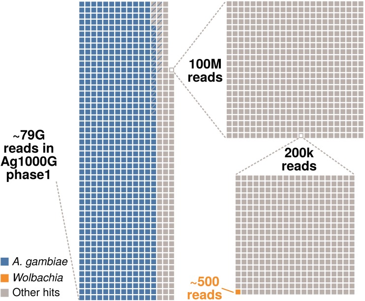
To identify additional Wolbachia sequences in A. gambiae, we screened data generated in the Ag1000G project, which investigates the genetic variance and population biology of A. gambiae (https://www.malariagen.net). We used the data released in the course of phase 1 AR3, namely, Illumina sequences of 765 wild-caught mosquitos from eight African countries (32). Reads for all samples were downloaded from the European Nucleotide Archive (ENA) and mapped to Wolbachia reference genomes. Using the criteria of Baldini et al. (26) (see Text S1 for details), we identified 446 reads from 96 libraries as matching Wolbachia. In total, there were ∼7.89 × 10¹⁰ reads across 765 libraries, so only 1 in ∼150 million reads maps to Wolbachia (Fig. 1), which corresponds to less than one Wolbachia read per sequencing library on average. Furthermore, for all investigated libraries, the reads not mapping to the A. gambiae genome were assembled, and the resulting contigs were subjected to a BLAST search against 54 currently available Wolbachia assemblies (Text S1). One out of 86,278,186 metagenomic contigs had Wolbachia as the best match. Two hundred sixty-four base pairs of this 330-bp contig had ∼91% similarity to the wCle reference genome. As wCle belongs to supergroup F and no other supergroup F sequences have been detected in A. gambiae so far, this sequence potentially belongs to Wolbachia from a filarial nematode and not to the putative symbiont of A. gambiae. Overall, based on a large and broad sampling, our analyses provide independent evidence for only a very sporadic presence, an extremely low titer, or even absence of Wolbachia in A. gambiae.
FIG 1.
Taxonomic composition of the reads generated in phase 1 of the Ag1000G project. In total, around 79 billion reads were generated from 765 A. gambiae mosquitos (32). Around 80% of these reads map to the A. gambiae host genome (represented by blue squares on the left). Panels on the right represent sequential magnifications of the portion of non-Anopheles reads to visualize the proportion of reads mapping to Wolbachia. The proportion of singletons (i.e., reads for which the mate did not map to the same chromosome) for each category are indicated by squares containing diagonal lines. Around 5% of all reads were classified as PCR duplicates but not removed prior to our analysis.
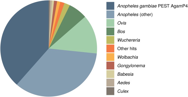
Contrasting with our findings, a recent in silico screen of archived arthropod short-read libraries extracted a highly covered Wolbachia supergroup B genome from a sample annotated as A. gambiae (33). To understand the reasons for this discrepancy, we inspected the sequencing libraries used by Pascar and Chandler (33) and discovered that they contain a mix of sequences of several other potential Wolbachia hosts (Fig. 2). Based on the analysis of the ITS2 and COI haplotypes of the most abundant sequences (36, 37), we conclude that the assembled Wolbachia genome likely originates from Anopheles “species A” and not A. gambiae (Fig. 2; Fig. S1; Text S1). Our interpretation is in line with a recent discovery of a highly prevalent supergroup B Wolbachia strain, distinct from other supergroup B strains, in Anopheles “species A” (31). Our phylogenomic reconstructions further support this, as they place the newly assembled Wolbachia genome (33) within supergroup B but separate from most other strains of this lineage (Fig. S1C). These analyses show that unambiguous identification of Anopheles species is an additional difficulty in detecting Wolbachia infections based on the sequencing data. Therefore, the newly reported genome does not contribute to the understanding of the elusive low-titer Wolbachia naturally associated with A. gambiae.
FIG 2.
Taxonomic classification of reads in the libraries from which the genome of a putative Wolbachia symbiont of A. gambiae was assembled (BioSample SAMEA3911293). For more details, refer to Text S1 and Fig. S1 in the supplemental material.
Phylogenetic assessment of SAMEA3911293 taxonomic composition. (A) Phylogenetic reconstruction of Anopheles species based on ITS2 alignment of previously published data and all ITS2 contigs present in the meta-assembly of all libraries from SAMEA3911293. Sequences recovered from this library are highlighted in blue. (B) Phylogenetic reconstruction of Anopheles based on mitochondrial COI. Sequences from the SAMEA3911293 meta-assembly are highlighted in blue. (C) Phylogeny of Wolbachia supergroup B based on concatenated core genome alignments of all strains with a (draft) genome in the NCBI database. Again, the strain isolated from SAMEA3911293 is highlighted in blue. Download FIG S1, PDF file, 0.04 MB (44.1KB, pdf) .
Copyright © 2019 Chrostek and Gerth.
This content is distributed under the terms of the Creative Commons Attribution 4.0 International license.
The putative low-titer Wolbachia infections required improved diagnostics. This prompted Shaw et al. to modify the wSpec PCR protocol by including a nested pair of primers and increasing the number of cycles to a total of 72 (nested PCR with 37 cycles in the 1st PCR and 35 cycles in the 2nd PCR, which uses the product of the 1st PCR as a template), potentially amplifying the initial 16S rRNA template over 1021 times (28). The protocol was used in several subsequent studies (29–31) but proved unreliable, as Gomes et al. reported 19% of the technical replicates yielding discordant results, even when the total number of cycles was increased to 80 (29). At the same time, the wSpec amplification protocol was sensitive enough to detect Wolbachia in a filarial nematode residing within one of the Anopheles coustani guts (30). Thus, this diagnostic test can detect Wolbachia in organisms interacting with Anopheles.
Meanwhile, Gomes et al. based their work on a 40-cycle quantitative PCR (qPCR) assay (29). The robustness of this test is not clear, as no raw data were included. Other methods routinely used to detect low-titer Wolbachia in insects, like PCR-Southern blotting or amplification of repeated sequences (e.g., the transposases with the highest coverage in the genomic data of Baldini et al. [26]) were never tested on Wolbachia sequences found in Anopheles (38, 39). Amplification of other Wolbachia sequences from putatively infected mosquitos, including Wolbachia surface protein and multilocus sequence typing (MLST) genes, has also been challenging (26, 27, 29–31), requiring protocol modifications (30) or the use of more than one mosquito sample (31), and was unsuccessful in some cases (26, 27). Overall, detection of Wolbachia sequences in A. gambiae by PCR-based methods remains challenging.
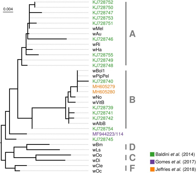
In summary, very few sequence data are available for the putative Wolbachia symbiont of A. gambiae, despite several attempts at generating and extracting such data. One common feature of all of them is an extremely low titer, at the limit of detection of PCR-based methods. Even from the few data available, it is obvious that there is no single Wolbachia strain associated with Anopheles gambiae (Fig. 3). In fact, almost every Wolbachia 16S rRNA amplicon and sequence attributed to A. gambiae is unique, and their diversity spans at least two Wolbachia supergroups (genetic lineages roughly equivalent to those of species in other bacterial genera) (Fig. 3) (40). In combination, we interpret the very low titers and the conflicting phylogenetic affiliations of the sequenced strains as incompatible with the notion of a stable, intraovarially transmitted Wolbachia symbiont in A. gambiae. However, this conclusion requires alternative explanations for the presence of Wolbachia DNA in these malaria mosquitos.
FIG 3.
Phylogenetic placement of Wolbachia sequences from Anopheles gambiae based on 16S rRNA sequences. Alignment was done with Mafft using the “–auto” option. A maximum-likelihood tree was inferred with automatic model selection in IQ-TREE version 1.62 (60). Origins of sequences are indicated by colors (see the key), and tip names correspond to NCBI accession numbers. All other sequences are reference Wolbachia strains. Tentative supergroup affiliations are denoted with capital letters. Please note that the two Wolbachia 16S rRNA sequences determined by Gomes et al. (29) overlap. Because the 117-bp overlap regions are 100% identical between these two sequences, we have merged them prior to phylogenetic analysis.
ORIGIN OF WOLBACHIA SEQUENCES IN ANOPHELES GAMBIAE
The presence of Wolbachia DNA in A. gambiae samples may be explained not only by a stable Wolbachia-Anopheles symbiosis but also in several alternative ways. First, the signal may stem from Wolbachia DNA insertions into an insect chromosome (26). Fragments of Wolbachia genomes are frequently found within insect genomes (41–43), and the most spectacular case includes a nearly complete genome insertion in Drosophila ananassae (44). This possibility was discussed by Baldini et al., but as the authors point out, the presence of the sequences in only some tissues and their very low titer argue against this hypothesis (26). The second possibility discussed by Baldini and colleagues is the insertion of a Wolbachia fragment into the chromosome of another, so-far-unidentified, mosquito-associated microorganism. However, this hypothesis does not help to explain the diversity of Wolbachia 16S rRNA sequences found in Anopheles.
Another hypothesis explaining the presence of Wolbachia sequences in A. gambiae tissues is contamination of the mosquito surface or gut. This contamination might come from several sources. First, ectoparasitic mites or midges and endoparasitic nematodes in Anopheles may contaminate whole-tissue DNA extracts, as shown by the detection of the Wolbachia symbiont of Dirofilaria immitis in an Anopheles coustani DNA preparation (30). However, the presence of unknown symbionts or parasites with novel Wolbachia strains is very challenging to test for.
The second possible source of Wolbachia contamination is plants. It has been shown that Wolbachia can persist in plants on which Wolbachia-infected insects feed and then be detected in previously uninfected insects reared on the same plants (reviewed in reference 45). As malaria vectors feed on plant nectar and fruits in the wild, Wolbachia DNA traces from these sources may accumulate in their guts. Feeding on Wolbachia-infected food may explain encountering Wolbachia 16S rRNA in the ovaries, as the adjacent gut can easily be perforated during dissections, releasing content and contaminating other tissues. Again, Wolbachia sequences from the gut may also explain the detection of Wolbachia sequences in larvae, as eggs and larval habitats may be contaminated with adult feces.
Another possible source of contamination is other insects cohabiting the collection sites. Culex, Aedes, and other Anopheles species can be found in sub-Saharan Africa, and all genera include natural Wolbachia hosts. This route of contamination seems especially plausible for mosquito larvae, which are avid predators, attacking other water-inhabiting insects. Moreover, Wolbachia 16S rRNA sequences can be detected in water storage containers inhabited by the larvae of various mosquito species (Text S1) and because of this may also be acquired by newly emerging adults and females during egg laying (46). Unfortunately, we have no data on the water composition of the breeding sites of the putative A. gambiae Wolbachia carriers, which may explain the Wolbachia sequence presence throughout the mosquito life cycle.
Part of the PCR signals observed throughout the studies reporting natural Wolbachia infections in A. gambiae may be purely technical and arise/spread at the level of DNA extraction and PCR. Although the original contamination likely originates from the field (as each sequenced amplicon has a different sequence), once amplified to high concentration in the lab, the contaminating templates may spread. This is especially true for extractions and PCR amplifications performed under field conditions and for labs that routinely amplify Wolbachia from other sources.
The data on natural Wolbachia infections in A. gambiae, together with similar reports suggesting Wolbachia infections in species previously considered uninfected, e.g., A. stephensi (47), Anopheles funestus (48), and A. aegypti (see references 47, 49, and 50 but also 51 and 52), should be carefully examined, as all have aquatic, detritus-feeding, and predatory larvae, while adults are terrestrial and can feed on nectar. Thus, bacteria and/or contaminating sequences may spread between these and other organisms sharing the same niches, necessitating studies designed to discern candidates for symbiotic taxa from transient and contaminating bacteria. Sampling of the mosquitos along with their environments and cohabiting species may help to reveal the origin and nature of Wolbachia sequences identified in A. gambiae.
Importantly, contamination from any of the mentioned sources cannot be ruled out with the data currently available. The previously mentioned sequencing of two Wolbachia-positive ovary samples resulted in 571 (out of ∼800,000,000) reads being classified as Wolbachia (0.000063%) (26). For a highly sensitive sequencing technique, such as Illumina sequencing, this falls well within the expected coverage of contaminants. Deep shotgun sequencing of eukaryotes usually results in some nontarget sequences from environmental contaminants, and it is unlikely that the A. gambiae libraries are an exception (53–55). Contamination stemming from nontarget microbial taxa is especially problematic in low-biomass samples (56), such as single mosquito ovaries. Adding to the difficulty, all of the studies reporting Wolbachia from amplicon or metagenomic sequencing do not present negative controls (e.g., sequencing of extraction or blank controls, quantification of microbial taxa, sequencing of mock communities [26, 27, 29–31]). This is not to say that the Wolbachia sequences definitely constitute contaminants, but they are simply not discernible from such. In general, the detection of very low-titer Wolbachia through highly sensitive methods (nested PCRs, Illumina sequencing) alone is not sufficient to conclude that an intracellular, inherited symbiont is present in a sample.
EXPECTED FEATURES OF NATURAL WOLBACHIA FROM ANOPHELES GAMBIAE
While sequence data alone are insufficient to determine whether Wolbachia is a symbiont of Anopheles gambiae and assembly of complete genomes has not been achieved due to low sequence abundance, other hallmarks of symbiotic interactions between the taxa can be used to support this claim.
First, intracellular localization is imperative for Wolbachia. The only published image of natural Wolbachia infections from A. gambiae is a combination of fluorescence in situ hybridization (FISH) and immunofluorescence. In this experiment, Wolbachia was detected with a combination of a Cy3-labeled probe, anti-Cy3 mouse antibody, and anti-mouse Alexa448 secondary antibody (see Fig. 1 in reference 28). The probe was designed to hybridize within the wSpec amplicon region. However, the low resolution of the image and the lack of host membrane staining do not allow us to confirm the wSpec intracellular localization (28). The indirect nature of the staining (the RNA probe was detected by primary and secondary antibodies) calls for additional controls acquired with the same microscope settings, and the nature of the findings calls for broader sampling and images at higher magnifications. Electron microscopy showing an immunogold-labeled Wolbachia cell or high-resolution FISH combined with membrane staining would provide unequivocal visual evidence for the existence of intracellular Wolbachia infections in A. gambiae.
Second, Wolbachia’s intracellular lifestyle is directly related to its mode of transmission, which is expected to occur from mother to offspring within the mother’s ovaries. In the first study on natural Wolbachia in A. gambiae, maternal transmission of the detected wSpec sequences was also examined. In this experiment, five wSpec-positive wild-caught gravid females oviposited in the lab, and their larval progeny was tested for wSpec amplification (detected in 56% to 100% of the offspring) (26). However, intraovarial transmission of Wolbachia was never explicitly addressed. Surface sterilization of eggs after oviposition would help to determine the transmission mode of these sequences, just as would testing for and excluding horizontal (between larvae or adult to larvae) and paternal wSpec sequence transmission. These experiments would help to confirm that A. gambiae is infected with an intracellular, transovarially transmitted symbiont and, together with the PCR evidence, diagnose a stable Wolbachia infection.
WOLBACHIA SYMBIONTS OF ANOPHELES GAMBIAE AND MALARIA
Wolbachia phenotypes similar to those observed in other insect hosts may have a huge impact on wild Anopheles populations and malaria transmission. Reproductive manipulations and fitness benefits may increase the proportion of biting females spreading the disease, while pathogen blocking may limit Plasmodium prevalence in the wild mosquito populations. Understanding Anopheles gambiae biology is crucial for the design of effective strategies aiming at limiting Plasmodium transmission.
Targeted Wolbachia-based Plasmodium control strategies, similar to the ones used for dengue and Zika virus control, are also exciting prospects. However, they are not reliant on Wolbachia symbionts naturally associated with Anopheles. Insect populations may equally well be suppressed by the release of males carrying incompatible Wolbachia strains by bidirectional CI on an infected population or by unidirectional CI on an uninfected one. The same applies to Wolbachia-induced pathogen blocking. Existing initiatives to control dengue and Zika virus with Wolbachia-conferred antiviral protection use naturally uninfected Aedes aegypti mosquitos that were artificially transinfected with Wolbachia from a different insect species (12). These mosquitos benefit not only from protection by the core and yet-unknown mechanism but also from immune system upregulation caused by a recent transinfection with Wolbachia (10). Thus, the Wolbachia-based population suppression and disease blocking can work in species not commonly infected with Wolbachia in the wild.
The presence of and, subsequently, the Plasmodium blocking properties of the presumed natural Wolbachia strains in A. gambiae remain to be confirmed. Given that Wolbachia detection in A. gambiae remains challenging (with PCR-based replicate experiments yielding discordant results [29]), it was surprising that two studies have reported negative correlations between the low-titer Wolbachia sequences and Plasmodium (28, 29). As pathogen protection has been shown to depend on the symbiont titer (57–59) and has so far been detected only in strains exhibiting relatively high bacterial load, it is likely to be absent from A. gambiae (31). However, the mechanism of Plasmodium blocking by Wolbachia may be different than the one characterized for viruses and requires further investigation. Moreover, CI necessary for the spread of Wolbachia in artificially infected vector populations was also not detected (28). Reliable protocols for the detection of Wolbachia in A. gambiae, together with independent repetition efforts, seem necessary to characterize the potential of the putative A. gambiae symbionts for their deployment in vector or disease control programs.
In summary, although using Wolbachia to fight malaria has been eagerly anticipated, naturally occurring Wolbachia strains in Anopheles were never an absolute requirement for this to be successful. Even now, their presence, phenotypes, and suitability for deployment in disease control remain to be confirmed. However, they should be studied, as understanding Anopheles gambiae biology and ecology, including its interactions with other micro- and macroscopic organisms, is crucial for designing effective malaria elimination programs.
CONCLUSIONS
The evidence for natural Wolbachia infections in Anopheles gambiae is currently limited to a small number of highly diverse, very low-titer DNA sequences detected in this important malaria vector. Further efforts toward characterization of the interaction between Wolbachia sequences and A. gambiae are required to establish that this is a true symbiotic association. Demonstrating the presence of intracellular bacterial cells and their intraovarian transmission are prerequisites to diagnose a symbiosis. Additionally, genomic data may shed light on the features of these Wolbachia and may reveal the origin of the sequences and the ecological interactions that caused their acquisition by A. gambiae mosquitos. Finally, ascertaining phenotypes associated with these Wolbachia sequence variants will improve our understanding of Anopheles gambiae biology, and as such inform future strategies aimed at limiting malaria spread and eventual disease eradication. Given that both Shaw et al. and Gomes et al. report the establishment of the wSpec-positive A. gambiae laboratory colonies (28, 29), the suggested conclusive experiments should be straightforward to perform.
The fact that Wolbachia sequences were encountered multiple times by independent groups of researchers clearly indicates present or past, direct or indirect ecological interactions between Wolbachia and Anopheles gambiae across Africa. While in-depth investigations of these interactions will be interesting from a basic biology, evolutionary, ecological, and disease control perspective, current data indicate that the postulated natural Wolbachia infections in Anopheles will be of limited use for application in fighting malaria with Wolbachia.
ACKNOWLEDGMENTS
We thank Francis Jiggins for a suggestion on using the Anopheles gambiae 1,000 genomes project data and for the critical reading of and helpful comments on the manuscript. We also thank Elena Levashina and Elves Heleno Duarte for comments on the draft of this work.
E.C. was supported by an EMBO Long-Term Fellowship (EMBO ALTF 1497-2015), cofunded by Marie Curie Actions and the European Commission (grants LTFCOFUND2013 and GA-2013-609409), and an FEBS Long-Term Fellowship. M.G. has received funding from the European Union’s Horizon 2020 Research and Innovation Program under Marie Sklodowska-Curie grant agreement 703379.
Footnotes
Citation Chrostek E, Gerth M. 2019. Is Anopheles gambiae a natural host of Wolbachia? mBio 10:e00784-19. https://doi.org/10.1128/mBio.00784-19.
REFERENCES
- 1.Weinert LA, Araujo-Jnr EV, Ahmed MZ, Welch JJ. 2015. The incidence of bacterial endosymbionts in terrestrial arthropods. Proc Biol Sci 282:20150249. doi: 10.1098/rspb.2015.0249. [DOI] [PMC free article] [PubMed] [Google Scholar]
- 2.Werren JH, Baldo L, Clark ME. 2008. Wolbachia: master manipulators of invertebrate biology. Nat Rev Microbiol 6:741–751. doi: 10.1038/nrmicro1969. [DOI] [PubMed] [Google Scholar]
- 3.Yen JH, Barr ARR. 1973. The etiological agent of cytoplasmic incompatibility in Culex pipiens. J Invertebr Pathol 22:242–250. doi: 10.1016/0022-2011(73)90141-9. [DOI] [PubMed] [Google Scholar]
- 4.Dobson SL, Fox CW, Jiggins FM. 2002. The effect of Wolbachia-induced cytoplasmic incompatibility on host population size in natural and manipulated systems. Proc Biol Sci 269:437–445. doi: 10.1098/rspb.2001.1876. [DOI] [PMC free article] [PubMed] [Google Scholar]
- 5.Laven H. 1967. Eradication of Culex pipiens fatigans through cytoplasmic incompatibility. Nature 216:383–384. doi: 10.1038/216383a0. [DOI] [PubMed] [Google Scholar]
- 6.Hosokawa T, Koga R, Kikuchi Y, Meng X-Y, Fukatsu T. 2010. Wolbachia as a bacteriocyte-associated nutritional mutualist. Proc Natl Acad Sci U S A 107:769–774. doi: 10.1073/pnas.0911476107. [DOI] [PMC free article] [PubMed] [Google Scholar]
- 7.Fast EM, Toomey ME, Panaram K, Desjardins D, Kolaczyk ED, Frydman HM. 2011. Wolbachia enhance Drosophila stem cell proliferation and target the germline stem cell niche. Science 334:990–992. doi: 10.1126/science.1209609. [DOI] [PMC free article] [PubMed] [Google Scholar]
- 8.Teixeira L, Ferreira A, Ashburner M. 2008. The bacterial symbiont Wolbachia induces resistance to RNA viral infections in Drosophila melanogaster. PLoS Biol 6:e1000002. doi: 10.1371/journal.pbio.1000002. [DOI] [PMC free article] [PubMed] [Google Scholar]
- 9.Hedges LM, Brownlie JC, O'Neill SL, Johnson KN. 2008. Wolbachia and virus protection in insects. Science 322:702. doi: 10.1126/science.1162418. [DOI] [PubMed] [Google Scholar]
- 10.Moreira LA, Iturbe-Ormaetxe I, Jeffery JA, Lu G, Pyke AT, Hedges LM, Rocha BC, Hall-Mendelin S, Day A, Riegler M, Hugo LE, Johnson KN, Kay BH, McGraw EA, van den Hurk AF, Ryan PA, O'Neill SL. 2009. A Wolbachia symbiont in Aedes aegypti limits infection with dengue, Chikungunya, and Plasmodium. Cell 139:1268–1278. doi: 10.1016/j.cell.2009.11.042. [DOI] [PubMed] [Google Scholar]
- 11.O’Neill SL. 2018. The use of Wolbachia by the World Mosquito Program to interrupt transmission of Aedes aegypti transmitted viruses In Hilgenfeld R, Vasudevan S (ed), Dengue and Zika: control and antiviral treatment strategies. Advances in experimental medicine and biology, vol 1062. Springer, Singapore. [DOI] [PubMed] [Google Scholar]
- 12.Walker T, Johnson PH, Moreira LA, Iturbe-Ormaetxe I, Frentiu FD, McMeniman CJ, Leong YS, Dong Y, Axford J, Kriesner P, Lloyd AL, Ritchie SA, O'Neill SL, Hoffmann AA. 2011. The wMel Wolbachia strain blocks dengue and invades caged Aedes aegypti populations. Nature 476:450–453. doi: 10.1038/nature10355. [DOI] [PubMed] [Google Scholar]
- 13.Frentiu FD, Zakir T, Walker T, Popovici J, Pyke AT, van den Hurk A, McGraw EA, O'Neill SL. 2014. Limited dengue virus replication in field-collected Aedes aegypti mosquitoes infected with Wolbachia. PLoS Negl Trop Dis 8:e2688. doi: 10.1371/journal.pntd.0002688. [DOI] [PMC free article] [PubMed] [Google Scholar]
- 14.Hoffmann AA, Iturbe-Ormaetxe I, Callahan AG, Phillips BL, Billington K, Axford JK, Montgomery B, Turley AP, O'Neill SL. 2014. Stability of the wMel Wolbachia infection following invasion into Aedes aegypti populations. PLoS Negl Trop Dis 8:e3115. doi: 10.1371/journal.pntd.0003115. [DOI] [PMC free article] [PubMed] [Google Scholar]
- 15.O’Neill SL. 2018. Scaled deployment of Wolbachia to protect the community from Aedes transmitted arboviruses. Gates Open Res 2:36. doi: 10.12688/gatesopenres.12844.1. [DOI] [PMC free article] [PubMed] [Google Scholar]
- 16.World Health Organization. 2015. World malaria report. World Health Organization, Geneva, Switzerland. [Google Scholar]
- 17.Kambris Z, Blagborough AM, Pinto SB, Blagrove MSC, Godfray HCJ, Sinden RE, Sinkins SP. 2010. Wolbachia stimulates immune gene expression and inhibits plasmodium development in Anopheles gambiae. PLoS Pathog 6:e1001143. doi: 10.1371/journal.ppat.1001143. [DOI] [PMC free article] [PubMed] [Google Scholar]
- 18.Ricci I, Cancrini G, Gabrielli S, D'amelio S, Favia G. 2002. Searching for Wolbachia (Rickettsiales: Rickettsiaceae) in mosquitoes (Diptera: Culicidae): large polymerase chain reaction survey and new identifications. J Med Entomol 39:562–567. doi: 10.1603/0022-2585-39.4.562. [DOI] [PubMed] [Google Scholar]
- 19.Kittayapong P, Baisley KJ, Baimai V, O’Neill SL. 2000. Distribution and diversity of Wolbachia infections in Southeast Asian mosquitoes (Diptera: Culicidae). J Med Entomol 37:340–345. doi: 10.1603/0022-2585(2000)037[0340:DADOWI]2.0.CO;2. [DOI] [PubMed] [Google Scholar]
- 20.Rasgon JL, Scott TW. 2004. An initial survey for Wolbachia (Rickettsiales: Rickettsiaceae) infections in selected California mosquitoes (Diptera: Culicidae). J Med Entomol 41:255–257. doi: 10.1603/0022-2585-41.2.255. [DOI] [PubMed] [Google Scholar]
- 21.Rasgon JL, Ren X, Petridis M. 2006. Can Anopheles gambiae be infected with Wolbachia pipientis? Insights from an in vitro system. Appl Environ Microbiol 72:7718–7722. doi: 10.1128/AEM.01578-06. [DOI] [PMC free article] [PubMed] [Google Scholar]
- 22.Jin C, Ren X, Rasgon JL. 2009. The virulent Wolbachia strain wMelPop efficiently establishes somatic infections in the malaria vector Anopheles gambiae. Appl Environ Microbiol 75:3373–3376. doi: 10.1128/AEM.00207-09. [DOI] [PMC free article] [PubMed] [Google Scholar]
- 23.Hughes GL, Koga R, Xue P, Fukatsu T, Rasgon JL. 2011. Wolbachia infections are virulent and inhibit the human malaria parasite Plasmodium falciparum in Anopheles gambiae. PLoS Pathog 7:e1002043. doi: 10.1371/journal.ppat.1002043. [DOI] [PMC free article] [PubMed] [Google Scholar]
- 24.Bian G, Joshi D, Dong Y, Lu P, Zhou G, Pan X, Xu Y, Dimopoulos G, Xi Z. 2013. Wolbachia invades Anopheles stephensi populations and induces refractoriness to Plasmodium infection. Science 340:748–751. doi: 10.1126/science.1236192. [DOI] [PubMed] [Google Scholar]
- 25.Hughes GL, Dodson BL, Johnson RM, Murdock CC, Tsujimoto H, Suzuki Y, Patt AA, Cui L, Nossa CW, Barry RM, Sakamoto JM, Hornett EA, Rasgon JL. 2014. Native microbiome impedes vertical transmission of Wolbachia in Anopheles mosquitoes. Proc Natl Acad Sci U S A 111:12498–12503. doi: 10.1073/pnas.1408888111. [DOI] [PMC free article] [PubMed] [Google Scholar]
- 26.Baldini F, Segata N, Pompon J, Marcenac P, Shaw WR, Dabiré RK, Diabaté A, Levashina EA, Catteruccia F. 2014. Evidence of natural Wolbachia infections in field populations of Anopheles gambiae. Nat Commun 5:3985. doi: 10.1038/ncomms4985. [DOI] [PMC free article] [PubMed] [Google Scholar]
- 27.Buck M. 2016. Bacterial associations reveal spatial population dynamics in Anopheles gambiae mosquitoes. Sci Rep 6:22806. doi: 10.1038/srep22806. [DOI] [PMC free article] [PubMed] [Google Scholar]
- 28.Shaw WR, Marcenac P, Childs LM, Buckee CO, Baldini F, Sawadogo SP, Dabiré RK, Diabaté A, Catteruccia F. 2016. Wolbachia infections in natural Anopheles populations affect egg laying and negatively correlate with Plasmodium development. Nat Commun 7:11772. doi: 10.1038/ncomms11772. [DOI] [PMC free article] [PubMed] [Google Scholar]
- 29.Gomes FM, Hixson BL, Tyner MDW, Ramirez JL, Canepa GE, Alves E Silva TL, Molina-Cruz A, Keita M, Kane F, Traoré B, Sogoba N, Barillas-Mury C. 2017. Effect of naturally occurring Wolbachia in Anopheles gambiae s.l. mosquitoes from Mali on Plasmodium falciparum malaria transmission. Proc Natl Acad Sci U S A 114:12566–12571. doi: 10.1073/pnas.1716181114. [DOI] [PMC free article] [PubMed] [Google Scholar]
- 30.Ayala D. 2018. Natural Wolbachia infections are common in the major malaria vectors in Central Africa. bioRxiv http://biorxiv.org/content/early/2018/06/11/343715.abstract. [DOI] [PMC free article] [PubMed]
- 31.Jeffries CL, Lawrence GG, Golovko G, Kristan M, Orsborne J, Spence K, Hurn E, Bandibabone J, Tantely LM, Raharimalala FN, Keita K, Camara D, Barry Y, Wat'senga F, Manzambi EZ, Afrane YA, Mohammed AR, Abeku TA, Hedge S, Khanipov K, Pimenova M, Fofanov Y, Boyer S, Irish SR, Hughes GL, Walker T. 2018. Novel Wolbachia strains in Anopheles malaria vectors from sub-Saharan Africa. Wellcome Open Res 3:113. doi: 10.12688/wellcomeopenres.14765.2. [DOI] [PMC free article] [PubMed] [Google Scholar]
- 32.Ag100G Consortium. 2015. Ag1000G phase 1 AR3 data release. MalariaGEN http://www.malariagen.net/data/ag1000g-phase1-AR3.
- 33.Pascar J, Chandler CH. 2018. A bioinformatics approach to identifying Wolbachia infections in arthropods. PeerJ 6:e5486. doi: 10.7717/peerj.5486. [DOI] [PMC free article] [PubMed] [Google Scholar]
- 34.Werren JH, Windsor DM. 2000. Wolbachia infection frequencies in insects: evidence of a global equilibrium? Proc Biol Sci 267:1277–1285. doi: 10.1098/rspb.2000.1139. [DOI] [PMC free article] [PubMed] [Google Scholar]
- 35.Richardson MF, Weinert LA, Welch JJ, Linheiro RS, Magwire MM, Jiggins FM, Bergman CM. 2012. Population genomics of the Wolbachia endosymbiont in Drosophila melanogaster. PLoS Genet 8:e1003129. doi: 10.1371/journal.pgen.1003129. [DOI] [PMC free article] [PubMed] [Google Scholar]
- 36.Lobo NF, St Laurent B, Sikaala CH, Hamainza B, Chanda J, Chinula D, Krishnankutty SM, Mueller JD, Deason NA, Hoang QT, Boldt HL, Thumloup J, Stevenson J, Seyoum A, Collins FH. 2015. Unexpected diversity of Anopheles species in Eastern Zambia: implications for evaluating vector behavior and interventions using molecular tools. Sci Rep 5:17952. doi: 10.1038/srep17952. [DOI] [PMC free article] [PubMed] [Google Scholar]
- 37.Stevenson JC, Norris DE. 2016. Implicating cryptic and novel anophelines as malaria vectors in Africa. Insects 8:1. doi: 10.3390/insects8010001. [DOI] [PMC free article] [PubMed] [Google Scholar]
- 38.Schneider DI, Klasson L, Lind AE, Miller WJ. 2014. More than fishing in the dark: PCR of a dispersed sequence produces simple but ultrasensitive Wolbachia detection. BMC Microbiol 14:121. doi: 10.1186/1471-2180-14-121. [DOI] [PMC free article] [PubMed] [Google Scholar]
- 39.Arthofer W, Riegler M, Schneider D, Krammer M, Miller WJ, Stauffer C. 2009. Hidden Wolbachia diversity in field populations of the European cherry fruit fly, Rhagoletis cerasi (Diptera, Tephritidae). Mol Ecol 18:3816–3830. doi: 10.1111/j.1365-294X.2009.04321.x. [DOI] [PubMed] [Google Scholar]
- 40.Chung M, Munro JB, Tettelin H, Dunning Hotopp JC. 2018. Using core genome alignments to assign bacterial species. mSystems 3:e00236-18. doi: 10.1128/mSystems.00236-18. [DOI] [PMC free article] [PubMed] [Google Scholar]
- 41.Nikoh N, Tanaka K, Shibata F, Kondo N, Hizume M, Shimada M, Fukatsu T. 2008. Wolbachia genome integrated in an insect chromosome: evolution and fate of laterally transferred endosymbiont genes. Genome Res 18:272–280. doi: 10.1101/gr.7144908. [DOI] [PMC free article] [PubMed] [Google Scholar]
- 42.Kondo N, Nikoh N, Ijichi N, Shimada M, Fukatsu T. 2002. Genome fragment of Wolbachia endosymbiont transferred to X chromosome of host insect. Proc Natl Acad Sci 99:14280–14285. doi: 10.1073/pnas.222228199. [DOI] [PMC free article] [PubMed] [Google Scholar]
- 43.Klasson L, Kambris Z, Cook PE, Walker T, Sinkins SP. 2009. Horizontal gene transfer between Wolbachia and the mosquito Aedes aegypti. BMC Genomics 10:33. doi: 10.1186/1471-2164-10-33. [DOI] [PMC free article] [PubMed] [Google Scholar]
- 44.Hotopp JCD, Clark ME, Oliveira DCSG, Foster JM, Fischer P, Torres MCM, Giebel JD, Kumar N, Ishmael N, Wang S, Ingram J, Nene RV, Shepard J, Tomkins J, Richards S, Spiro DJ, Ghedin E, Slatko BE, Tettelin H, Werren JH. 2007. Widespread lateral gene transfer from intracellular bacteria to multicellular eukaryotes. Science 317:1753–1756. doi: 10.1126/science.1142490. [DOI] [PubMed] [Google Scholar]
- 45.Chrostek E, Pelz-Stelinski K, Hurst GDD, Hughes GL. 2017. Horizontal transmission of intracellular insect symbionts via plants. Front Microbiol 8:2237. doi: 10.3389/fmicb.2017.02237. [DOI] [PMC free article] [PubMed] [Google Scholar]
- 46.Nilsson LKJ, Sharma A, Bhatnagar RK, Bertilsson S, Terenius O. 2018. Presence of Aedes and Anopheles mosquito larvae is correlated to bacteria found in domestic water-storage containers. FEMS Microbiol Ecol 94:fiy058. doi: 10.1093/femsec/fiy058. [DOI] [PubMed] [Google Scholar]
- 47.Soni M, Bhattacharya C, Sharma J, Khan SA, Dutta P. 2017. Molecular typing and phylogeny of Wolbachia: a study from Assam, north-eastern part of India. Acta Trop 176:421–426. doi: 10.1016/j.actatropica.2017.09.005. [DOI] [PubMed] [Google Scholar]
- 48.Niang EHA, Bassene H, Makoundou P, Fenollar F, Weill M, Mediannikov O. 2018. First report of natural Wolbachia infection in wild Anopheles funestus population in Senegal. Malar J 17:408. doi: 10.1186/s12936-018-2559-z. [DOI] [PMC free article] [PubMed] [Google Scholar]
- 49.Coon KL, Brown MR, Strand MR. 2016. Mosquitoes host communities of bacteria that are essential for development but vary greatly between local habitats. Mol Ecol 25:5806–5826. doi: 10.1111/mec.13877. [DOI] [PMC free article] [PubMed] [Google Scholar]
- 50.Carvajal T, Hashimoto K, Harnandika RK, Amalin D, Watanabe K. 2018. Detection of Wolbachia in field-collected mosquito vector, Aedes aegypti. bioRxiv http://biorxiv.org/content/early/2018/09/08/408856.abstract. [DOI] [PMC free article] [PubMed]
- 51.Gloria-Soria A, Chiodo TG, Powell JR. 2018. Lack of evidence for natural Wolbachia infections in Aedes aegypti (Diptera: Culicidae). J Med Entomol 55:1354–1356. doi: 10.1093/jme/tjy084. [DOI] [PMC free article] [PubMed] [Google Scholar]
- 52.Hegde S. 2018. Microbiome interaction networks and community structure from laboratory-reared and field-collected Aedes aegypti, Aedes albopictus, and Culex quinquefasciatus mosquito vectors. Front Microbiol 9:2160. doi: 10.3389/fmicb.2018.02160. [DOI] [PMC free article] [PubMed] [Google Scholar]
- 53.Gerth M, Hurst GDD. 2017. Short reads from honey bee (Apis sp.) sequencing projects reflect microbial associate diversity. PeerJ 5:e3529. doi: 10.7717/peerj.3529. [DOI] [PMC free article] [PubMed] [Google Scholar]
- 54.Salter SJ, Cox MJ, Turek EM, Calus ST, Cookson WO, Moffatt MF, Turner P, Parkhill J, Loman NJ, Walker AW. 2014. Reagent and laboratory contamination can critically impact sequence-based microbiome analyses. BMC Biol 12:87. doi: 10.1186/s12915-014-0087-z. [DOI] [PMC free article] [PubMed] [Google Scholar]
- 55.Lusk RW. 2014. Diverse and widespread contamination evident in the unmapped depths of high throughput sequencing data. PLoS One 9:e110808. doi: 10.1371/journal.pone.0110808. [DOI] [PMC free article] [PubMed] [Google Scholar]
- 56.de Goffau MC, Lager S, Salter SJ, Wagner J, Kronbichler A, Charnock-Jones DS, Peacock SJ, Smith GCS, Parkhill J. 2018. Recognizing the reagent microbiome. Nat Microbiol 3:851–853. doi: 10.1038/s41564-018-0202-y. [DOI] [PubMed] [Google Scholar]
- 57.Chrostek E, Marialva MSP, Esteves SS, Weinert LA, Martinez J, Jiggins FM, Teixeira L. 2013. Wolbachia variants induce differential protection to viruses in Drosophila melanogaster: a phenotypic and phylogenomic analysis. PLoS Genet 9:e1003896. doi: 10.1371/journal.pgen.1003896. [DOI] [PMC free article] [PubMed] [Google Scholar]
- 58.Osborne SE, Iturbe-Ormaetxe I, Brownlie JC, O'Neill SL, Johnson KN. 2012. Antiviral protection and the importance of Wolbachia density and tissue tropism in Drosophila simulans. Appl Environ Microbiol 78:6922–6929. doi: 10.1128/AEM.01727-12. [DOI] [PMC free article] [PubMed] [Google Scholar]
- 59.Martinez J, Longdon B, Bauer S, Chan Y-S, Miller WJ, Bourtzis K, Teixeira L, Jiggins FM. 2014. Symbionts commonly provide broad spectrum resistance to viruses in insects: a comparative analysis of Wolbachia strains. PLoS Pathog 10:e1004369. doi: 10.1371/journal.ppat.1004369. [DOI] [PMC free article] [PubMed] [Google Scholar]
- 60.Nguyen LT, Schmidt HA, Von Haeseler A, Minh BQ. 2015. IQ-TREE: a fast and effective stochastic algorithm for estimating maximum-likelihood phylogenies. Mol Biol Evol 32:268–274. doi: 10.1093/molbev/msu300. [DOI] [PMC free article] [PubMed] [Google Scholar]
Associated Data
This section collects any data citations, data availability statements, or supplementary materials included in this article.
Supplementary Materials
Additional text and methods. Download Text S1, PDF file, 0.1 MB (126.9KB, pdf) .
Copyright © 2019 Chrostek and Gerth.
This content is distributed under the terms of the Creative Commons Attribution 4.0 International license.
Phylogenetic assessment of SAMEA3911293 taxonomic composition. (A) Phylogenetic reconstruction of Anopheles species based on ITS2 alignment of previously published data and all ITS2 contigs present in the meta-assembly of all libraries from SAMEA3911293. Sequences recovered from this library are highlighted in blue. (B) Phylogenetic reconstruction of Anopheles based on mitochondrial COI. Sequences from the SAMEA3911293 meta-assembly are highlighted in blue. (C) Phylogeny of Wolbachia supergroup B based on concatenated core genome alignments of all strains with a (draft) genome in the NCBI database. Again, the strain isolated from SAMEA3911293 is highlighted in blue. Download FIG S1, PDF file, 0.04 MB (44.1KB, pdf) .
Copyright © 2019 Chrostek and Gerth.
This content is distributed under the terms of the Creative Commons Attribution 4.0 International license.


