Abstract
Obesity is associated with increased incidence and worse prognosis of more than one dozen tumor types; however, the molecular mechanisms for this association remain under debate. We hypothesized that insulin, which is elevated in obesity-driven insulin resistance, would increase tumor glucose oxidation in obesity-associated tumors. To test this hypothesis, we applied and validated a stable isotope method to measure the ratio of pyruvate dehydrogenase flux to citrate synthase flux (VPDH/VCS, i.e. the percent of total mitochondrial oxidation fueled by glucose) in tumor cells. Using this method, we found that three tumor cell lines associated with obesity (colon cancer [MC38], breast cancer [4T1], and prostate cancer [TRAMP-C3] cells) increase VPDH/VCS in response to physiologic concentrations of insulin. In contrast, three tumor cell lines that are not associated with obesity (melanoma [YUMM1.7], B cell lymphoma [BCL1 clone 5B1b], and small cell lung cancer [NCI-H69] cells) exhibited no oxidative response to insulin. The observed increase in glucose oxidation in response to insulin correlated with a dose-dependent increase in cell division in obesity-associated tumor cell lines when grown in insulin, whereas no alteration in cell division was seen in tumor types not associated with obesity. These data reveal that a shift in substrate preference in the setting of physiologic insulin may comprise a metabolic signature of obesity-associated tumors that differs from that of those not associated with obesity.
Introduction
Obesity is well-known to increase the prevalence and mortality of more than one dozen tumor types. In spite of the prevalence of obesity and its privileged place in public health discourse, the metabolic and molecular mechanisms underpinning the relationship between obesity and cancer remain contentious. Hyperinsulinemia has emerged as a focal point of research on obesity-related tumors, with increased plasma insulin concentrations independently predicting increased risk and mortality in prostate [1, 2], colon [3–7], breast [8–13], endometrial [12, 14, 15], and pancreatic cancer [16, 17], as well as several other tumor types. The idea that hyperinsulinemia may promote cancer risk is bolstered by the fact that biguanides such as metformin and phenformin, the most commonly prescribed class of diabetes drug worldwide, slow tumor growth associated with reductions in plasma insulin concentrations [18–30], although this class of agents has also shown efficacy in vivo independent of changes in plasma insulin concentrations in a minority of studies [31–33]. We recently showed that both metformin and a novel insulin sensitizer, a controlled-release mitochondrial protonophore, slows tumor growth in two models of colon cancer, and that the tumor-slowing effects of both agents were dependent on reversal of hyperinsulinemia [21], demonstrating a causative role for hyperinsulinemia in these mouse models.
While the association between hyperinsulinemia and obesity-related cancer progression is well established, the mechanisms by which hyperinsulinemia may promote tumor growth remain under debate. High doses of dichloroacetate, an indirect activator of pyruvate dehydrogenase and therefore of mitochondrial glucose oxidation, were shown to inhibit proliferation of colorectal cancer cells, particularly under hypoxic conditions [34]; however because these studies were performed in unphysiologic media containing glucose but without pyruvate, lactate, amino acids, or fatty acids, it is difficult to draw strong conclusions regarding the impact of a shift in substrate utilization from glycolytic to oxidative metabolism on tumor cell division under physiologic conditions. To that end, we show here that insulin increases mitochondrial glucose oxidation and augments cell division in cells from obesity-associated tumors, while obesity-independent cell lines show no alteration of substrate preference. These data break with the conventional stance that glucose oxidation is constitutively high in cancer cells, revealing a shift in substrate preference which may comprise a metabolic signature of obesity-related tumors.
Materials and methods
Cells
MC38 cells (ENH204) were obtained from Kerafast and YUMM1.7 (CRL-3362), TRAMP-C3 (CRL-2732), BCL1 clone 5B1b (TIB-197), 4T1 (CRL-2539), NCI-H69 (HTB-119), HCT 116 (CCL-247), DLD-1 (CCL-221), B16-F10 (CRL-6475), and COLO 829 (CRL-1974) cells from ATCC. All cells were cultured in the manufacturer’s recommended media, supplemented with penicillin/streptomycin, and were trypsinized and split 2–3 times weekly. Cells were plated in 6 well plates (5x10⁵ cells per well) one day prior to each in vitro experiment, and on the day of the experiment were washed twice with warmed PBS prior to the study. For the cell division studies, two insulin doses were chosen: 0.1 nM (the approximate plasma insulin concentration previously measured in vivo in overnight fasted rodents [30, 35, 36] and utilized in in vitro tumor studies [37–40]) and 100 nM (a dose previously used extensively in in vitro studies to assess the impact of insulin on tumor cells [38, 41–45]). Cells were plated in 6 well plates (1x10⁵ cells per well), incubated in the manufacturer’s recommended media with or without insulin (0.1 or 100 μM), dichloroacetate (25 mM in 0.1% DMSO), or 6,8-bis(benzylthio)octanoic acid (1 μM in 0.1% DMSO), and counted by a blinded investigator three days later. These data are presented normalized to controls (without insulin/6,8-bis(benzylthio)octanoic acid) from the same cell line.
In vitro 13C labeling studies
The base media used for the 13C labeling studies was Dulbecco’s Modified Eagle’s Medium (DMEM) containing 5 mM [13C6] glucose, nonessential amino acids, 2 mM glutamine, 1 mM lactate, 1 mM palmitate, and 0.1 mM β-OHB. In certain cases, insulin (100 nM), etomoxir (10 μM in 0.1% DMSO), dichloroacetate (25 mM in 0.1% DMSO), or 6,8-bis(benzylthio)octanoic acid (1 μM in 0.1% DMSO) was added, with 0.1% DMSO added to the media in the control wells. Cells were incubated for 2 hours in this media, after which the media was aspirated and cells were washed three times with warmed PBS, after which they were quenched with ice-cold 50% methanol, scraped from the cell plate, and frozen at -80°C pending further analysis.
Measurement of glucose uptake and oxidation
Glucose uptake was measured by incubating 105 cells per replicate in DMEM containing 5 mM glucose, nonessential amino acids, 2 mM glutamine, 1 mM lactate, 1 mM palmitate, and 0.1 mM β-OHB, and [1-14C] 2-deoxyglucose (PerkinElmer) (0.5 μCi per replicate) for 120 min, after which cells were washed three times in PBS, scraped, and collected into scintillation vials. The 14C specific activity was quantified using a scintillation counter, and glucose uptake was calculated by assuming a constant isotopically labeled precursor and a constant rate of glucose uptake over the 120 min incubation period.
VPDH/VCS was measured in cells incubated in [13C6] glucose as [35, 46]. Briefly, this method employs measurement of [4,5-13C2] glutamate as equivalent to [13C2] acetyl-CoA, the product of PDH (S1 Fig), whereas [13C3] alanine serves as a reciprocal pool for [13C3] pyruvate, the latter of which is found at lower concentrations and is much more labile, rendering it difficult to reliably measure under these conditions. Cell samples quenched in 50% methanol were prepared for LC-MS/MS analysis of [4,5-13C2] glutamate enrichment and GC/MS analysis of [13C3] alanine enrichment as we have described [35].
Absolute rates of glucose oxidation were determined in MC38 and YUMM1.7 cells by incubating 5x10⁵ cells in sealed flasks for 30 min in DMEM culture media (5 mM glucose, nonessential amino acids, 2 mM glutamine, 1 mM lactate, 1 mM palmitate, and 0.1 mM β-OHB) containing 0.2 μCi [14C6] glucose. The [14CO2] produced was trapped on Whatman paper in a holder suspending it in the air above the cells and, after 30 min of incubation in 14C media, the 14C activity was determined using a scintillation counter.
Assessment of lactate production
To measure lactate production, 105 cells were washed three times with warmed PBS and placed in DMEM containing 5 mM glucose, nonessential amino acids, 2 mM glutamine, 1 mM palmitate, and 1 mM β-OHB, but without lactate or pyruvate. After 120 min, the media was collected and spiked with 13C3 lactate (3 ng). The concentration of lactate in the media was measured by determining the ratio of 13C3 to 12C lactate by GC/MS using the same method as we have previously published to examine alanine concentrations/enrichment in plasma and tissues [35]. The rate of net lactate production was calculated by assuming linear accumulation of lactate in the media over time, and assuming an unchanged concentration of lactate in the cells.
Measurement of Vβ-OHB-ox/VCS
Vβ-OHB-ox/VCS was measured by incubating cells in DMEM containing 1 mM [13C4] β-OHB, 5 mM glucose, nonessential amino acids, 2 mM glutamine, 1 mM lactate, and 1 mM palmitate. Using this tracer, Vβ-OHB-ox/VCS is given as .
Cell samples quenched in 50% methanol were prepared for LC-MS/MS analysis of [4,5-13C2] glutamate enrichment and GC/MS analysis of [13C3] alanine enrichment as described above and in our previous report [35]; [13C4] β-OHB enrichment was measured using the same GC/MS method as was used for [13C3] alanine.
Assessment of insulin receptor expression and phosphorylation
Insulin receptor expression and pTyr1162 phosphorylation was measured by Western blot using antibodies from Cell Signaling (catalog numbers 3025 and 3918, respectively) and normalized to beta-actin expression (Cell Signaling #9457).
Statistical analysis
Statistical analysis was performed using Prism 7.0. Groups were compared by the 2-tailed unpaired Student’s t-test (for comparisons of two groups) or by ANOVA with Bonferroni’s multiple comparisons test (for comparisons of three or more groups) after verifying that the data met the assumptions of the statistical test employed.
Results
Insulin activates the insulin receptor in all tumor cell lines
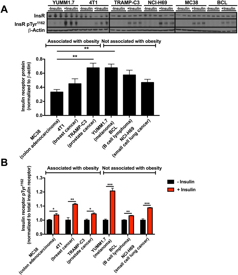
All cell lines employed in this study robustly expressed the insulin receptor, and differences in insulin receptor expression between cell lines did not correlate with obesity association or lack thereof (Fig 1A). Insulin activated the insulin receptor, as indicated by increased insulin receptor Tyr1162 phosphorylation [47] in all cell lines regardless of their correlation, or lack thereof, with obesity (Fig 1B).
Fig 1. Insulin receptor expression and insulin-stimulated insulin receptor phosphorylation do not correlate with tumor cell lines’ association with obesity.
(A) Insulin receptor expression. The data are the mean ± S.E.M. of six replicates from each cell line, including cells with and without insulin. **P<0.01 by ANOVA with Bonferroni’s multiple comparisons test. (B) Insulin receptor Tyr1162 phosphorylation. The data are presented as fold change from baseline, representing cells from the same cell line without insulin. *P<0.05, **P<0.01, ***P<0.001 by the 2-tailed unpaired Student’s t-test. Data are the mean ± S.E.M. of n = 3 per condition.
Insulin increases glucose oxidation in obesity-related, but not obesity-independent, cancer cell lines
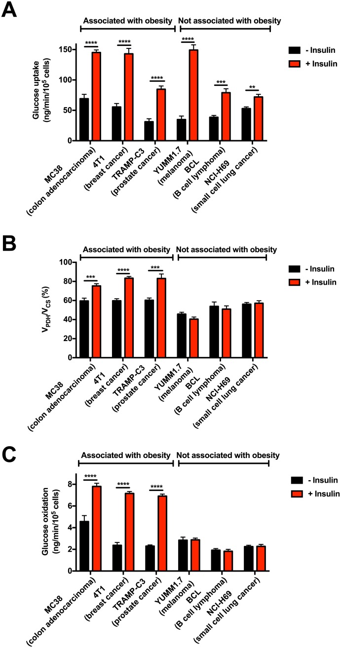
To identify differences in tumor metabolism in obesity-related cancers, as opposed to obesity-independent cancers, we investigated substrate preference in three cell lines from obesity-associated cancer types, each of which exhibits accelerated tumor growth associated with obesity: MC38, a colon adenocarcinoma [19, 30, 48–50], 4TI, a triple-negative breast cancer [51, 52], and TRAMP-C3, a prostate adenocarcinoma [53]. These cell lines were compared with three cancer cell lines from tumor types not associated with obesity: YUMM1.7, melanoma; BCL1 clone 5B1b, B-cell lymphoma; and NCI-H69, small-cell lung cancer. Insulin increased glucose uptake in all cell lines, independent of their association, or lack thereof, with obesity (Fig 2A). However, the ratio of pyruvate dehydrogenase flux to citrate synthase flux (VPDH/VCS, i.e. the percent of total mitochondrial oxidation fueled by glucose oxidation, S1 Fig) increased in all obesity-associated tumor types in the presence of insulin (100 nM); in contrast, all three obesity-independent cell lines showed no increase in glucose oxidation in response to insulin (Fig 2B). Instead, YUMM1.7 and BCL cells increased and NCI-H69 tended to increase lactate production after insulin treatment (S2A Fig), indicating that glucose taken up in response to insulin is diverted into lactate production if it is not shuttled into mitochondrial oxidation in response to insulin. Finally we confirmed the differential ability of insulin to stimulate glucose oxidation in obesity-associated versus obesity-independent tumor cells in studies of absolute glucose oxidation measured by trapping 14CO2 generated by oxidizing 14C6 glucose (Fig 2C): incubation with insulin doubles colon cancer cell glucose oxidation, while melanoma cells show no change in glucose oxidation rates when insulin is added to media.
Fig 2. Tumor types associated with obesity, but not those not associated with obesity, increase mitochondrial glucose oxidation in the presence of insulin.
(A) Glucose uptake. (B) VPDH/VCS. (C) Absolute rates of glucose oxidation. In all panels, data are the mean ± S.E.M. of n = 5–9 replicates per condition, with cells from the same line ± insulin compared using the 2-tailed unpaired Student’s t-test.
Utilization of glutamine and ketones is minimal in MC38 and YUMM1.7 cells
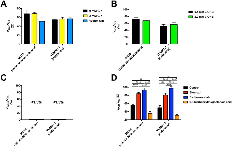
To ensure that physiologic levels of glutamine do not significantly affect our measurements of VPDH/VCS by dilution of glutamate, MC38 and YUMM1.7 cells were incubated in increasing levels of glutamine. No significant change in VPDH/VCS was found between within the physiologic range of glutamine (Fig 3A), indicating that glutamine utilization has minimal impact on the measured VPDH/VCS ratio.
Fig 3. The stable isotope method applied in this study is sensitive to detect the expected differences in VPDH/VCS with physiologic alterations in these fluxes, and is not affected by physiologically relevant glutamine or ketone concentrations.
(A) Glutamine in the physiologic range (0–10 mM) does not significantly affect the measured VPDH/VCS in MC38 or YUMM1.7 cells. n = 4 replicates per condition, with comparisons by ANOVA with Bonferroni’s multiple comparisons test. (B) β-OHB in the physiologic range (0–2 mM) does not significantly alter the measured VPDH/VCS in MC38 or YUMM1.7 cells. Conditions were compared by the 2-tailed unpaired Student’s t-test. In panels (B)-(D), n = 6–9 replicates per condition. (C) Vβ-OHB-ox/VCS is minimal in MC38 and YUMM1.7 cells. (D) VPDH/VCS is increased with inhibition of fatty acid oxidation (etomoxir) or stimulation of PDH (dichloroacetate), and decreased with inhibition of PDH (6,8-bis(benzylthio)octanoic acid). Groups were compared by ANOVA with Bonferroni’s multiple comparisons test. In all panels, data are the mean ± S.E.M.
Next, to determine the potential contribution of ketone oxidation to tumor metabolism, we incubated both MC38 and YUMM1.7 cells in low and high concentrations of β-hydroxybutyrate (β-OHB) and found that increased β-OHB concentrations have no impact upon VPDH/VCS (Fig 3B). To confirm this, we measured the contribution of ketone oxidation to total mitochondrial oxidation, Vβ-OHB/VCS (S1 Fig), and found that ketone oxidation is minimal in both MC38 and YUMM1.7 cells in vitro (Fig 3C). These findings indicate a central role for glucose oxidation and, to a lesser extent, fatty acid oxidation in cancer metabolism, in the absence of a key role for ketone oxidation.
VPDH/VCS is altered as expected with pharmacologic manipulation of fatty acid and glucose oxidation
To validate the sensitivity of our method to detect differences in VPDH/VCS, we employed three pharmacologic agents to alter substrate oxidation: etomoxir, an inhibitor of fatty acid oxidation; dichloroacetate, a PDH activator, and 6,8-bis(benzylthio)octanoic acid, a PDH inhibitor (Fig 3D). Inhibition of fatty acid oxidation led to a relative increase in VPDH/VCS in both MC38 and YUMM1.7 cells, reflecting a decrease in fatty acid oxidation relative to citrate synthase flux. As expected, activation of PDH with dichloroacetate increased the measured VPDH/VCS ratio in both cell lines, whereas PDH inhibition with 6,8-bis(benzylthio)octanoic acid reduced this ratio. The response of MC38 and YUMM1.7 cells to each small molecule glucose or fat oxidation modulator was similar, demonstrating that the differing responses of the cell lines to insulin was not attributable to inherent alterations in mitochondrial function. Taken together, these data demonstrate that our stable isotope method has the necessary sensitivity to detect expected differences in glucose or fatty acid oxidation under these conditions in vitro.
Insulin increases cell division in obesity-associated, but not obesity-independent, tumor cell types
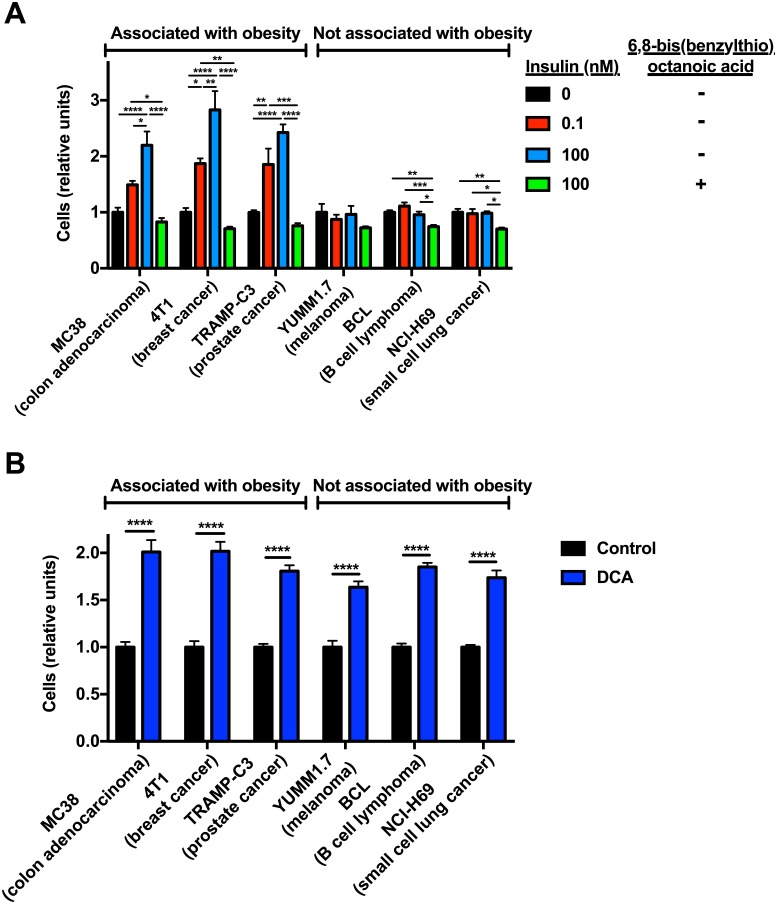
To determine whether the observed increases in VPDH/VCS in response to insulin affected cell division, we incubated all cell types in insulin (0.1 and 100 nM) and found that insulin increased rates of cell division in all three obesity-associated cell lines, but in none of the cell lines from tumors not associated with obesity (Fig 4A). By examining two additional colon cancer cell lines and two additional melanoma cell lines, we then confirmed that the insulin response is conserved across tumor types: all three colon cancer cell lines increased cell division in response to incubation in 0.1 nM insulin, whereas none of the melanoma cell lines exhibited this response (S2B Fig). However, inhibition of PDH using 6,8-bis(benzylthio)octanoic acid reduced cell division in both obesity-associated and -independent cell lines. Conversely, activation of PDH with DCA promoted tumor cell division in all cell lines (Fig 4B). Taken together, these data demonstrate that PDH-mediated glucose oxidation, which increases in response to insulin in obesity-associated but not obesity-independent tumor cell lines, drives cell division in these six tumor models, at least in in vitro culture.
Fig 4. PDH activity promotes tumor growth in both obesity-associated and obesity-independent tumor cell lines.
(A) Cell division in tumor cell lines incubated with or without insulin, with or without a PDH inhibitor. (B) Cell division in tumor cell lines incubated with or without the PDH activator dichloroacetate. In both panels, n = 6 replicates per condition, normalized to controls from the same cell line. *P<0.05, **P<0.01, ***P<0.001, ****P<0.0001 by ANOVA with Bonferroni’s multiple comparisons test (panel (A)) or by the 2-tailed unpaired Student’s t-test (panel (B)). Data are the mean± S.E.M.
Discussion
Hyperinsulinemia has been discussed as a potential mediator of obesity-related cancer growth: activating mutations in the PI3K/Akt pathway are common (10–30% incidence) and confer a poorer prognosis in humans with colon cancer [54, 55], breast cancer [56, 57], and prostate cancer [58, 59]. Activating mutations in the PI3K/Akt pathway have also been observed–albeit at a lower incidence (2–9%)–in melanoma [60, 61] and small cell lung cancer [62, 63], although, to our knowledge, they have not been observed in B cell lymphoma [64, 65]. Mutations in this pathway do not divide as clearly along obesity-associated versus independent lines in the cell lines examined in this study: whereas mutations in this pathway have been described in 4T1 [66] and NCI-H69 cells [67], they have not been described in the other obesity-associated or -independent cell lines studied here. Evidence for a critical role for insulin in directly promoting tumor cell division is provided by data showing that while insulin promotes tumor cell division in vivo [30, 68–71], pharmacologic agents which reverse hyperinsulinemia slow tumor growth unless exogenous insulin replacement is provided [30]. However, the mechanisms undergirding insulin’s effect upon certain tumors have remained unclear, in part due to a lack of methods to assess tumor substrate preference and the impact of variations in hormones and substrates on tumor fuel preference. To that end, we adapted a stable isotope method which we have previously applied in vivo for tissue-specific measurements of glucose oxidation via pyruvate dehydrogenase relative to total citrate synthase flux [30, 35]. Validation studies demonstrate the sensitivity of this method to alterations in glucose and fatty acid oxidation, and further demonstrate that glucose oxidation is not maximized under these conditions; therefore the lack of an oxidative response to insulin in obesity-independent cell lines does not reflect an inherent limitation in mitochondrial glucose utilization in these cells (Fig 3D).
Somewhat unexpectedly, we found that VPDH/VCS measured as the ratio of [4,5-13C2] glutamate/[13C3] alanine was unaltered by the presence or absence of physiologic (2 and 10 mM) concentrations of glutamine. These data stand in contrast to studies, which have previously been extensively reviewed [72–74], indicating that glutamine metabolism plays a critical role in cancer cell metabolism. It is possible that glutamine may be used preferentially for nucleotide synthesis rather than mitochondrial oxidation when the preferred glycolytic metabolites (glucose, lactate, pyruvate) are available, and may be required for oxidative metabolism only when substrate supply is limiting due to inadequate vascularization. Future tracer studies utilizing 13C glutamate as a surrogate for 13C acetyl-CoA, the immediate product of PDH, would need to confirm a minimal contribution of glutamine to oxidative metabolism; otherwise, [13C2] acetyl-CoA would need to replace [4,5-13C2] glutamate in the numerator of this ratio.
Using our stable isotope method, which we have now validated in multiple tumor cell types, we show here that three obesity-associated tumor cell lines respond to insulin by increasing glucose oxidation relative to total mitochondrial oxidation. This effect suggests that the mechanism by which obesity-related tumors capitalize upon their hyperinsulinemic environment is conserved across tumor types. These data were validated by use of an entirely independent method, trapping [14CO2] generated by the oxidation of [14C] glucose, which showed that rates of absolute glucose oxidation increase similarly to the change in VPDH/VCS upon insulin stimulation in MC38 colon cancer, 4T1 breast cancer, and TRAMPC3 prostate cancer cells, but are unaltered by insulin in YUMM1.7 melanoma, BCL B cell lymphoma, and NCI-H69 small cell lung cancer cells. This method of measuring absolute rates of glucose oxidation, which has been applied extensively in various cell types including neurons and astroglia [75, 76], oocytes [77], hepatocytes [78], intestine [79], and heart [80, 81], as well as tumor cells [82, 83], relies upon a set of assumptions that differ significantly from those of the VPDH/VCS method; the similar impact of insulin upon absolute glucose oxidation and VPDH/VCS lends further credence to the latter method. In contrast to the three obesity-associated tumor types, melanoma, B cell lymphoma, and small cell lung cancer cell lines showed no mitochondrial oxidative response to insulin. The oxidative response to insulin did not correlate with insulin receptor expression or activation: insulin receptor expression did not differ between cell types. However while insulin-stimulated glucose uptake correlated with insulin receptor activation, neither parameter was associated with the oxidative response to insulin: insulin increased both insulin receptor phosphorylation (Fig 1B) and glucose uptake (Fig 2A) in all cell lines to varying degrees, insulin promoted glucose oxidation only in obesity-associated tumors (Fig 2B and 2C). However, insulin did not decrease lactate production in obesity-associated cells, consistent with the fact that while the insulin-stimulated increase in glucose uptake outpaced insulin-stimulated increases in mitochondrial oxidation in all cells, excess glucose may be used for a variety of synthetic pathways (including glycogen synthesis and/or the energetic requirements of cell division) as well as lactate production. These increases in tumor glucose oxidation correlated with accelerated cell division in response to insulin: whereas there was no difference in cell division whether or not melanoma, small cell lung cancer, or B cell lymphoma cells were incubated in insulin, the three obesity-associated tumor types each showed a dose-dependent increase in cell division with insulin (Fig 4A, S2B Fig). Importantly, due to their differing doubling times, the magnitude of the impact of insulin on cell division should not be compared across different tumor types; however, this study does show that insulin promotes cell division in each of five obesity-associated tumor types, but not in any of the five obesity-independent tumor cell lines studied. However, activation of PDH increased and inhibition of PDH inhibited cell division in all six cell lines tested, regardless of their association, or lack thereof, with obesity (Fig 4B). These data indicate that the idea that a switch from glycolytic to oxidative glucose metabolism will slow tumor cell proliferation may be an oversimplification and that under conditions in which substrate limitation is not present, oxidative metabolism may actually promote tumor cell division. Taken together, this study underscores the possibility that upregulated glucose oxidation and increased VPDH/VCS under conditions of hyperinsulinemia may constitute a metabolic signature of obesity-related cancer and recommends further studies to explore the link between hyperinsulinemia, increased glucose uptake, and tumor cell division in vivo.
Supporting information
(A) Isotopomers generated on the first turn (solid red circles) and second turn (dashed red circles) of the tricarboxylic acid cycle during incubation in [13C6] glucose. VPDH, pyruvate dehydrogenase flux. VPC, pyruvate carboxylase flux. VCS, citrate synthase flux. OAA, oxaloacetate. α-KG, alpha-ketoglutarate. (B) Isotopomers generated on the first turn (solid red circles) and second turn (dashed red circles) of the tricarboxylic acid cycle during incubation in [13C4] β-hydroxybutyrate. Vβ-OHB-ox, ketone (β-hydroxybutyrate) oxidation.
(PDF)
(A) Rate of lactate production. (B) Impact of insulin (0.1 nM) on cell division. Data for MC38 and YUMM1.7 cells are duplicated from Fig 4A. In both panels, n = 6 replicates per condition. *P<0.05, **P<0.01, ****P<0.0001 by the 2-tailed unpaired Student’s t-test.
(PDF)
Acknowledgments
The authors thank Dr. Gerald Shulman and Dr. Curtis Perry for helpful discussions and Gina Butrico for her expert technical assistance. This study was funded by a grant from the National Institutes of Health (R00 CA-215315), a Yale Cancer Innovators Award, a Yale SPORE in Melanoma award, and a Yale Diabetes Research Center pilot award (to R.J.P.). The funders had no role in study design, data collection and analysis, decision to publish, or preparation of the manuscript.
Data Availability
All relevant data are within the manuscript and its Supporting Information files.
Funding Statement
This study was funded by a grant from the National Institutes of Health (R00 CA-215315), a Yale Cancer Innovators Award, and a Yale SPORE in Melanoma award (all to R.J.P.). The funders had no role in study design, data collection and analysis, decision to publish, or preparation of the manuscript.
References
- 1.Saboori S, Rad EY, Birjandi M, Mohiti S, Falahi E. Serum insulin level, HOMA-IR and prostate cancer risk: A systematic review and meta-analysis. Diabetes Metab Syndr. 2019;13(1):110–5. Epub 2019/01/16. 10.1016/j.dsx.2018.08.031 . [DOI] [PubMed] [Google Scholar]
- 2.Albanes D, Weinstein SJ, Wright ME, Mannisto S, Limburg PJ, Snyder K, et al. Serum insulin, glucose, indices of insulin resistance, and risk of prostate cancer. J Natl Cancer Inst. 2009;101(18):1272–9. Epub 2009/08/25. 10.1093/jnci/djp260 [DOI] [PMC free article] [PubMed] [Google Scholar]
- 3.Xu J, Ye Y, Wu H, Duerksen-Hughes P, Zhang H, Li P, et al. Association between markers of glucose metabolism and risk of colorectal cancer. BMJ Open. 2016;6(6):e011430 Epub 2016/06/30. 10.1136/bmjopen-2016-011430 [DOI] [PMC free article] [PubMed] [Google Scholar]
- 4.Limburg PJ, Stolzenberg-Solomon RZ, Vierkant RA, Roberts K, Sellers TA, Taylor PR, et al. Insulin, glucose, insulin resistance, and incident colorectal cancer in male smokers. Clinical gastroenterology and hepatology: the official clinical practice journal of the American Gastroenterological Association. 2006;4(12):1514–21. 10.1016/j.cgh.2006.09.014 [DOI] [PMC free article] [PubMed] [Google Scholar]
- 5.Wolpin BM, Meyerhardt JA, Chan AT, Ng K, Chan JA, Wu K, et al. Insulin, the insulin-like growth factor axis, and mortality in patients with nonmetastatic colorectal cancer. J Clin Oncol. 2009;27(2):176–85. 10.1200/JCO.2008.17.9945 [DOI] [PMC free article] [PubMed] [Google Scholar]
- 6.Cohen DH, LeRoith D. Obesity, type 2 diabetes, and cancer: the insulin and IGF connection. Endocr Relat Cancer. 2012;19(5):F27–45. 10.1530/ERC-11-0374 . [DOI] [PubMed] [Google Scholar]
- 7.Pollak M. Insulin-like growth factor-related signaling and cancer development. Recent Results Cancer Res. 2007;174:49–53. . [DOI] [PubMed] [Google Scholar]
- 8.Gunter MJ, Xie X, Xue X, Kabat GC, Rohan TE, Wassertheil-Smoller S, et al. Breast cancer risk in metabolically healthy but overweight postmenopausal women. Cancer Res. 2015;75(2):270–4. Epub 2015/01/17. 10.1158/0008-5472.CAN-14-2317 [DOI] [PMC free article] [PubMed] [Google Scholar]
- 9.Gunter MJ, Hoover DR, Yu H, Wassertheil-Smoller S, Rohan TE, Manson JE, et al. Insulin, insulin-like growth factor-I, and risk of breast cancer in postmenopausal women. J Natl Cancer Inst. 2009;101(1):48–60. Epub 2009/01/01. 10.1093/jnci/djn415 [DOI] [PMC free article] [PubMed] [Google Scholar]
- 10.Kabat GC, Kim M, Caan BJ, Chlebowski RT, Gunter MJ, Ho GY, et al. Repeated measures of serum glucose and insulin in relation to postmenopausal breast cancer. Int J Cancer. 2009;125(11):2704–10. Epub 2009/07/10. 10.1002/ijc.24609 . [DOI] [PubMed] [Google Scholar]
- 11.Shu X, Wu L, Khankari NK, Shu XO, Wang TJ, Michailidou K, et al. Associations of obesity and circulating insulin and glucose with breast cancer risk: a Mendelian randomization analysis. Int J Epidemiol. 2018. Epub 2018/10/03. 10.1093/ije/dyy201 . [DOI] [PMC free article] [PubMed] [Google Scholar]
- 12.Kabat GC, Kim MY, Lane DS, Zaslavsky O, Ho GYF, Luo J, et al. Serum glucose and insulin and risk of cancers of the breast, endometrium, and ovary in postmenopausal women. Eur J Cancer Prev. 2018;27(3):261–8. Epub 2018/02/14. 10.1097/CEJ.0000000000000435 . [DOI] [PubMed] [Google Scholar]
- 13.Lawlor DA, Smith GD, Ebrahim S. Hyperinsulinaemia and increased risk of breast cancer: findings from the British Women’s Heart and Health Study. Cancer Causes Control. 2004;15(3):267–75. Epub 2004/04/20. 10.1023/B:CACO.0000024225.14618.a8 . [DOI] [PubMed] [Google Scholar]
- 14.Hernandez AV, Pasupuleti V, Benites-Zapata VA, Thota P, Deshpande A, Perez-Lopez FR. Insulin resistance and endometrial cancer risk: A systematic review and meta-analysis. Eur J Cancer. 2015;51(18):2747–58. Epub 2015/11/26. 10.1016/j.ejca.2015.08.031 . [DOI] [PubMed] [Google Scholar]
- 15.Nead KT, Sharp SJ, Thompson DJ, Painter JN, Savage DB, Semple RK, et al. Evidence of a Causal Association Between Insulinemia and Endometrial Cancer: A Mendelian Randomization Analysis. J Natl Cancer Inst. 2015;107(9). Epub 2015/07/03. 10.1093/jnci/djv178 [DOI] [PMC free article] [PubMed] [Google Scholar]
- 16.Stolzenberg-Solomon RZ, Graubard BI, Chari S, Limburg P, Taylor PR, Virtamo J, et al. Insulin, glucose, insulin resistance, and pancreatic cancer in male smokers. JAMA. 2005;294(22):2872–8. Epub 2005/12/15. 10.1001/jama.294.22.2872 . [DOI] [PubMed] [Google Scholar]
- 17.Michaud DS, Wolpin B, Giovannucci E, Liu S, Cochrane B, Manson JE, et al. Prediagnostic plasma C-peptide and pancreatic cancer risk in men and women. Cancer Epidemiol Biomarkers Prev. 2007;16(10):2101–9. Epub 2007/10/02. 10.1158/1055-9965.EPI-07-0182 . [DOI] [PubMed] [Google Scholar]
- 18.Algire C, Amrein L, Bazile M, David S, Zakikhani M, Pollak M. Diet and tumor LKB1 expression interact to determine sensitivity to anti-neoplastic effects of metformin in vivo. Oncogene. 2011;30(10):1174–82. Epub 2010/11/26. 10.1038/onc.2010.483 . [DOI] [PubMed] [Google Scholar]
- 19.Algire C, Amrein L, Zakikhani M, Panasci L, Pollak M. Metformin blocks the stimulative effect of a high-energy diet on colon carcinoma growth in vivo and is associated with reduced expression of fatty acid synthase. Endocr Relat Cancer. 2010;17(2):351–60. Epub 2010/03/17. 10.1677/ERC-09-0252 . [DOI] [PubMed] [Google Scholar]
- 20.Zhuang S, Jian Y, Sun Y. Metformin Inhibits N-Methyl-N-Nitrosourea Induced Gastric Tumorigenesis in db/db Mice. Exp Clin Endocrinol Diabetes. 2017;125(6):392–9. Epub 2017/04/14. 10.1055/s-0043-100118 . [DOI] [PubMed] [Google Scholar]
- 21.Perry RJ, Zhang D, Zhang XM, Boyer JL, Shulman GI. Controlled-release mitochondrial protonophore reverses diabetes and steatohepatitis in rats. Science. 2015;347(6227):1253–6. Epub 2015/02/28. 10.1126/science.aaa0672 [DOI] [PMC free article] [PubMed] [Google Scholar]
- 22.Sarmento-Cabral A, LL F, Gahete MD, Castano JP, Luque RM. Metformin Reduces Prostate Tumor Growth, in a Diet-Dependent Manner, by Modulating Multiple Signaling Pathways. Mol Cancer Res. 2017;15(7):862–74. Epub 2017/04/08. 10.1158/1541-7786.MCR-16-0493 . [DOI] [PubMed] [Google Scholar]
- 23.Incio J, Tam J, Rahbari NN, Suboj P, McManus DT, Chin SM, et al. PlGF/VEGFR-1 Signaling Promotes Macrophage Polarization and Accelerated Tumor Progression in Obesity. Clin Cancer Res. 2016;22(12):2993–3004. Epub 2016/02/11. 10.1158/1078-0432.CCR-15-1839 [DOI] [PMC free article] [PubMed] [Google Scholar]
- 24.Al-Wahab Z, Mert I, Tebbe C, Chhina J, Hijaz M, Morris RT, et al. Metformin prevents aggressive ovarian cancer growth driven by high-energy diet: similarity with calorie restriction. Oncotarget. 2015;6(13):10908–23. Epub 2015/04/22. 10.18632/oncotarget.3434 [DOI] [PMC free article] [PubMed] [Google Scholar]
- 25.Ohno T, Shimizu M, Shirakami Y, Baba A, Kochi T, Kubota M, et al. Metformin suppresses diethylnitrosamine-induced liver tumorigenesis in obese and diabetic C57BL/KsJ-+Leprdb/+Leprdb mice. PLoS One. 2015;10(4):e0124081 Epub 2015/04/17. 10.1371/journal.pone.0124081 [DOI] [PMC free article] [PubMed] [Google Scholar]
- 26.Cifarelli V, Lashinger LM, Devlin KL, Dunlap SM, Huang J, Kaaks R, et al. Metformin and Rapamycin Reduce Pancreatic Cancer Growth in Obese Prediabetic Mice by Distinct MicroRNA-Regulated Mechanisms. Diabetes. 2015;64(5):1632–42. Epub 2015/01/13. 10.2337/db14-1132 [DOI] [PMC free article] [PubMed] [Google Scholar]
- 27.Zaafar DK, Zaitone SA, Moustafa YM. Role of metformin in suppressing 1,2-dimethylhydrazine-induced colon cancer in diabetic and non-diabetic mice: effect on tumor angiogenesis and cell proliferation. PLoS One. 2014;9(6):e100562 Epub 2014/06/28. 10.1371/journal.pone.0100562 [DOI] [PMC free article] [PubMed] [Google Scholar] [Retracted]
- 28.Checkley LA, Rho O, Angel JM, Cho J, Blando J, Beltran L, et al. Metformin inhibits skin tumor promotion in overweight and obese mice. Cancer Prev Res (Phila). 2014;7(1):54–64. Epub 2013/11/08. 10.1158/1940-6207.CAPR-13-0110 [DOI] [PMC free article] [PubMed] [Google Scholar]
- 29.Quinn BJ, Dallos M, Kitagawa H, Kunnumakkara AB, Memmott RM, Hollander MC, et al. Inhibition of lung tumorigenesis by metformin is associated with decreased plasma IGF-I and diminished receptor tyrosine kinase signaling. Cancer Prev Res (Phila). 2013;6(8):801–10. Epub 2013/06/19. 10.1158/1940-6207.CAPR-13-0058-T [DOI] [PMC free article] [PubMed] [Google Scholar]
- 30.Wang Y, Nasiri AR, Damsky WE, Perry CJ, Zhang XM, Rabin-Court A, et al. Uncoupling Hepatic Oxidative Phosphorylation Reduces Tumor Growth in Two Murine Models of Colon Cancer. Cell Rep. 2018;24(1):47–55. Epub 2018/07/05. 10.1016/j.celrep.2018.06.008 [DOI] [PMC free article] [PubMed] [Google Scholar]
- 31.Shackelford DB, Abt E, Gerken L, Vasquez DS, Seki A, Leblanc M, et al. LKB1 inactivation dictates therapeutic response of non-small cell lung cancer to the metabolism drug phenformin. Cancer Cell. 2013;23(2):143–58. Epub 2013/01/29. 10.1016/j.ccr.2012.12.008 [DOI] [PMC free article] [PubMed] [Google Scholar]
- 32.Tomimoto A, Endo H, Sugiyama M, Fujisawa T, Hosono K, Takahashi H, et al. Metformin suppresses intestinal polyp growth in ApcMin/+ mice. Cancer Sci. 2008;99(11):2136–41. Epub 2008/09/23. 10.1111/j.1349-7006.2008.00933.x . [DOI] [PMC free article] [PubMed] [Google Scholar]
- 33.Memmott RM, Mercado JR, Maier CR, Kawabata S, Fox SD, Dennis PA. Metformin prevents tobacco carcinogen—induced lung tumorigenesis. Cancer Prev Res (Phila). 2010;3(9):1066–76. Epub 2010/09/03. 10.1158/1940-6207.CAPR-10-0055 [DOI] [PMC free article] [PubMed] [Google Scholar]
- 34.Madhok BM, Yeluri S, Perry SL, Hughes TA, Jayne DG. Dichloroacetate induces apoptosis and cell-cycle arrest in colorectal cancer cells. Br J Cancer. 2010;102(12):1746–52. Epub 2010/05/21. 10.1038/sj.bjc.6605701 [DOI] [PMC free article] [PubMed] [Google Scholar]
- 35.Perry RJ, Wang Y, Cline GW, Rabin-Court A, Song JD, Dufour S, et al. Leptin Mediates a Glucose-Fatty Acid Cycle to Maintain Glucose Homeostasis in Starvation. Cell. 2018;172(1–2):234–48 e17. Epub 2018/01/09. 10.1016/j.cell.2017.12.001 [DOI] [PMC free article] [PubMed] [Google Scholar]
- 36.Perry RJ, Camporez JG, Kursawe R, Titchenell PM, Zhang D, Perry CJ, et al. Hepatic acetyl CoA links adipose tissue inflammation to hepatic insulin resistance and type 2 diabetes. Cell. 2015;160(4):745–58. Epub 2015/02/11. 10.1016/j.cell.2015.01.012 [DOI] [PMC free article] [PubMed] [Google Scholar]
- 37.Iqbal MA, Siddiqui FA, Gupta V, Chattopadhyay S, Gopinath P, Kumar B, et al. Insulin enhances metabolic capacities of cancer cells by dual regulation of glycolytic enzyme pyruvate kinase M2. Mol Cancer. 2013;12:72 Epub 2013/07/11. 10.1186/1476-4598-12-72 [DOI] [PMC free article] [PubMed] [Google Scholar]
- 38.Costantino A, Milazzo G, Giorgino F, Russo P, Goldfine ID, Vigneri R, et al. Insulin-resistant MDA-MB231 human breast cancer cells contain a tyrosine kinase inhibiting activity. Mol Endocrinol. 1993;7(12):1667–76. Epub 1993/12/01. 10.1210/mend.7.12.8145772 . [DOI] [PubMed] [Google Scholar]
- 39.Gross GE, Boldt DH, Osborne CK. Perturbation by insulin of human breast cancer cell cycle kinetics. Cancer Res. 1984;44(8):3570–5. Epub 1984/08/01. . [PubMed] [Google Scholar]
- 40.Kalli KR, Falowo OI, Bale LK, Zschunke MA, Roche PC, Conover CA. Functional insulin receptors on human epithelial ovarian carcinoma cells: implications for IGF-II mitogenic signaling. Endocrinology. 2002;143(9):3259–67. Epub 2002/08/24. 10.1210/en.2001-211408 . [DOI] [PubMed] [Google Scholar]
- 41.Chappell J, Leitner JW, Solomon S, Golovchenko I, Goalstone ML, Draznin B. Effect of insulin on cell cycle progression in MCF-7 breast cancer cells. Direct and potentiating influence. J Biol Chem. 2001;276(41):38023–8. Epub 2001/08/14. 10.1074/jbc.M104416200 . [DOI] [PubMed] [Google Scholar]
- 42.Weichhaus M, Broom J, Wahle K, Bermano G. A novel role for insulin resistance in the connection between obesity and postmenopausal breast cancer. Int J Oncol. 2012;41(2):745–52. Epub 2012/05/23. 10.3892/ijo.2012.1480 . [DOI] [PubMed] [Google Scholar]
- 43.Lu CC, Chu PY, Hsia SM, Wu CH, Tung YT, Yen GC. Insulin induction instigates cell proliferation and metastasis in human colorectal cancer cells. Int J Oncol. 2017;50(2):736–44. Epub 2017/01/20. 10.3892/ijo.2017.3844 . [DOI] [PubMed] [Google Scholar]
- 44.Hampton KK, Anderson K, Frazier H, Thibault O, Craven RJ. Insulin Receptor Plasma Membrane Levels Increased by the Progesterone Receptor Membrane Component 1. Mol Pharmacol. 2018;94(1):665–73. Epub 2018/04/21. 10.1124/mol.117.110510 [DOI] [PMC free article] [PubMed] [Google Scholar]
- 45.Chen X, Liang H, Song Q, Xu X, Cao D. Insulin promotes progression of colon cancer by upregulation of ACAT1. Lipids Health Dis. 2018;17(1):122 Epub 2018/05/26. 10.1186/s12944-018-0773-x [DOI] [PMC free article] [PubMed] [Google Scholar]
- 46.Shulman GI, Rossetti L, Rothman DL, Blair JB, Smith D. Quantitative analysis of glycogen repletion by nuclear magnetic resonance spectroscopy in the conscious rat. J Clin Invest. 1987;80(2):387–93. Epub 1987/08/01. 10.1172/JCI113084 [DOI] [PMC free article] [PubMed] [Google Scholar]
- 47.Petersen MC, Madiraju AK, Gassaway BM, Marcel M, Nasiri AR, Butrico G, et al. Insulin receptor Thr1160 phosphorylation mediates lipid-induced hepatic insulin resistance. J Clin Invest. 2016;126(11):4361–71. Epub 2016/11/02. 10.1172/JCI86013 [DOI] [PMC free article] [PubMed] [Google Scholar]
- 48.Yakar S, Nunez NP, Pennisi P, Brodt P, Sun H, Fallavollita L, et al. Increased tumor growth in mice with diet-induced obesity: impact of ovarian hormones. Endocrinology. 2006;147(12):5826–34. Epub 2006/09/09. 10.1210/en.2006-0311 . [DOI] [PubMed] [Google Scholar]
- 49.Nimri L, Saadi J, Peri I, Yehuda-Shnaidman E, Schwartz B. Mechanisms linking obesity to altered metabolism in mice colon carcinogenesis. Oncotarget. 2015;6(35):38195–209. Epub 2015/10/17. 10.18632/oncotarget.5561 [DOI] [PMC free article] [PubMed] [Google Scholar]
- 50.Wheatley KE, Williams EA, Smith NC, Dillard A, Park EY, Nunez NP, et al. Low-carbohydrate diet versus caloric restriction: effects on weight loss, hormones, and colon tumor growth in obese mice. Nutr Cancer. 2008;60(1):61–8. Epub 2008/04/30. 10.1080/01635580701510150 . [DOI] [PubMed] [Google Scholar]
- 51.Kim EJ, Choi MR, Park H, Kim M, Hong JE, Lee JY, et al. Dietary fat increases solid tumor growth and metastasis of 4T1 murine mammary carcinoma cells and mortality in obesity-resistant BALB/c mice. Breast Cancer Res. 2011;13(4):R78 Epub 2011/08/13. 10.1186/bcr2927 [DOI] [PMC free article] [PubMed] [Google Scholar]
- 52.Zhu Y, Aupperlee MD, Haslam SZ, Schwartz RC. Pubertally Initiated High-Fat Diet Promotes Mammary Tumorigenesis in Obesity-Prone FVB Mice Similarly to Obesity-Resistant BALB/c Mice. Transl Oncol. 2017;10(6):928–35. Epub 2017/10/13. 10.1016/j.tranon.2017.09.004 [DOI] [PMC free article] [PubMed] [Google Scholar]
- 53.Cho HJ, Kwon GT, Park H, Song H, Lee KW, Kim JI, et al. A high-fat diet containing lard accelerates prostate cancer progression and reduces survival rate in mice: possible contribution of adipose tissue-derived cytokines. Nutrients. 2015;7(4):2539–61. Epub 2015/04/29. 10.3390/nu7042539 [DOI] [PMC free article] [PubMed] [Google Scholar]
- 54.Liao X, Morikawa T, Lochhead P, Imamura Y, Kuchiba A, Yamauchi M, et al. Prognostic role of PIK3CA mutation in colorectal cancer: cohort study and literature review. Clin Cancer Res. 2012;18(8):2257–68. Epub 2012/02/24. 10.1158/1078-0432.CCR-11-2410 [DOI] [PMC free article] [PubMed] [Google Scholar]
- 55.Mei ZB, Duan CY, Li CB, Cui L, Ogino S. Prognostic role of tumor PIK3CA mutation in colorectal cancer: a systematic review and meta-analysis. Ann Oncol. 2016;27(10):1836–48. Epub 2016/07/21. 10.1093/annonc/mdw264 [DOI] [PMC free article] [PubMed] [Google Scholar]
- 56.Sobhani N, Roviello G, Corona SP, Scaltriti M, Ianza A, Bortul M, et al. The prognostic value of PI3K mutational status in breast cancer: A meta-analysis. J Cell Biochem. 2018;119(6):4287–92. Epub 2018/01/19. 10.1002/jcb.26687 [DOI] [PMC free article] [PubMed] [Google Scholar]
- 57.Baselga J. Targeting the phosphoinositide-3 (PI3) kinase pathway in breast cancer. Oncologist. 2011;16 Suppl 1:12–9. Epub 2011/02/10. 10.1634/theoncologist.2011-S1-12 . [DOI] [PubMed] [Google Scholar]
- 58.Bitting RL, Armstrong AJ. Targeting the PI3K/Akt/mTOR pathway in castration-resistant prostate cancer. Endocr Relat Cancer. 2013;20(3):R83–99. Epub 2013/03/05. 10.1530/ERC-12-0394 . [DOI] [PubMed] [Google Scholar]
- 59.Crumbaker M, Khoja L, Joshua AM. AR Signaling and the PI3K Pathway in Prostate Cancer. Cancers (Basel). 2017;9(4). Epub 2017/04/20. 10.3390/cancers9040034 [DOI] [PMC free article] [PubMed] [Google Scholar]
- 60.Millis SZ, Ikeda S, Reddy S, Gatalica Z, Kurzrock R. Landscape of Phosphatidylinositol-3-Kinase Pathway Alterations Across 19784 Diverse Solid Tumors. JAMA Oncol. 2016;2(12):1565–73. Epub 2016/07/09. 10.1001/jamaoncol.2016.0891 . [DOI] [PubMed] [Google Scholar]
- 61.Shull AY, Latham-Schwark A, Ramasamy P, Leskoske K, Oroian D, Birtwistle MR, et al. Novel somatic mutations to PI3K pathway genes in metastatic melanoma. PLoS One. 2012;7(8):e43369 Epub 2012/08/23. 10.1371/journal.pone.0043369 [DOI] [PMC free article] [PubMed] [Google Scholar]
- 62.Umemura S, Mimaki S, Makinoshima H, Tada S, Ishii G, Ohmatsu H, et al. Therapeutic priority of the PI3K/AKT/mTOR pathway in small cell lung cancers as revealed by a comprehensive genomic analysis. J Thorac Oncol. 2014;9(9):1324–31. Epub 2014/08/15. 10.1097/JTO.0000000000000250 [DOI] [PMC free article] [PubMed] [Google Scholar]
- 63.Sarris EG, Saif MW, Syrigos KN. The Biological Role of PI3K Pathway in Lung Cancer. Pharmaceuticals (Basel). 2012;5(11):1236–64. Epub 2012/01/01. 10.3390/ph5111236 [DOI] [PMC free article] [PubMed] [Google Scholar]
- 64.Cerami E, Gao J, Dogrusoz U, Gross BE, Sumer SO, Aksoy BA, et al. The cBio cancer genomics portal: an open platform for exploring multidimensional cancer genomics data. Cancer Discov. 2012;2(5):401–4. Epub 2012/05/17. 10.1158/2159-8290.CD-12-0095 [DOI] [PMC free article] [PubMed] [Google Scholar]
- 65.Baohua Y, Xiaoyan Z, Tiecheng Z, Tao Q, Daren S. Mutations of the PIK3CA gene in diffuse large B cell lymphoma. Diagn Mol Pathol. 2008;17(3):159–65. Epub 2008/04/03. 10.1097/PDM.0b013e31815d0588 . [DOI] [PubMed] [Google Scholar]
- 66.Castle JC, Loewer M, Boegel S, Tadmor AD, Boisguerin V, de Graaf J, et al. Mutated tumor alleles are expressed according to their DNA frequency. Sci Rep. 2014;4:4743 Epub 2014/04/23. 10.1038/srep04743 [DOI] [PMC free article] [PubMed] [Google Scholar]
- 67.Walls M, Baxi SM, Mehta PP, Liu KK, Zhu J, Estrella H, et al. Targeting small cell lung cancer harboring PIK3CA mutation with a selective oral PI3K inhibitor PF-4989216. Clin Cancer Res. 2014;20(3):631–43. Epub 2013/11/19. 10.1158/1078-0432.CCR-13-1663 . [DOI] [PubMed] [Google Scholar]
- 68.Beck SA, Tisdale MJ. Effect of insulin on weight loss and tumour growth in a cachexia model. Br J Cancer. 1989;59(5):677–81. Epub 1989/05/01. 10.1038/bjc.1989.140 [DOI] [PMC free article] [PubMed] [Google Scholar]
- 69.Heuson JC, Legros N, Heimann R. Influence of insulin administration on growth of the 7,12-dimethylbenz(a)anthracene-induced mammary carcinoma in intact, oophorectomized, and hypophysectomized rats. Cancer Res. 1972;32(2):233–8. Epub 1972/02/01. . [PubMed] [Google Scholar]
- 70.Hvid H, Fendt SM, Blouin MJ, Birman E, Voisin G, Svendsen AM, et al. Stimulation of MC38 tumor growth by insulin analog X10 involves the serine synthesis pathway. Endocr Relat Cancer. 2012;19(4):557–74. Epub 2012/06/12. 10.1530/ERC-12-0125 . [DOI] [PubMed] [Google Scholar]
- 71.Hvid H, Blouin MJ, Birman E, Damgaard J, Poulsen F, Fels JJ, et al. Treatment with insulin analog X10 and IGF-1 increases growth of colon cancer allografts. PLoS One. 2013;8(11):e79710 Epub 2013/11/22. 10.1371/journal.pone.0079710 [DOI] [PMC free article] [PubMed] [Google Scholar]
- 72.DeBerardinis RJ, Cheng T. Q’s next: the diverse functions of glutamine in metabolism, cell biology and cancer. Oncogene. 2010;29(3):313–24. Epub 2009/11/03. 10.1038/onc.2009.358 [DOI] [PMC free article] [PubMed] [Google Scholar]
- 73.Hensley CT, Wasti AT, DeBerardinis RJ. Glutamine and cancer: cell biology, physiology, and clinical opportunities. J Clin Invest. 2013;123(9):3678–84. Epub 2013/09/04. 10.1172/JCI69600 [DOI] [PMC free article] [PubMed] [Google Scholar]
- 74.Altman BJ, Stine ZE, Dang CV. From Krebs to clinic: glutamine metabolism to cancer therapy. Nat Rev Cancer. 2016;16(11):749 Epub 2017/07/14. 10.1038/nrc.2016.114 . [DOI] [PubMed] [Google Scholar]
- 75.Itoh Y, Esaki T, Shimoji K, Cook M, Law MJ, Kaufman E, et al. Dichloroacetate effects on glucose and lactate oxidation by neurons and astroglia in vitro and on glucose utilization by brain in vivo. Proc Natl Acad Sci U S A. 2003;100(8):4879–84. Epub 2003/04/02. 10.1073/pnas.0831078100 [DOI] [PMC free article] [PubMed] [Google Scholar]
- 76.Rodriguez-Rodriguez P, Fernandez E, Bolanos JP. Underestimation of the pentose-phosphate pathway in intact primary neurons as revealed by metabolic flux analysis. J Cereb Blood Flow Metab. 2013;33(12):1843–5. Epub 2013/09/26. 10.1038/jcbfm.2013.168 [DOI] [PMC free article] [PubMed] [Google Scholar]
- 77.Urner F, Sakkas D. Characterization of glycolysis and pentose phosphate pathway activity during sperm entry into the mouse oocyte. Biol Reprod. 1999;60(4):973–8. Epub 1999/03/20. 10.1095/biolreprod60.4.973 . [DOI] [PubMed] [Google Scholar]
- 78.Bissell DM, Levine GA, Bissell MJ. Glucose metabolism by adult hepatocytes in primary culture and by cell lines from rat liver. Am J Physiol. 1978;234(3):C122–30. Epub 1978/03/01. 10.1152/ajpcell.1978.234.3.C122 . [DOI] [PubMed] [Google Scholar]
- 79.Kight CE, Fleming SE. Oxidation of glucose carbon entering the TCA cycle is reduced by glutamine in small intestine epithelial cells. Am J Physiol. 1995;268(6 Pt 1):G879–88. Epub 1995/06/01. 10.1152/ajpgi.1995.268.6.G879 . [DOI] [PubMed] [Google Scholar]
- 80.Bakrania B, Granger JP, Harmancey R. Methods for the Determination of Rates of Glucose and Fatty Acid Oxidation in the Isolated Working Rat Heart. J Vis Exp. 2016;(115). Epub 2016/10/22. 10.3791/54497 [DOI] [PMC free article] [PubMed] [Google Scholar]
- 81.Goodwin GW, Cohen DM, Taegtmeyer H. [5-3H]glucose overestimates glycolytic flux in isolated working rat heart: role of the pentose phosphate pathway. Am J Physiol Endocrinol Metab. 2001;280(3):E502–8. Epub 2001/02/15. 10.1152/ajpendo.2001.280.3.E502 . [DOI] [PubMed] [Google Scholar]
- 82.Fanciulli M, Bruno T, Giovannelli A, Gentile FP, Di Padova M, Rubiu O, et al. Energy metabolism of human LoVo colon carcinoma cells: correlation to drug resistance and influence of lonidamine. Clin Cancer Res. 2000;6(4):1590–7. Epub 2000/04/25. . [PubMed] [Google Scholar]
- 83.Blouin JM, Penot G, Collinet M, Nacfer M, Forest C, Laurent-Puig P, et al. Butyrate elicits a metabolic switch in human colon cancer cells by targeting the pyruvate dehydrogenase complex. Int J Cancer. 2011;128(11):2591–601. Epub 2010/08/18. 10.1002/ijc.25599 . [DOI] [PubMed] [Google Scholar]
Associated Data
This section collects any data citations, data availability statements, or supplementary materials included in this article.
Supplementary Materials
(A) Isotopomers generated on the first turn (solid red circles) and second turn (dashed red circles) of the tricarboxylic acid cycle during incubation in [13C6] glucose. VPDH, pyruvate dehydrogenase flux. VPC, pyruvate carboxylase flux. VCS, citrate synthase flux. OAA, oxaloacetate. α-KG, alpha-ketoglutarate. (B) Isotopomers generated on the first turn (solid red circles) and second turn (dashed red circles) of the tricarboxylic acid cycle during incubation in [13C4] β-hydroxybutyrate. Vβ-OHB-ox, ketone (β-hydroxybutyrate) oxidation.
(PDF)
(A) Rate of lactate production. (B) Impact of insulin (0.1 nM) on cell division. Data for MC38 and YUMM1.7 cells are duplicated from Fig 4A. In both panels, n = 6 replicates per condition. *P<0.05, **P<0.01, ****P<0.0001 by the 2-tailed unpaired Student’s t-test.
(PDF)
Data Availability Statement
All relevant data are within the manuscript and its Supporting Information files.


