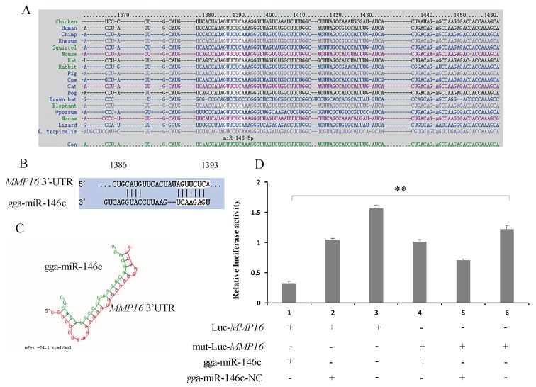
Figure 2.
MMP16 is a target of gga-miR-146c. (A) Alignment of MMP16 3′-UTR from different species. The highlighted sequence is conserved region; (B) Alignments of gga-miR-146c and the target site in MMP16 3′-UTR. The highlighted part is gga-miR146c seed sequence; (C) Structure of gga-miR-146c and MMP16 3′-UTR target site. Red strand represents target sequence and green represents gga-miR146c; (D) Luc-MMP16 (3′-UTR) and either gga-miR-146c or gga-miR-146c-NC were co-transfected into cells. The dual-luciferase glow assay was performed 24 h transfection later. Data from three experiment results were mean ± SD. Two-tailed Student’s t-test, ** p < 0.01.

