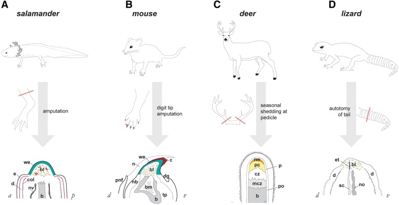
Fig. 2.
Comparative anatomy of regenerating appendages in salamander, mouse, deer, and lizard. A In axolotl, amputation at the mid-humerus level produces a mid-bud-sized blastema (bl, blue cells) within 7–23 days post-amputation, depending upon animal size. Intermingled with blastema cells are blood cells (shown in red). Overlying the blastema is wound epidermis, also known as apical epidermal cap. Note that more proximal epidermis (e) shows distinctly visible basal lamina (magenta), and dermis (d) is bound by a thick collagen mesh (black hatch marks). These features are absent beneath wound epidermis. Nerve: nv; bone: b. Adapted from Payzin-Dogru and Whited, 2018. B In mouse, amputation through the distal-most phalange at the level of the nail bed, produces a blastema (bl) growth at the distal tip beneath both a clot (c) and a wound epidermis (we), shown around day 10 post-amputation. The nail (n) has already grown past these structures by this time. Histolysing bone (b) is shown with bone marrow (bm). Nail bed: nb; toe pad: tp; proximal nail fold: pnf; distal groove: dg. Adapted from Lehoczky et al., 2011, Fernando et al., 2011, and Payzin-Dogru and Whited, 2018. C In deer, antlers are shed from pedicles. Regrowth occurs in zones distal to the bone (b). Mineralized cartilage zone: mcz; cartilage zone: cz; precartilage zone: pc; reserve mesenchyme: rm; periosteum: po; perichondrium: pc. Adapted from Kierdorf et al. [55]. D In lizards, autotomy of the tail produces a blastema-like structure (bl) encasing ependymal tube (et) at the tip of the stump spinal cord (sc). Notochord: no; dermis: d. Adapted from Gilbert et al. [13]

