Abstract
Endometriosis is an inflammatory gynecological disease of reproductive-age women. The prevalence of endometriosis is 5~10% in reproductive-age women. Modern medical treatments are directed to inhibit the action of estrogen in endometriotic cells. However, hormonal therapies targeting estrogen can be prescribed only for a short time because of their undesirable side effects. Recent studies from our laboratory, using endometriotic epithelial cell line 12Z and stromal cell line 22B derived from red lesion, discovered that selective inhibition of prostaglandin E2 (PGE2) receptors EP2 and EP4 inhibits adhesion, invasion, growth, and survival of 12Z and 22B cells by modulating integrins, MMPs and TIMPs, cell cycle, survival, and intrinsic apoptotic pathways, suggesting multiple epigenetic mechanisms. The novel findings of the present study indicate that selective pharmacological inhibition of EP2 and EP4: (i) decreases expression of DNMT3a, DNMT3b, H3K9me3, H3K27me3, SUV39H1, HP1a, H3K27, EZH2, JMJD2a, HDAC1, HDAC3, MeCP2, CoREST and Sin3A; (ii) increases expression of H3K4me3, H3H9ac, H3K27ac; and (iii) does not modulate the expression of DNMT1, hSET1, LSD1, MBD1, p300, HDAC2, and JMJD3 epigenetic machinery proteins in an epithelial and stromal cell specific manner. In this study, we report for the first time that inhibition of PGE2-EP2/EP4 signaling modulates DNA methylation, H3 Histone methylation and acetylation, and epigenetic memory machinery proteins in human endometriotic epithelial cells and stromal cells. Thus, targeting EP2 and EP4 receptors may emerge as long-term nonsteroidal therapy for treatment of active endometriotic lesions in women.
Keywords: PGE2, EP2 and EP4, DNA Methylation, Histone Modification, Endometriosis, Epigenetics
INTRODUCTION
Endometriosis is an estrogen-dependent and progesterone-resistant inflammatory gynecological disease of reproductive-age women. It is characterized by the presence of functional endometrium outside the uterine cavity. The prevalence of endometriosis is 5~10% in reproductive-age women, increases to 20–30% in women with subfertility, and to 40–60% in women with pain and infertility [1,2]. Modern medical treatments are directed to inhibit the action of estrogen in endometriotic cells through suppression of ovarian estrogen production via oral contraceptives, aromatase inhibitors, androgenic agents, and GnRH analogues [1–4]. However, these hormonal therapies can be prescribed only for a short time (~6–9 months) because of their undesirable side effects [1–4]. In addition, endometriosis reestablishes (~50–60%) within a year after cessation of anti-estrogen hormonal therapies [3,4].
Concentrations of PGE2 in the peritoneal fluid are higher in women with endometriosis compared to that of endometriosis-free women, and this increased PGE2 plays important role in survival and growth of endometriotic lesions [5–11]. Inhibition of PGE2 biosynthesis impedes growth of endometriosis [11] and endometriosis-associated pelvic pain in women [8], and decreases growth and survival of experimental endometriosis in animal models [7,9,10]. Cytosolic phospholipase A2 liberates arachidonic acid (AA) from phospholipids. Cyclooxygenases COX-1 and COX-2 convert AA into PGH2 [12]. Prostaglandin E synthases covert PGH2 into PGE2. PGE2 exerts its biological effects via seven-transmembrane G-protein coupled receptors EP1, EP2, EP3, and EP4 by integrating multiple cell signaling pathways [13–17]. EP1 activates PKC and Ca2+ pathways. EP2 and EP4 activate PKA pathway. Activation of EP3A-D produces a wide range of complex and opposite actions [18]. Recent studies indicate that PGE2 transactivates ERK1/2, AKT, NFκB, and β-catenin pathways through EP2 and EP4 in cancer cells [13–17]. PGE2 has been shown to increase the survival of normal lung fibroblast and colon cancer cells through DNA methylation and histone modifications [19,20].
Clinical, cellular, and molecular evidence support a stagewise phenotypic progression of peritoneal endometriotic lesions which include red vesicular, black powder-burn, and fibrotic phenotypes [21–29]. The red lesions are the earliest and most biochemically active phenotype of endometriosis. Importantly, the glandular epithelial and stromal cells of these red lesions secrete large amounts of estradiol, PGE2, cytokines, growth factors, and bleed in response to menstrual hormones [21–29] and thus possess greater adhesion, invasion, proliferation, and survival potential compared to other lesion phenotypes [21–23,26–28,30]. Recent studies from our laboratory, using endometriotic epithelial cell line 12Z and stromal cell line 22B derived from a red lesion, discovered that selective inhibition of EP2 and EP4 receptors: (i) inhibits adhesion, invasion, growth, and survival of endometriotic epithelial cells 12Z and stromal cells 22B by modulating integrins, MMPs and TIMPs, cell cycle, survival, and intrinsic apoptotic pathways [31–37]. These results indicate that multifactorial effects of selective inhibition of EP2 and EP4 may be due to epigenetic mechanisms such as DNA methylation and histone modifications.
DNA methylation is established and maintained by three active DNA methyltransferases, DNMT3a, DNMT3b and DNMT1 [38]. DNMT3a and DNMT3b are involved in de novo DNA methylation [38]. DNA methylation recruits methyl CpG binding domain proteins, HDACs, and co-repressors and thus establishes silenced heterochromatin and transcriptional repression of genes through histone modifications [38–42]. On the other hand, histone methylation is a prerequisite for DNA methylation [43–47]. Multiple histone acetyltransferases [48,49], deacetylases [50,51], methyltransferases [52,53], and demethylases [54–57] have been discovered. Methylation of H3K9me3 and H3K27me3 leads to the formation of closed chromatin structure (heterochromatin) and thus marks transcriptional repression [43–45]. DNMT interacts with H3K9me3 and H3K27me3 through multiple mechanisms and results in DNA methylation [43–45]. In contrast, acetylation of H3K9ac and H3K27ac and methylation of H3K4me3 leads to the formation of an open chromatin structure (euchromatin) and thus marks transcriptional activation [43–45]. Together, cross-talk between these two epigenetic pathways reinforces long-term transcriptional repression or activation [43–45].
While this epigenetic cross-talk pathway has been well studied in embryogenesis and tumorigenesis in the last decade, these processes are largely unknown in endometriosis. Emerging evidence suggests that epigenetics plays a definite role in the pathogenesis of endometriosis [58,59]. DNMT1, DNMT3a and DNMT3b mRNAs are overexpressed in endometriosis [60]. HOXA10 and PR-B genes are hypermethylated in endometriosis [61–64]. Trichostatin A (HDAC pan inhibitor) inhibits NFκB signaling, COX-2 expression, and cell proliferation; by contrast, increases expression of PR-B and E-cadherin in endometriotic cells in vitro [64–67]. The DNA methylation inhibitor 5-Aza differentially regulates expression of ERβ and SF-1 in endometrial and endometriotic cells [68–70]. Although PGE2 plays an important role in the pathogenesis of endometriosis [5–11,32], the underlying epigenetic mechanisms of PGE2 action are largely unknown. The objective of the present study was to identify the effects of selective inhibition of EP2 and EP4 on regulation of DNA methylation, H3K4, H3K9, and H3K29 Histone modifications (methylation and acetylation), and transcriptional activation or suppression machinery proteins in human endometriotic epithelial and stromal cells.
MATERIALS AND METHODS
Materials:
The reagents used in this study were purchased from the following suppliers: Prestained protein markers and Bio-Rad assay reagents and standards (Bio-Rad Laboratories, Hercules, CA); Protran BA83 Nitrocellulose membrane (Whatman Inc, Sanford, ME); Pierce ECL (Pierce Biotechnology, Rockford, IL); protease inhibitor cocktail tablets complete EDTA-free and PhosStop (Roche Applied Biosciences, Indianapolis, IN); antibiotic-antimycotic, and trypsin–EDTA (Invitrogen Life Technologies Inc, Carlsbad, CA); Blue X-Ray film (Phenix Research Products, Hayward, CA); fetal bovine serum (HyClone, Logan, UT); and tissue culture dishes and plates (Corning Inc, Corning, NY). Antagonists/inhibitors for EP2 (AH6809), EP4 (AH23848) were purchased from Sigma (Sigma-Aldrich, St-Louis, MO). All other antibodies used in this study were purchased from Cell Signaling Technology (Danvers, MA), Chemicon International (Billerica, MA), or Santa Cruz Biotechnology (Santa Cruz, CA) except β-actin monoclonal antibody (Sigma-Aldrich), goat anti-rabbit or anti-mouse IgG conjugated with horseradish peroxidase (Kirkegaard & Perry Laboratories, Gaithersburg, MA). The chemicals used were molecular biological grade from Fisher Scientific (Pittsburgh, PA) or Sigma-Aldrich (St. Louis, MO).
Human Endometriotic Cells:
Immortalized endometriotic epithelial cell line 12Z and stromal cell line 22B used in this study were derived from active red peritoneal endometriosis lesions during the proliferative phase of the menstrual cycle from women [30]. These 12Z and 22B cells share several phenotypic and molecular characteristics of primary cultured endometriotic cells [30]. Accumulating information from our and other laboratories indicates that 12Z and 22B cells mimic the active/progressive phase of endometriosis. [30,31,36,37,67]. Importantly, xenograft of a mixed population of these 12Z and 22B cells into the peritoneal cavity of nude mice is able to proliferate, attach, invade, reorganize and establish peritoneal endometriosis-like lesions and that histomorphology are similar to that of spontaneous peritoneal endometriosis in women [34]. We have shown that 12Z and 22B cells produce large amounts of PGE2 at basal conditions, and therefore, inhibition of EP2 and EP4 is the best approach rather than treating the cells with PGE2 to investigate PGE2-EP2/EP4 signaling [31,35–37].
Treatment:
These well-characterized 12Z and 22B cells were cultured in DMEM/F12 without special steroid treatment containing 10% fetal bovine serum (FBS) and penicillin (100 U/ml), streptomycin (100 μg/ml) and amphotericin-B 2.5 μg/ml in a humidified 5% CO2 and 95% air at 37°C as we described previously [31,35–37]. At 70–80% confluency the cells were cultured in DMEM/F12 with 2% dextran-charcoal-treated fetal bovine serum (DC-FBS) and treated with EP2 and EP4 inhibitors (EP-I) for EP2 (AH6809–75 μM) and EP4 (AH23848–50 μM) for 24h. These inhibitors competitively bind with the respective EP2 or EP4 receptors and inhibit their activations [71–73] but not their expressions [31]. The doses for these inhibitors were selected based on the dose response experiments as we published previously [34,36,37].
Protein Extraction:
Total protein was isolated from endometriotic cells and immunoblotting/western blotting was performed as we described previously [31,32]. Briefly, the cells were harvested using 1% Trypsin-EDTA and pelleted. The cell lysates were sonicated in sonication buffer consisting of 20mM Tris-Hcl, 0.5mM EDTA, 100 μM DEDTC, 1% Tween, 1 mM phenylmethylsulfonyl fluoride, and protease inhibitor cocktail tablets: complete EDTA-free (1 tablet/50 ml) and PhosStop (1 tablet/10 ml). Sonication was performed using a Microson ultrasonic cell disruptor (Microsonix Incorporated, Farmingdale, NY). Protein concentration was determined using the Bradford method [74] and a Bio-Rad Protein Assay kit.
Western Blot:
Protein samples (75 μg) were resolved using 7.5%, 10% or 12.5% SDS-PAGE, as we described previously [31,32]. A total of 24 epigenetics modifier proteins were measured. The primary and secondary antibodies used and concentrations are given in Table-1. Briefly, the western blots were incubated with primary antibody overnight at 4°C and with the secondary antibody for 1h at room temperature. Chemiluminescent substrate was applied according to the manufacturer’s instructions (Pierce Biotechnology). The blots were exposed to Blue X-Ray film and densitometry of autoradiograms was performed using an Alpha Imager (Alpha Innotech Corporation, San Leandro, CA).
Table 1:
Details of Antibodies used.
| Antibodies | Manufacturer | Cat # | Concentration used in western blot |
|---|---|---|---|
| DNMT1 | Abcam | ab13537 | 1:200 |
| DNMT3a | Abcam | ab2850 | 1:400 |
| DNMT3b | Abcam | ab13604 | 1:200 |
| H3K4me3 | Abcam | ab8580 | 1:1000 |
| H3K9ac | Abcam | ab4441 | 1:500 |
| H3K9me3 | Abcam | ab8898 | 1:1000 |
| H3K27ac | Abcam | ab4729 | 1:1000 |
| H3K27me3 | Abcam | ab6002 | 1:1000 |
| hSET 1 | Abcam | ab70378 | 1:1000 |
| HP1a | Abcam | ab77256 | 1:2000 |
| SUV39H1 | Cell Signaling | 8729 | 1:1000 |
| EZH2 | Abcam | 3748 | 1:200 |
| LSD1 | Cell Signaling | 2139 | 1:1000 |
| JMJD2A | Cell Signaling | 3393 | 1:1000 |
| JMJD3 | Cell Signaling | 3457 | 1:1000 |
| HDAC1 | Cell Signaling | 5356 | 1:1000 |
| HDAC2 | Cell Signaling | 5113 | 1:1000 |
| HDAC3 | Cell Signaling | 3949 | 1:1000 |
| p300 | Santa Cruz | sc-585 | 1:500 |
| Pol II | Santa Cruz | sc-899 | 1:500 |
| MeCP2 | Abcam | ab2828 | 1:1000 |
| MBD1 | Abcam | ab2846 | 1:500 |
| CoREST | Millipore | 07–455 | 1:2000 |
| Sin3A | Abcam | ab3479 | 1:2000 |
| β-actin | Sigma | A2228 | 1:8000 |
| Goat anti-rabbit IgG | Kirkegaard & Perry Laboratories | 474–1506 | 1:10000 |
| Goat anti-mouse IgG | Kirkegaard & Perry Laboratories | 474–1806 | 1:10000 |
| Rabbit anti-goat IgG | Kirkegaard & Perry Laboratories | 14–13-06 | 1:5000 |
Statistical Analyses:
Statistical analyses were performed using general linear models of Statistical Analysis System (SAS, Cary, NC). Effects of inhibition of EP2 and EP4 on expression levels of different proteins in 12Z and 22B cells were analyzed by one-way analysis of variance (ANOVA) followed by Tukey-Kramer HSD test. The numerical data are expressed as the mean ± SEM. Statistical significance was considered at P<0.05.
RESULTS
EP2 and EP4 signaling and DNMTs in human endometriotic cells:
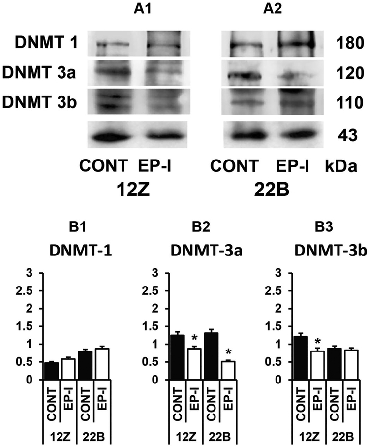
First, we investigated the effects of selective inhibition of EP2/EP4 signaling on DNA methylation machinery proteins in endometriotic epithelial cells 12Z and stromal cells 22B. Results (Fig-1) indicated that inhibition of EP2/EP4 decreased (p<0.05) expression of DNMT3a protein in 12Z and 22B cells, decreased (p<0.05) DNMT3b protein in 12Z cells but not in 22B cells, and did not modulate the expression of DNMT1 in both 12Z and 22B cells. These results indicate that inhibition of EP2/EP4 selectively regulates the DNMT3a and DNMT3b proteins in an epithelial-stromal cell specific manner in endometriosis.
Figure 1: Effects of inhibition of EP2 and EP4 receptors on expression of DNA methyltransferases (DNMTs) in human endometriotic epithelial cells 12Z and stromal cells 22B.
(A1-A2) Western blot analysis and densitometry of (B1) DNMT1, (B2) DNMT3a, and (B3) DNMT3b proteins in 12Z and 22B cells. β-actin protein was measured as an internal control. The 12Z and 22B cells were treated with EP2 and EP4 inhibitors (EP-I) EP2 (AH6809–75 μM) and EP4 (AH23848- μM) for 24h. The experiments were performed as we described in the “Materials and Methods”. *-Control vs. EP2-I/EP4-I, P<0.05. Numerical data are expressed as Mean ± SEM of three (n=3) experiments.
EP2 and EP4 signaling and H3 histone modification in human endometriotic cells:
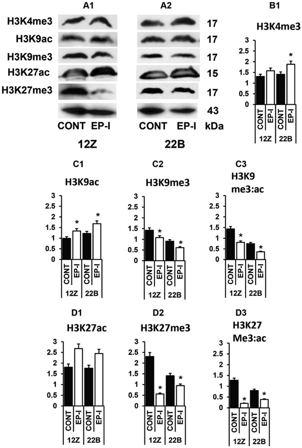
Next, we investigated the effects of selective inhibition of EP2/EP4 signaling on acetylation and methylation of H3 histone at K4, K9 and K27 in endometriotic epithelial cells 12Z and stromal cells 22B. Results (Fig-2) indicated that inhibition of EP2/EP4 increased (p<0.05) methylation of H3K4 protein in 22B cells but not in 12Z cells. Inhibition of EP2 and EP4 decreased (p<0.05) methylation of H3K9 and H3K27 and concomitantly increased (p<0.05) acetylation of H3K9 and H3K27 in both 12Z and 22B cells. These results together indicate that inhibition of EP2 and EP4 selectively regulates acetylation and methylation of H3K9 and H3K27 proteins and sustains methylation H3K4 in an epithelial-stromal cell specific manner in endometriosis.
Figure 2: Effects of inhibition of EP2 and EP4 receptors on H3K4, H3K9, and H3K27 methylation and acetylation in human endometriotic epithelial cells 12Z and stromal cells 22B.
(A1-A2) Western blot analysis and densitometry of proteins (B1) H3K4me3, (C1) H3K9ac, (C2) H3K9me3, (C3) H3K9me3: H3K9ac ratio, (D1) H3K27ac, (D2) H3K27me3, and (D3) H3K9me3: H3K9ac ratio in 12Z and 22B cells. β-actin protein was measured as an internal control. The 12Z and 22B cells were treated with EP2 and EP4 inhibitors (EP-I) EP2 (AH6809–75 μM) and EP4 (AH23848- μM) for 24h. The experiments were performed as we described in the “Materials and Methods”. *-Control vs. EP2-I/EP4-I, P<0.05. Numerical data are expressed as Mean ± SEM of three (n=3) experiments.
EP2 and EP4 signaling and H3 histone methylation machinery in human endometriotic cells:
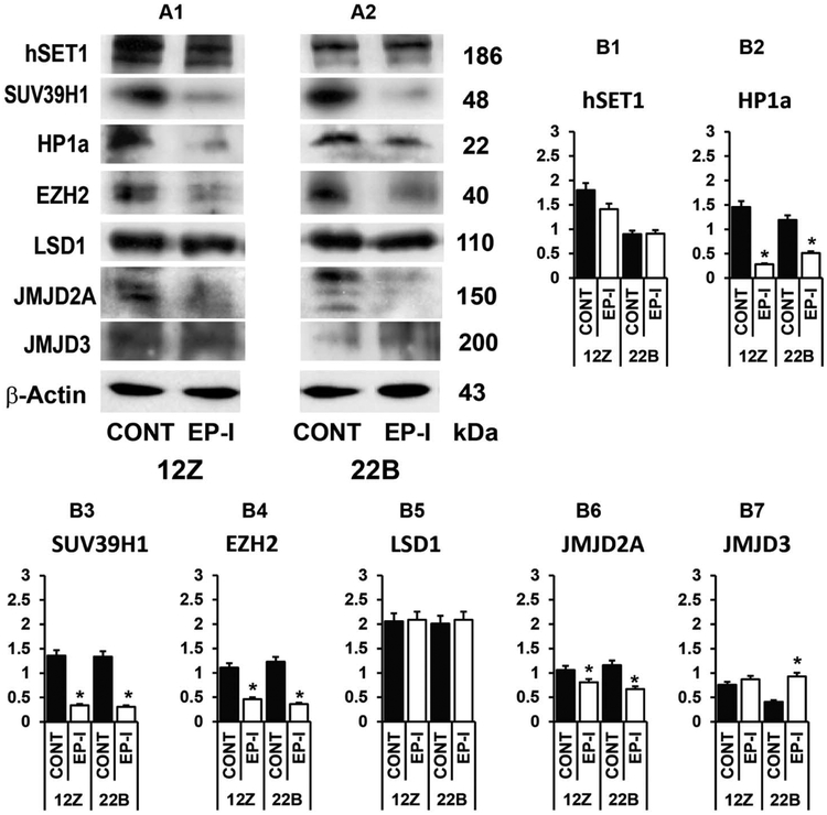
Modulation in methylation status of H3K4, H3K9 and H3K27 in 12Z and 22B cells lead us to investigate the effects of selective inhibition of EP2/EP4 signaling on H3 histone methylation and demethylation machinery proteins. Results (Fig-3) indicated that inhibition of EP2/EP4 did not modulate expression of H3K4 methyltransferase hSET1 and demethylase LSD1 proteins in 12Z and 22B cells. Inhibition of EP2/EP4 decreased (p<0.05) H3K9 methyltransferase SUV39H1 protein and its adapter protein heterochromatin protein 1a (HP1a) and demethylase JMJD2A in 12Z and 22B cells. Inhibition of EP2/EP4 decreased (p<0.05) H3K27 methyltransferase EZH2 protein but not demethylase JMJD3 protein in 12Z and 22B cells. These results together indicate that inhibition of EP2/EP4 does not modulate methylation of H3K4 but regulates methylation of H3K9 and H3K27 through key important machinery proteins in an epithelial-stromal cell specific manner in endometriosis.
Figure 3: Effects of inhibition of EP2 and EP4 receptors on H3 histone methylation and demethylation machinery proteins in human endometriotic epithelial cells 12Z and stromal cells 22B.
(A1-A2) Western blot analysis and densitometry of proteins (B1) hSET1, (B2) HP1a, (B3) SUV39H1, (B4) EZH2, (B5) LSD1, (B6) JMJD2A, and (B7) JMJD3 in 12Z and 22B cells. β-actin protein was measured as an internal control. The 12Z and 22B cells were treated with EP2 and EP4 inhibitors (EP-I) EP2 (AH6809–75 μM) and EP4 (AH23848- μM) for 24h. The experiments were performed as we described in the “Materials and Methods”. *-Control vs. EP2-I/EP4-I, P<0.05. Numerical data are expressed as Mean ± SEM of three (n=3) experiments.
EP2 and EP4 signaling and H3 histone acetylation machinery in human endometriotic cells:
Modulation in acetylation status of H3K9 and H3K27 in 12Z and 22B cells lead us to investigate effects of selective inhibition of EP2/EP4 signaling on H3 histone acetylation and deacetylation machinery proteins. Results (Fig-4) indicated that inhibition of EP2/EP4 decreased (p<0.05) expression of HDAC1 protein in both 12Z and 22B cells, decreased (p<0.05) expression of HDAC3 only in 12Z cells, and did not modulate expression of HDAC2 in both 12Z and 22B cells. Importantly, inhibition of EP2/EP4 did not decrease expression of acetyltransferase p300 in 12Z and 22B cells. These results together indicate that inhibition of EP2/EP4 sustains acetylation whereas selectively suppresses deacetylation machinery proteins in an epithelial-stromal cell specific manner in endometriosis.
Figure 4: Effects of inhibition of EP2 and EP4 receptors on H3 histone acetylation and deacetylation machinery proteins in human endometriotic epithelial cells 12Z and stromal cells 22B.
(A1-A2) Western blot analysis and densitometry of proteins (B1) HDAC1, (B2) HDAC2, (B3) HDAC3, and (B4) p300 in endometriotic epithelial cells 12Z and stromal cells 22B. β-actin protein was measured as an internal control. The 12Z and 22B cells were treated with EP2 and EP4 inhibitors (EP-I) EP2 (AH6809–75 μM) and EP4 (AH23848- μM) for 24h. The experiments were performed as we described in the “Materials and Methods”. *-Control vs. EP2-I/EP4-I, P<0.05. Numerical data are expressed as Mean ± SEM of three (n=3) experiments.
EP2 and EP4 signaling and transcriptional activation and suppression machinery in human endometriotic cells:
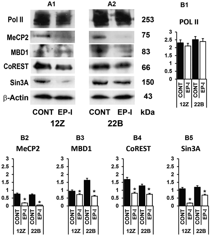
Finally, we investigated effects of selective inhibition of EP2/EP4 signaling on transcriptional activation and suppression machinery proteins in 12Z and 22B cells. Results (Fig-5) indicated that inhibition of EP2/EP4 decreased (p<0.05) expression of transcriptional repression machinery proteins MeCP2, MBD1, CoREST and Sin3A in both 12Z and 22B cells. Importantly in contrast, inhibition of EP2/EP4 did not decrease expression of transcription activation machinery protein Pol II in both 12Z and 22B cells. These results together indicate that inhibition of EP2/EP4 sustains transcriptional activation whereas selectively decreases transcriptional suppression machinery proteins in an epithelial-stromal cell specific manner in endometriosis.
Figure 5: Effects of inhibition of EP2 and EP4 receptors on transcriptional activation and suppression machinery proteins in human endometriotic epithelial cells 12Z and stromal cells 22B.
(A1-A2) Western blot analysis and densitometry of proteins (B1) Pol II, (B2) MeCP2, (B3) MBD1, (B4) CoREST, and (B5) Sin3A in endometriotic epithelial cells 12Z and stromal cells 22B. β-actin protein was measured as an internal control. The 12Z and 22B cells were treated with EP2 and EP4 inhibitors (EP-I) EP2 (AH6809–75 μM) and EP4 (AH23848- μM) for 24h. The experiments were performed as we described in the “Materials and Methods”. *-Control vs. EP2-I/EP4-I, P<0.05. Numerical data are expressed as Mean ± SEM of three (n=3) experiments.
DISCUSSION
Although PGE2 plays important roles in the pathogenesis of endometriosis [5–11], its epigenetic actions in endometriosis is not known. In this study, we report for the first time that inhibition of PGE2-EP2/EP4 signaling regulates several epigenetics machinery proteins associated with DNA methylation and histone modification in human endometriotic epithelial cells 12Z and stromal cells 22B.
DNA methylation is a covalent chemical modification of DNA resulting in addition of a methyl group at the carbon 5-position of the cytosine within CpG [38]. DNA methylation is established and maintained by three active DNA methyltransferases, DNMT3a, DNMT3b and DNMT1. DNMT3a and DNMT3b are involved in de novo DNA methylation and DNMT1 is primarily involved in propagating heritable DNA methylation patterns [38]. Results of the present study indicate that inhibition of EP2/EP4 decreases expression de novo DNMAT3a and DNMT3c in an epithelial and stromal cell specific manner in endometriosis. These results suggest that inhibition PGE2-EP2/EP4 signaling may alter DNA methylation state of epigenetically silenced gens in endometriosis through suppression of DNMT3a and DNMT3b.
Recent studies indicate that histone methylation is a prerequisite for DNA methylation [43–47]. The core histone octomer comprises histone proteins H2A, H2B, H3, and H4 [43–47]. Among the histone modifications implicated in gene activation and silencing, the best characterized to date are acetylation or methylation of lysine (K) at Histone 3 such as H3K9, H3K27 and H3K4 [43–47]. Acetylation of H3K9 and H3K27 and tri-methylation of H3K4 leads to the formation of an open chromatin structure (euchromatin) and thus transcriptional activation [43–45]. By contrast, tri-methylation of H3K9 and H3K27 leads to the formation of closed chromatin structure (heterochromatin) and thus marks transcriptional repression [43–45]. Results of the present study indicate that Inhibition of EP2/EP4 decreases methylation of H3K9 and H3K27 and concomitantly increases acetylation of H3K9 and H3K27 in epithelial and stromal cell specific manner in endometriosis. These results suggest that inhibition of PGE2-EP2/EP4 signaling selectively suppresses methylation of H3K9 and H3K27 and thereby inhibits heterochromatin and establishes euchromatin states in endometriosis.
Addition or removal of a tri-methyl group (CH3) into Lysine (K) at H3 histone is catalyzed by histone methyltransferases [52,53] and histone/lysine demethylases [54–57]. Addition or removal of an acetyl group (COCH3) into Lysine (K) at H3 histone is catalyzed by histone acetyltransferases [48,49] and histone deacetylases (HDACs) [50,51], respectively. Multiple enzymes are discovered until today and we have examined some of the important and well characterized enzymes in the preset study. Addition of CH3 into H3K9 is catalyzed by histone methyltransferase SUV39H1 protein through its adapter protein HP1a and removal of CH3 from H3K9 is catalyzed by histone demethylase JMJD2A. Addition of CH3 into H3K27 is catalyzed by histone methyltransferase EZH2 and removal of CH3 from H3K27 is catalyzed by histone demethylase JMJD3 [43–47]. Results of the present study indicate that inhibition of EP2/EP4 decreases H3K9 methylation machinery proteins SUV39H1 and HP1a and JMJD2A, and decreases H3K27 methylation machinery proteins EZH2 but not JMJD3 in an epithelial and stromal cell specific manner in endometriosis. In addition, our results indicate that inhibition of EP2/EP4 decreases expression of HDAC1, HDAC3 but not HDAC2 and acetyltransferase p300 in an epithelial and stromal cell specific manner in endometriosis. These results suggest that inhibition of PGE2-EP2/EP4 decreases methylation of H3K9 and H3K27 primarily through down-regulation of methyltransferases SUV39H1 and EZH2, respectively, and concomitantly increases acetylation of H3K9 and H3K27 through down-regulation of HDAC1 and HDAC3 in endometriosis.
Methylation of H3K4 is a classical transcription activation mark. Addition of CH3 group into H3K4 is catalyzed by hSET1 protein and removal of CH3 group from H3K4 is catalyzed by LSD1 protein [52,53]. Results of the present study indicate that inhibition of EP2/EP4 did not modulate methylation of H3K4 and expression of hSET1 and LSD1 proteins in endometriotic epithelial and stromal cells. These results suggest that inhibition of PGE2-EP2/EP4 sustains methylation of H3K4 and thus maintains H3K4me3-mediated transcriptional activation memory in endometriosis.
It is well known that hypermethylated CpG recruit methyl CpG binding domain proteins (MBDs) such as MeCp2 and MBD1–4 and these MBD proteins in turn recruit a variety of protein complexes that contain HDACs and co-repressors sin3A and Co-REST and thus establish silenced heterochromatin and transcriptional repression of genes [38–42]. In contrast, open euchromatin allows Poll II and other transcriptional activation factors to promote transcriptional activation [75,76]. Results of the present study indicate that inhibition of EP2/EP4 decreases expression of transcriptional repression machinery proteins MeCP2, MBD1, CoREST and Sin3A but do not modulate the expression of the important transcriptional activation machinery protein Pol II in endometriotic epithelial and stromal cells. These results together indicate that inhibition of PGE2-EP2/EP4 sustains transcriptional activation whereas selectively decreases transcriptional suppression machinery proteins in an epithelial-stromal cell specific manner in endometriosis.
Many recent findings highlight the intimate link between DNA methylation and histone modifications [43–47]. DNMT interacts with H3K9 through SUV39h1 and HP1a. Loss of function of SUV39h1 and loss of methylation of H3K9 decreases DNMT3b-dependent CpG methylation at major pericentric satellites [43,46,47]. Poly-comb repressive complex 2 (PRC2) is composed of EZH2, EED, and SUZ12 [44,45]. The set domain EZH2 directly catalyzes methylation of H3K27, and its indirect interaction with DNMTs may result in DNA methylation [43–45]. These land-mark studies together indicate that epigenetic information can flow from histones to DNA through and results in subsequent DNA methylation; concomitantly the DNA methylation can exert a positive feedback on histone acetylation and/or methylation. Interaction between these two distinct epigenetic pathways results in a self-propagating cycle that promotes long-term epigenetic marks. Results of the present study and along with these previous studies suggest that PGE2-EP2/EP4 signaling may regulate these two epigenetic pathways in the pathogenesis of endometriosis.
Using endometriotic epithelial cells 12Z and stromal cells 22B model system, we have shown that selective inhibition of EP2/EP4 inhibits adhesions, migration, invasion, growth and survival of endometriotic epithelial and stromal cells by modulating up to 75 proteins associated with these pathways. In this study, we have shown that inhibition of EP2 and EP4 modulates up to 24 proteins involved in DNA methylation, H3K4, H3K9 and H3K9 methylation and acetylation, transcriptional activation and suppression in endometriotic cells. Our unpublished data indicate that inhibition of EP2/EP4 signaling upregulates epigenetically silenced proapoptotic miRNAs in 12Z and 22B cells. Ongoing studies in our laboratory will identify the interaction among PGE2-EP2/EP4 signaling and restoration of epigenetically silenced proapoptotic miRNAs in endometriosis using in vitro and in vivo model systems. In addition, our future studies will be directed to dissect interaction between EP2/EP4 signaling and loss of PR-B (progesterone resistance) in the endometriosis.
In conclusion, results of the present study indicate that EP2/EP4-mediated PGE2 signaling regulates epigenetics of endometriosis. The novel findings are that selective inhibition of EP2/EP4: (i) inhibits DNMT3a and DNMT3b; (ii) decrease methylations and increase acetylation of H3K9 and H3K27 by modulating the epigenetic modifier/memory proteins; (iii) sustains methylation of H3K4 and its methylation machinery proteins; and (iv) inhibits transcriptional suppression machinery proteins without modulating transcriptional activation machinery proteins in endometriotic epithelial and stromal cells. Targeting EP2 and EP4 receptors may emerge as long-term nonsteroidal therapy for treatment of active red endometriotic lesions in women.
Grant Support:
This work is partially supported by Eunice Kennedy Shriver National Institute of Child Health & Human Development (NICHD) and NIH Office of Research on Women’s Health (1R21HD065138 and 1R21HD066248 to JAA).
Footnotes
Publisher's Disclaimer: This is a PDF file of an unedited manuscript that has been accepted for publication. As a service to our customers we are providing this early version of the manuscript. The manuscript will undergo copyediting, typesetting, and review of the resulting proof before it is published in its final citable form. Please note that during the production process errors may be discovered which could affect the content, and all legal disclaimers that apply to the journal pertain.
Disclosure Statement: The authors have nothing to disclose.
REFERENCES
- [1].Bulun SE (2009) Endometriosis. N Engl J Med 360, 268–79. [DOI] [PubMed] [Google Scholar]
- [2].Giudice LC and Kao LC (2004) Endometriosis. Lancet 364, 1789–99. [DOI] [PubMed] [Google Scholar]
- [3].Guo SW and Olive DL (2007) Two unsuccessful clinical trials on endometriosis and a few lessons learned. Gynecol Obstet Invest 64, 24–35. [DOI] [PubMed] [Google Scholar]
- [4].Kyama CM, Mihalyi A, Simsa P, Mwenda JM, Tomassetti C, Meuleman C and D’Hooghe TM (2008) Non-steroidal targets in the diagnosis and treatment of endometriosis. Curr Med Chem 15, 1006–17. [DOI] [PubMed] [Google Scholar]
- [5].Wu MH, Shoji Y, Chuang PC and Tsai SJ (2007) Endometriosis: disease pathophysiology and the role of prostaglandins. Expert Rev Mol Med 9, 1–20. [DOI] [PubMed] [Google Scholar]
- [6].Wu MH, Lu CW, Chuang PC and Tsai SJ (2010) Prostaglandin E2: the master of endometriosis? Exp Biol Med (Maywood) 235, 668–77. [DOI] [PubMed] [Google Scholar]
- [7].Chuang PC, Lin YJ, Wu MH, Wing LY, Shoji Y and Tsai SJ (2010) Inhibition of CD36-dependent phagocytosis by prostaglandin E2 contributes to the development of endometriosis. Am J Pathol 176, 850–60. [DOI] [PMC free article] [PubMed] [Google Scholar]
- [8].Cobellis L, Razzi S, De Simone S, Sartini A, Fava A, Danero S, Gioffre W, Mazzini M and Petraglia F (2004) The treatment with a COX-2 specific inhibitor is effective in the management of pain related to endometriosis. Eur J Obstet Gynecol Reprod Biol 116, 100–2. [DOI] [PubMed] [Google Scholar]
- [9].Laschke MW, Elitzsch A, Scheuer C, Vollmar B and Menger MD (2007) Selective cyclo-oxygenase-2 inhibition induces regression of autologous endometrial grafts by down-regulation of vascular endothelial growth factor-mediated angiogenesis and stimulation of caspase-3-dependent apoptosis. Fertil Steril 87, 163–71. [DOI] [PubMed] [Google Scholar]
- [10].Matsuzaki S, Canis M, Darcha C, Dallel R, Okamura K and Mage G (2004) Cyclooxygenase-2 selective inhibitor prevents implantation of eutopic endometrium to ectopic sites in rats. Fertil Steril 82, 1609–15. [DOI] [PubMed] [Google Scholar]
- [11].Olivares C, Bilotas M, Buquet R, Borghi M, Sueldo C, Tesone M and Meresman G (2008) Effects of a selective cyclooxygenase-2 inhibitor on endometrial epithelial cells from patients with endometriosis. Hum Reprod 23, 2701–8. [DOI] [PubMed] [Google Scholar]
- [12].Smith WL and Dewitt DL (1996) Prostaglandin endoperoxide H synthases-1 and −2. Adv Immunol 62, 167–215. [DOI] [PubMed] [Google Scholar]
- [13].Cha YI and DuBois RN (2007) NSAIDs and cancer prevention: targets downstream of COX-2. Annu Rev Med 58, 239–52. [DOI] [PubMed] [Google Scholar]
- [14].Fujino H and Regan JW (2003) Prostaglandin F(2alpha) stimulation of cyclooxygenase-2 promoter activity by the FP(B) prostanoid receptor. Eur J Pharmacol 465, 39–41. [DOI] [PubMed] [Google Scholar]
- [15].Jabbour HN and Sales KJ (2004) Prostaglandin receptor signalling and function in human endometrial pathology. Trends Endocrinol Metab 15, 398–404. [DOI] [PubMed] [Google Scholar]
- [16].Buchanan FG, Gorden DL, Matta P, Shi Q, Matrisian LM and DuBois RN (2006) Role of beta-arrestin 1 in the metastatic progression of colorectal cancer. Proc Natl Acad Sci U S A 103, 1492–7. [DOI] [PMC free article] [PubMed] [Google Scholar]
- [17].Castellone MD, Teramoto H, Williams BO, Druey KM and Gutkind JS (2005) Prostaglandin E2 promotes colon cancer cell growth through a Gs-axin-beta-catenin signaling axis. Science 310, 1504–10. [DOI] [PubMed] [Google Scholar]
- [18].Narumiya S, Sugimoto Y and Ushikubi F (1999) Prostanoid receptors: structures, properties, and functions. Physiol Rev 79, 1193–226. [DOI] [PubMed] [Google Scholar]
- [19].Huang SK, Scruggs AM, Donaghy J, McEachin RC, Fisher AS, Richardson BC and Peters-Golden M (2012) Prostaglandin E(2) increases fibroblast gene-specific and global DNA methylation via increased DNA methyltransferase expression. Faseb J 26, 3703–14. [DOI] [PMC free article] [PubMed] [Google Scholar]
- [20].Xia D, Wang D, Kim SH, Katoh H and DuBois RN (2012) Prostaglandin E2 promotes intestinal tumor growth via DNA methylation. Nat Med 18, 224–6. [DOI] [PMC free article] [PubMed] [Google Scholar]
- [21].Burney RO and Giudice LC (2012) Pathogenesis and pathophysiology of endometriosis. Fertil Steril 98, 511–9. [DOI] [PMC free article] [PubMed] [Google Scholar]
- [22].Burney RO and Lathi RB (2009) Menstrual bleeding from an endometriotic lesion. Fertil Steril 91, 1926–7. [DOI] [PubMed] [Google Scholar]
- [23].Chatman DL (1981) Pelvic peritoneal defects and endometriosis: Allen-Masters syndrome revisited. Fertil Steril 36, 751–6. [DOI] [PubMed] [Google Scholar]
- [24].Donnez J, Nisolle M and Casanas-Roux F (1992) Three-dimensional architectures of peritoneal endometriosis. Fertil Steril 57, 980–3. [PubMed] [Google Scholar]
- [25].Kokorine I, Nisolle M, Donnez J, Eeckhout Y, Courtoy PJ and Marbaix E (1997) Expression of interstitial collagenase (matrix metalloproteinase-1) is related to the activity of human endometriotic lesions. Fertil Steril 68, 246–51. [DOI] [PubMed] [Google Scholar]
- [26].Koninckx PR, Meuleman C, Demeyere S, Lesaffre E and Cornillie FJ (1991) Suggestive evidence that pelvic endometriosis is a progressive disease, whereas deeply infiltrating endometriosis is associated with pelvic pain. Fertil Steril 55, 759–65. [DOI] [PubMed] [Google Scholar]
- [27].Nieminen U (1963) On the Vasculature of Ectopic Endometrium with Decidual Reaction. Acta Obstet Gynecol Scand 42, 151–9. [DOI] [PubMed] [Google Scholar]
- [28].Vernon MW, Beard JS, Graves K and Wilson EA (1986) Classification of endometriotic implants by morphologic appearance and capacity to synthesize prostaglandin F. Fertil Steril 46, 801–6. [PubMed] [Google Scholar]
- [29].Vipond MN, Whawell SA, Thompson JN and Dudley HA (1990) Peritoneal fibrinolytic activity and intra-abdominal adhesions. Lancet 335, 1120–2. [DOI] [PubMed] [Google Scholar]
- [30].Zeitvogel A, Baumann R and Starzinski-Powitz A (2001) Identification of an invasive, N-cadherin-expressing epithelial cell type in endometriosis using a new cell culture model. Am J Pathol 159, 1839–52. [DOI] [PMC free article] [PubMed] [Google Scholar]
- [31].Banu SK, Lee J, Speights VO Jr, Starzinski-Powitz A and Arosh JA (2009) Selective inhibition of prostaglandin E2 receptors EP2 and EP4 induces apoptosis of human endometriotic cells through suppression of ERK1/2, AKT, NFkB and b-catenin pathways and activation of intrinsic apoptotic mechanisms. Molecular Endocrinology 23, 1291–1305. [DOI] [PMC free article] [PubMed] [Google Scholar]
- [32].Banu SK, Lee J, Speights VO, Starzinski-Powitz A and Arosh JA (2008) Cyclooxygenase-2 regulates survival, migration and invasion of human endometriotic cells through multiple mechanisms. Endocrinology 149, 1180–1189. [DOI] [PubMed] [Google Scholar]
- [33].Banu SK, Lee J, Starzinski-Powitz A and Arosh JA (2008) Gene expression profiles and functional characterization of human immortalized endometriotic epithelial and stromal cells. Fertility and Sterlity 90, 972–987. [DOI] [PubMed] [Google Scholar]
- [34].Banu SK, Starzinski-Powitz A, Speights VO, Burghardt RC and Arosh JA (2009) Induction of peritoneal endometriosis in nude mice using human immortalized endometriosis epithelial and stromal cells: A potential experimental tool to study molecular pathogenesis of endometriosis in human. Fertil Steril 91, 2199–2209. [DOI] [PubMed] [Google Scholar]
- [35].Lee J, Banu SK, Burghardt RC, Starzinski-Powitz A and Arosh JA (2013) Selective inhibition of prostaglandin E2 receptors EP2 and EP4 inhibits adhesion of human endometriotic epithelial and stromal cells through suppression of integrin-mediated mechanisms. Biol Reprod 88, 77. [DOI] [PMC free article] [PubMed] [Google Scholar]
- [36].Lee J, Banu SK, Rodriguez R, Starzinski-Powitz A and Arosh JA (2010) Selective blockade of prostaglandin E2 receptors EP2 and EP4 signaling inhibits proliferation of human endometriotic epithelial cells and stromal cells through distinct cell cycle arrest. Fertil Steril 93, 2498–506. [DOI] [PubMed] [Google Scholar]
- [37].Lee J, Banu SK, Subbarao T, Starzinski-Powitz A and Arosh JA (2011) Selective inhibition of prostaglandin E2 receptors EP2 and EP4 inhibits invasion of human immortalized endometriotic epithelial and stromal cells through suppression of metalloproteinases. Mol Cell Endocrinol 332, 306–13. [DOI] [PubMed] [Google Scholar]
- [38].Klose RJ and Bird AP (2006) Genomic DNA methylation: the mark and its mediators. Trends Biochem Sci 31, 89–97. [DOI] [PubMed] [Google Scholar]
- [39].Esteller M (2002) CpG island hypermethylation and tumor suppressor genes: a booming present, a brighter future. Oncogene 21, 5427–40. [DOI] [PubMed] [Google Scholar]
- [40].Hendrich B and Tweedie S (2003) The methyl-CpG binding domain and the evolving role of DNA methylation in animals. Trends Genet 19, 269–77. [DOI] [PubMed] [Google Scholar]
- [41].Fuks F, Hurd PJ, Wolf D, Nan X, Bird AP and Kouzarides T (2003) The methyl-CpG-binding protein MeCP2 links DNA methylation to histone methylation. J Biol Chem 278, 4035–40. [DOI] [PubMed] [Google Scholar]
- [42].Hashimoto H, Vertino PM and Cheng X (2010) Molecular coupling of DNA methylation and histone methylation. Epigenomics 2, 657–69. [DOI] [PMC free article] [PubMed] [Google Scholar]
- [43].Fuks F (2005) DNA methylation and histone modifications: teaming up to silence genes. Curr Opin Genet Dev 15, 490–5. [DOI] [PubMed] [Google Scholar]
- [44].Vire E, Brenner C, Deplus R, Blanchon L, Fraga M, Didelot C, Morey L, Van Eynde A, Bernard D, Vanderwinden JM, Bollen M, Esteller M, Di Croce L, de Launoit Y and Fuks F (2006) The Polycomb group protein EZH2 directly controls DNA methylation. Nature 439, 871–4. [DOI] [PubMed] [Google Scholar]
- [45].Kondo Y (2009) Epigenetic cross-talk between DNA methylation and histone modifications in human cancers. Yonsei Med J 50, 455–63. [DOI] [PMC free article] [PubMed] [Google Scholar]
- [46].Fuks F, Hurd PJ, Deplus R and Kouzarides T (2003) The DNA methyltransferases associate with HP1 and the SUV39H1 histone methyltransferase. Nucleic Acids Res 31, 2305–12. [DOI] [PMC free article] [PubMed] [Google Scholar]
- [47].Lehnertz B, Ueda Y, Derijck AA, Braunschweig U, Perez-Burgos L, Kubicek S, Chen T, Li E, Jenuwein T and Peters AH (2003) Suv39h-mediated histone H3 lysine 9 methylation directs DNA methylation to major satellite repeats at pericentric heterochromatin. Curr Biol 13, 1192–200. [DOI] [PubMed] [Google Scholar]
- [48].Lee KK and Workman JL (2007) Histone acetyltransferase complexes: one size doesn’t fit all. Nat Rev Mol Cell Biol 8, 284–95. [DOI] [PubMed] [Google Scholar]
- [49].Roth SY, Denu JM and Allis CD (2001) Histone acetyltransferases. Annu Rev Biochem 70, 81–120. [DOI] [PubMed] [Google Scholar]
- [50].Clayton AL, Hazzalin CA and Mahadevan LC (2006) Enhanced histone acetylation and transcription: a dynamic perspective. Mol Cell 23, 289–96. [DOI] [PubMed] [Google Scholar]
- [51].Mehnert JM and Kelly WK (2007) Histone deacetylase inhibitors: biology and mechanism of action. Cancer J 13, 23–9. [DOI] [PubMed] [Google Scholar]
- [52].Greer EL and Shi Y (2012) Histone methylation: a dynamic mark in health, disease and inheritance. Nat Rev Genet 13, 343–57. [DOI] [PMC free article] [PubMed] [Google Scholar]
- [53].Teperino R, Schoonjans K and Auwerx J (2010) Histone methyl transferases and demethylases; can they link metabolism and transcription? Cell Metab 12, 321–7. [DOI] [PMC free article] [PubMed] [Google Scholar]
- [54].Chen S, Ma J, Wu F, Xiong LJ, Ma H, Xu W, Lv R, Li X, Villen J, Gygi SP, Liu XS and Shi Y (2012) The histone H3 Lys 27 demethylase JMJD3 regulates gene expression by impacting transcriptional elongation. Genes Dev 26, 1364–75. [DOI] [PMC free article] [PubMed] [Google Scholar]
- [55].Cloos PA, Christensen J, Agger K and Helin K (2008) Erasing the methyl mark: histone demethylases at the center of cellular differentiation and disease. Genes Dev 22, 1115–40. [DOI] [PMC free article] [PubMed] [Google Scholar]
- [56].Lee MG, Wynder C, Bochar DA, Hakimi MA, Cooch N and Shiekhattar R (2006) Functional interplay between histone demethylase and deacetylase enzymes. Mol Cell Biol 26, 6395–402. [DOI] [PMC free article] [PubMed] [Google Scholar]
- [57].Wilson JR (2007) Targeting the JMJD2A histone lysine demethylase. Nat Struct Mol Biol 14, 682–4. [DOI] [PubMed] [Google Scholar]
- [58].Guo SW (2009) Epigenetics of endometriosis. Mol Hum Reprod 15, 587–607. [DOI] [PubMed] [Google Scholar]
- [59].Guo SW, Wu Y, Strawn E, Basir Z, Wang Y, Halverson G, Montgomery K and Kajdacsy-Balla A (2004) Genomic alterations in the endometrium may be a proximate cause for endometriosis. Eur J Obstet Gynecol Reprod Biol 116, 89–99. [DOI] [PubMed] [Google Scholar]
- [60].Wu Y, Strawn E, Basir Z, Halverson G and Guo SW (2007) Aberrant expression of deoxyribonucleic acid methyltransferases DNMT1, DNMT3A, and DNMT3B in women with endometriosis. Fertil Steril 87, 24–32. [DOI] [PubMed] [Google Scholar]
- [61].Wu Y, Halverson G, Basir Z, Strawn E, Yan P and Guo SW (2005) Aberrant methylation at HOXA10 may be responsible for its aberrant expression in the endometrium of patients with endometriosis. Am J Obstet Gynecol 193, 371–80. [DOI] [PubMed] [Google Scholar]
- [62].Wu Y, Strawn E, Basir Z, Halverson G and Guo SW (2006) Promoter hypermethylation of progesterone receptor isoform B (PR-B) in endometriosis. Epigenetics 1, 106–11. [DOI] [PubMed] [Google Scholar]
- [63].Wu Y, Shi X and Guo SW (2008) The knockdown of progesterone receptor isoform B (PR-B) promotes proliferation in immortalized endometrial stromal cells. Fertil Steril 90, 1320–3. [DOI] [PubMed] [Google Scholar]
- [64].Wu Y, Starzinski-Powitz A and Guo SW (2008) Prolonged stimulation with tumor necrosis factor-alpha induced partial methylation at PR-B promoter in immortalized epithelial-like endometriotic cells. Fertil Steril 90, 234–7. [DOI] [PubMed] [Google Scholar]
- [65].Wu Y and Guo SW (2007) Suppression of IL-1beta-induced COX-2 expression by trichostatin A (TSA) in human endometrial stromal cells. Eur J Obstet Gynecol Reprod Biol 135, 88–93. [DOI] [PubMed] [Google Scholar]
- [66].Wu Y and Guo SW (2008) Histone deacetylase inhibitors trichostatin A and valproic acid induce cell cycle arrest and p21 expression in immortalized human endometrial stromal cells. Eur J Obstet Gynecol Reprod Biol 137, 198–203. [DOI] [PubMed] [Google Scholar]
- [67].Wu Y, Starzinski-Powitz A and Guo SW (2007) Trichostatin A, a histone deacetylase inhibitor, attenuates invasiveness and reactivates E-cadherin expression in immortalized endometriotic cells. Reprod Sci 14, 374–82. [DOI] [PubMed] [Google Scholar]
- [68].Izawa M, Harada T, Taniguchi F, Ohama Y, Takenaka Y and Terakawa N (2008) An epigenetic disorder may cause aberrant expression of aromatase gene in endometriotic stromal cells. Fertil Steril 89, 1390–6. [DOI] [PubMed] [Google Scholar]
- [69].Xue Q, Lin Z, Cheng YH, Huang CC, Marsh E, Yin P, Milad MP, Confino E, Reierstad S, Innes J and Bulun SE (2007) Promoter methylation regulates estrogen receptor 2 in human endometrium and endometriosis. Biol Reprod 77, 681–7. [DOI] [PubMed] [Google Scholar]
- [70].Xue Q, Lin Z, Yin P, Milad MP, Cheng YH, Confino E, Reierstad S and Bulun SE (2007) Transcriptional activation of steroidogenic factor-1 by hypomethylation of the 5’ CpG island in endometriosis. J Clin Endocrinol Metab 92, 3261–7. [DOI] [PubMed] [Google Scholar]
- [71].Coleman RA, Smith WL and Narumiya S (1994) International Union of Pharmacology classification of prostanoid receptors: properties, distribution, and structure of the receptors and their subtypes. Pharmacol Rev 46, 205–29. [PubMed] [Google Scholar]
- [72].Woodward DF, Pepperl DJ, Burkey TH and Regan JW (1995) 6-Isopropoxy-9-oxoxanthene-2-carboxylic acid (AH 6809), a human EP2 receptor antagonist. Biochem Pharmacol 50, 1731–3. [DOI] [PubMed] [Google Scholar]
- [73].Crider JY, Griffin BW and Sharif NA (2000) Endogenous EP4 prostaglandin receptors coupled positively to adenylyl cyclase in Chinese hamster ovary cells: pharmacological characterization. Prostaglandins Leukot Essent Fatty Acids 62, 21–6. [DOI] [PubMed] [Google Scholar]
- [74].Bradford MM (1976) A rapid and sensitive method for the quantitation of microgram quantities of protein utilizing the principle of protein-dye binding. Anal Biochem 72, 248–54. [DOI] [PubMed] [Google Scholar]
- [75].Grunberg S and Hahn S (2013) Structural insights into transcription initiation by RNA polymerase II. Trends Biochem Sci 38, 603–11. [DOI] [PMC free article] [PubMed] [Google Scholar]
- [76].Shilatifard A, Conaway RC and Conaway JW (2003) The RNA polymerase II elongation complex. Annu Rev Biochem 72, 693–715. [DOI] [PubMed] [Google Scholar]


