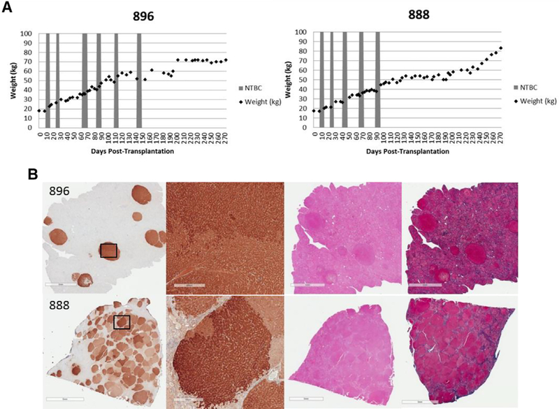
Fig. 6.

Weight gain and corresponding histology. (A) Weight stabilization of pigs 896 (single cell) and 888 (spheroid), demonstrating similar growth and NTBC-independence patterns. (B) Representative liver tissues stained for FAH from pigs 896 (top) and 888 (bottom ) at 9–10 months post-transplantation, showing near-complete liver repopulation with FAH-positive hepatocytes in pig 888. H&E-stained and Masson’s trichrome-stained serial liver sections are also presented. Scale bars, 5 mm (low magnification) and 500 pm (high magnification).
