Abstract
Hyperexcitable reflex blinks are a cardinal sign of Parkinson’s disease. We investigated the neural circuit through which a loss of dopamine in the substantia nigra pars compacta (SNc) leads to increased reflex blink excitability. Through its inhibitory inputs to the thalamus, the basal ganglia could modulate the brainstem reflex blink circuits via descending cortical projections. Alternatively, with its inhibitory input to the superior colliculus, the basal ganglia could regulate brainstem reflex blink circuits via tecto-reticular projections. Our study demonstrated that the basal ganglia utilizes its GABAergic input to the superior colliculus to modulate reflex blinks. In rats with previous unilateral 6-hydroxydopamine (6-OHDA) lesions of the dopamine neurons of the SNc, we found that microinjections of bicuculline, a GABA antagonist, into the superior colliculus of both alert and anesthetized rats eliminated the reflex blink hyperexcitability associated with dopamine depletion. In normal, alert rats, decreasing the basal ganglia output to the superior colliculus by injecting muscimol, a GABA agonist, into the substantia nigra pars reticulata (SNr) markedly reduced blink amplitude. Finally, brief trains of microstimulation to the superior colliculusreduced blink amplitude. Histological analysis revealed that effective muscimol microinjection and microstimulation sites in the superior colliculus overlapped the nigro-tectal projection from the basal ganglia. These data support models of Parkinsonian symtomatology that rely on changes in the inhibitory drive from basal ganglia output structures. Moreover, they support a model of Parkinsonian reflex blink hyperexcitability in which the SNr–SC target projection is critical.
Keywords: Parkinson’s disease, blink reflex, 6-hydroxydopamine, superior colliculus, rats
Experimental Parkinsonism in animal models, as well as Parkinson’s disease in humans, increases reflex blink excitability. For example, lesions of the ascending dopaminergic pathway with 6-hydroxydopamine (6-OHDA) dramatically increase excitability and reduce habituation of the blink reflex in rats (Shallert et al., 1989; Basso et al., 1993). Similar increases in reflex blink excitability and loss of habituation occur in humans with Parkinson’s disease (Pearce et al., 1968; Penders and Delwaide, 1971;Messina et al., 1972; Kimura, 1973; Esteban et al., 1981; Caliguiri et al., 1987; Masumoto et al., 1992).
The basal ganglia could modify reflex blink excitability through at least two routes. Changes in the activity of substantia nigra pars reticulata (SNr) and globus pallidus internal (GPi) neurons could alter thalamic input to the cerebral cortex and thereby modify descending cortical control of brainstem reflex blink circuits. Alternatively, changes in the activity of SNr neurons inhibiting the superior colliculus (SC) could change control of reflex blinks circuits via tecto-bulbar projections.
One reason to expect that the basal ganglia (BG) modulate reflex blink excitability through the nigro-collicular projection is the linkage of blinking and saccadic gaze shifts. In normal humans, a blink accompanies saccadic gaze shifts larger than 20°, and a reflex blink decreases the latency of saccadic gaze shifts (Evinger et al., 1994). In diseases with pathologically slow saccades, combining a voluntary blink with a saccade significantly increases maximum saccadic eye velocity (Leigh et al., 1983; Zee et al., 1983). Given the importance of the SC in controlling saccadic gaze shifts (Grantyn, 1988), it is reasonable that the SC could modulate reflex blinks by acting on circuits shared by saccadic gaze shifts and blinking.
In both normal and dopamine-depleted animals, it is possible to test both the importance of the nigro-collicular path in the BG modulation of blinking and the predictions of Parkinsonian models. Loss of dopamine-containing cells in the substantia nigra pars compacta (SNc) increases the activity of the monkey GPi neurons (Filion and Tremblay, 1991; Filion et al., 1991; DeLong and Wichman, 1993) and decreases the activity of rat globus pallidus external neurons (Pan and Walters, 1988). Therefore, it is likely that in both monkeys and rats, dopamine depletion leads to an increase in the inhibitory output of the BG. To test this hypothesis, blocking the expected increase in SC inhibition should reduce or eliminate reflex blink hyperexcitability in animals with experimental Parkinsonism. In normal animals, reducing the activity of SNr cells with the GABAergic agonist muscimol should also reduce reflex blinking. Similarly, inhibiting the SC in a dopamine intact animal shouldincrease reflex blink excitability, whereas activation of the SC should decrease reflex blink excitability. The present experiments confirm these predictions and strongly support the hypothesis that the level of inhibition from BG output structures modulates the excitability of brainstem reflex blink circuitry. Further, these data support a model of Parkinsonian reflex blink hyperexcitability mediated through the SNr–SC projection rather than via the pallido–thalamo–cortical loop.
MATERIALS AND METHODS
Subjects. Sprague Dawley rats weighing between 150 and 400 gm served as subjects. Animals were maintained on a 12 hr light/dark cycle and fed ad libitum. All procedures in this experiment strictly adhered to federal, state, and university guidelines concerning the use of animals in research.
Chronic preparation. Under general anesthesia (90 mg/kg ketamine and 10 mg/kg xylazine, i.m.) and using aseptic techniques, the supraorbital branch of the trigeminal nerve (SO) in five animals was exposed and encased in a Teflon nerve cuff. Electromyographic recordings of the lid-closing, orbicularis oculi muscle (OOemg) were made through a pair of Teflon-coated stainless steel wires (0.011 cm coated diameter) bared 1 mm at the tip and implanted into the medial and lateral margins of the OO muscle. All wires were led subcutaneously to a dental acrylic base on the top of the skull, which was secured by 3–4 3/16 inch self tapping stainless steel screws. For later microinjection of muscimol in the SC or SNr, four rats were implanted with guide tube cannulae placed 3 mm above the microinjection site. The guide tube was 26-gauge stainless steel tubing embedded in the dental acrylic base attached to the skull. Guide tubes were occluded before and after the microinjections with stylets. In one animal, which previously received a unilateral 6-OHDA lesion of the ascending mesotelencephalic dopamine system (Basso et al., 1993), a cannula was similarly implanted above the SC ipsilateral to the 6-OHDA lesion for subsequent microinjections of bicuculline.
Acute preparation. Initially, animals were injected with xylazine (10 mg/kg) intramuscularly, followed by urethane (1.2 gm/kg) intraperitoneally. After the skull was placed in a stereotaxic instrument, holes were drilled in the skull overlying the microstimulation or microinjection sites. A pair of silver ball electrodes was placed on the cornea to evoke blinks electrically, and Teflon-coated stainless steel wires, bared 1 mm at the tip, identical to those used in the chronic preparation, were implanted into the margins of the OO muscle to record the OOemg. In most animals, this procedure was performed bilaterally. If only one eye was prepared, it was the eye contralateral to the tested SC. Stimulation parameters for evoking a blink were determined by adjusting the intensity and duration of the electrical pulse to the cornea to evoke a consistent OOemg response for each animal. For all rats in this and the companion study [see Basso and Evinger (1996) in this issue], the corneal stimulus current intensities ranged from 0.1 to 3.0 mA and the stimulus durations ranged from 10 to 100 μS. These parameters remained constant for each animal throughout the duration of the testing session. Three animals in these acute experiments received 6-OHDA lesions at least 10 d before testing (Basso et al., 1993).
Experimental procedures. In both acute and chronic experiments, the general testing procedure was identical. Data were collected to establish baseline blink amplitude. In two chronic animals, the SNr was injected with muscimol. In two additional chronic animals and eight acute animals, the SC was injected with muscimol. In one chronic and three acute 6-OHDA-lesioned animals, the SC ipsilateral to the 6-OHDA lesion was injected with bicuculline. After drug microinjection, blinks were evoked with the same parameters as were used before the drug microinjections. The SC of five acutely prepared animals was microstimulated. In this procedure, blinks with and without preceding SC stimulation were alternately evoked. To avoid habituation, the time between blinks was 50 ± 5 sec for experiments on anesthetized rats and 25 ± 5 sec for experiments on alert animals.
Microstimulation. Glass microelectrodes (5–10 μm tip diameter) filled with 2 m sodium acetate saturated with fast green were used to microstimulate the SC in anesthetized animals. We delivered a 70 msec train of 80 μS duration cathodal pulses at 200 Hz. This train ended 5 msec before the onset of the corneal stimulus. Except where specifically stated, the maximum current intensity never exceeded 50 μA. At the end of all experiments, fast green was deposited at the microstimulation sites by passing 12–15 μA cathodal current through the electrode for 15 min (Thomas and Wilson, 1965).
Microinjections. A 30-gauge, 5 μl syringe mounted to a stereotaxic instrument was used to deliver the muscimol or bicuculline into the SC of anesthetized animals. Both muscimol and bicuculline were dissolved in 0.9% saline at concentrations of 0.01 or 1.0 μg/μl immediately before the microinjection. The injected volumes were between 0.2 and 1.0 μl. Vehicle microinjections of saline served as controls.
After collecting premicroinjection data from alert rats, the chronically prepared animals were lightly anesthetized with halothane and 1.0 μl containing either 0.01 or 0.1 μg of muscimol or bicuculline was injected through a 30-gauge cannula placed 3 mm below the previously implanted guide tube. Data collection began at least 5 min after the microinjection, when the rats had recovered fully from the halothane.
Reflex excitability paradigm. In addition to assessing reflex excitability by the changes in the amplitude of the evoked blink reflex, we quantified blink reflex excitability using the paired-stimulus paradigm. In this procedure, two identical blink-evoking stimuli were presented with interstimulus intervals between 100 and 800 msec. To avoid habituation, particularly in anesthetized rats, the time between stimulus pairs was 50 ± 5 sec.
Data acquisition and analysis. OOemg signals were acquired and stored on a computer (4000 Hz, 12-bit A/D resolution) and analyzed off-line using an interactive computer program that integrated rectified OOemg records and determined latencies of averaged OOemg records. For statistical analysis, the integrated OOemg amplitude measured in A/D units was normalized to the mean blink amplitude of all animals.
The corneal blink reflex amplitude before and after the muscimol microinjections from nine microinjections in eight anesthetized animals was analyzed with a Wilcoxon signed rank test. Amplitudes of the blink reflex were compared before and after the microinjections in each animal individually. Each animal contributed two points to each cell of the analysis. Each point was the mean of at least five blinks and normalized to the mean blink amplitude for all animals. Blink reflex amplitudes from the five anesthetized animals that received unilateral SC microstimulation and bilateral OOemg recording were analyzed with a 2 × 2 repeated-measures ANOVA that compared the left and right corneal blink reflex amplitude with and without SC microstimulation. Every animal contributed five blinks to each cell of the analysis. The OOemg data were normalized to the mean amplitude without microstimulation for all animals. The data acquired from three 6-OHDA-lesioned animals that received bicuculline microinjections into the SC were also normalized to the mean preinjection blink amplitude of all animals and were analyzed with a one-way ANOVA. This analysis compared the effect between conditions, before the microinjection, after the microinjection, and after recovery. Every cell contained three points, and each point was the mean of at least 10 blinks from each animal.
Histology. At the end of the experiments, animals were deeply anesthetized with ketamine (90 mg/kg, i.m.) and xylazine (10 mg/kg, i.m.) and perfused intracardially with a warm solution of 6.0% dextran in phosphate buffer (PB; pH 7.4), with cold 10% formalin in PB to localize fast green deposits, or with cold 4.0% paraformaldehyde in PB to localize tyrosine hydroxylase. The brains were post-fixed in fixative for 2 hr and stored in a 30% sucrose solution in PB. To localize fast green deposits, 100 μm sections were cut on a freezing microtome and counterstained with cresyl violet. The extent of the 6-OHDA lesion was assessed by processing the tissue immunohistochemically with an antibody against tyrosine hydroxylase (Eugene Tech). Forty micron sections cut on a freezing microtome were preincubated in 1.0% normal goat serum diluted in 0.3% Triton X-100 in PBS (PBSGT). The tissue was rinsed in PBS and incubated overnight at 4°C in primary antibody against tyrosine hydroxylase raised in rabbit and diluted 1:5000 in PBSGT. After incubation, the tissue was rinsed in PBS and then incubated in secondary antibody (dilution 1:200) for 2 hr. The avidin–biotin method using DAB was used for visualization.
RESULTS
Modifying the inhibitory SNr input to the SC
Previous studies suggest that dopamine depletion in the SNc results in increased SNr and GPi activity. Most Parkinsonian symptoms are explained by these changes and, in particular, by alterations in the pallidal–thalamo–cortical loop (for review, see DeLong and Wichmann, 1993). To establish that modifications of the blink reflex in Parkinsonism also result from changes in BG output nuclei and, in particular, from alterations in the nigro-collicular pathway, we demonstrated that blocking GABA receptors in the SC eliminated reflex blink hyperexcitability in rats with 6-OHDA lesions and showed that reducing SNr activity decreased reflex blink amplitude in normal rats.
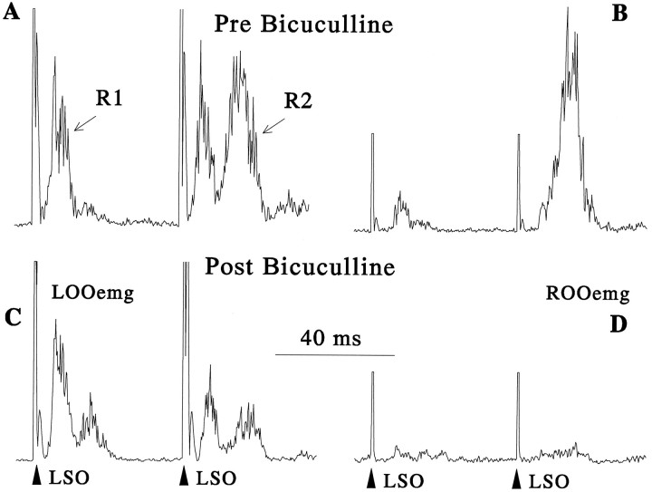
Stimulation of the supraorbital branch of the trigeminal nerve (SO) evoked two components of OOemg activity, R1 and R2, in normal (Evinger et al., 1993) and 6-OHDA-lesioned alert animals (Basso et al., 1993) (Fig. 1A). The first component, R1, arose at a mean latency of 5.12 msec after the stimulus, whereas the second component, R2, occurred at a mean latency of 15.93 msec (40 blinks from 4 animals). 6-OHDA lesions dramatically augmented the excitability of the SO-evoked blinks (Basso et al., 1993). First, the paired stimulus paradigm with a 50 msec interstimulus interval produced an increase in the amplitude of the R2 component evoked by the second stimulus (Fig. 1A) instead of the usual 50–80% decrease in R2 amplitude exhibited by unlesioned rats (Basso et al., 1993). Second, 6-OHDA lesions enhanced consensual reflex blinks. For instance, stimulating the left SO evoked a strong, consensual response in the right OOemg of 6-OHDA-lesioned rats (Fig.1B) that frequently was not present in unlesioned rats. A large (1.0 μg) bicuculline microinjection into the right SC of one chronically prepared animal eliminated reflex blink excitability caused by the 6-OHDA lesion, returning blink reflex excitability responses to near normal values in the paired stimulus paradigm (Fig.1C) and virtually abolishing the consensual response (Fig.1D).
Fig. 1.
Elimination of blink reflex hyperexcitability by a microinjection of bicuculline into the superior colliculus of an alert rat with a 6-OHDA lesion of the right ascending mesotelencephalic dopamine system. Each trace is the average of 10 rectified OOemg responses to a pair of stimuli to the left supraorbital branch of the trigeminal nerve (LSO) separated by 50 msec. A, C, The left records are from the left lid OOemg (LOOemg), the direct response, and the (B, D)right records are consensual, right lid OOemg (ROOemg) responses. A, B, The top records are before microinjection of bicuculline (Pre Bicuculline). C, D, The bottom records are after a microinjection of 1 μg of bicuculline into the right superior colliculus.LOOemg, Left orbicularis oculi response;LSO, left supraorbital nerve stimulation;R1, short-latency response; R2, long-latency response; ROOemg, right orbicularis oculi emg.
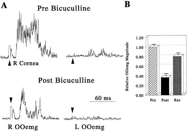
Bicuculline microinjections into the superior colliculus of three anesthetized, 6-OHDA-lesioned rats also eliminated blink reflex excitability. Electrical stimulation of the cornea in urethane-anesthetized rats evoked a single component of OOemg activity in normal (Evinger et al., 1993) and 6-OHDA-lesioned rats (Fig.2). In unlesioned rats, corneal stimulation only evoked an ipsilateral response with a mean latency of 15 msec. Consistent with the increased reflex blink excitability generated by dopamine depletion in alert rats (Basso et al., 1993) (Fig. 1), anesthetized 6-OHDA-lesioned rats exhibited reflex blink hyperexcitability evidenced by the appearance of a consensual corneal response (Fig.2A, top right trace). A bicuculline microinjection (0.5 μg) into the SC ipsilateral to the lesioned SNc eliminated the consensual blink response (Fig. 2A,bottom right trace) and significantly reduced the amplitude of the ipsilateral corneally evoked reflex blink [Fig.2A (bottom left trace), 2B; ANOVA F(3,5) = 5.43, p < 0.05]. These bicuculline microinjections into the SC also precipitated contralateral whisker movement, tail pointing ipsilateral to the microinjection site, and ipsilateral hindlimb flexion, i.e., behaviors consistent with orienting. After ∼2 hr, the ipsilateral reflex blink returned to its normal amplitude (Fig.2B, Rec).
Fig. 2.
Elimination of blink reflex hyperexcitability by a microinjection of bicuculline into the superior colliculus of a urethane-anesthetized rat with a 6-OHDA lesion of the ascending mesotelencephalic dopamine system. A, Each trace is the average of 10 rectified OOemg responses to stimulation of the right cornea (R Cornea). Left traces show the right OOemg (R OOemg), the direct response, and theright traces display the left OOemg (L OOemg), the consensual response, not typically seen in normal rats. The top traces illustrate the direct and consensual response before microinjection of bicuculline (Pre Bicuculline), and the bottom traces show the same responses after microinjection of 0.5 μg of bicuculline into the superior colliculus (Post Bicuculline).B, Pooled data from three anesthetized rats with 6-OHDA lesions showing the corneally evoked OOemg amplitude before (Pre), after (Post), and at least 2 hr after (Rec) microinjections of bicuculline into the superior colliculus ipsilateral to the 6-OHDA lesion. All integrated OOemg data were normalized to the mean preinjection OOemg amplitude for all animals, and error bars are 1 SEM.
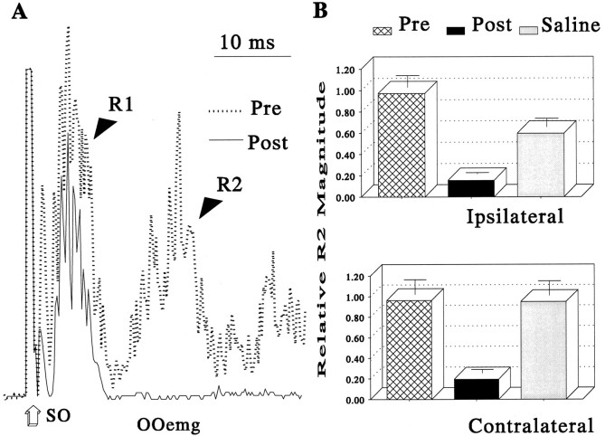
To demonstrate that changes in SNr activity could modify blink amplitude in normal animals, we microinjected 0.5 μg of muscimol into the SNr of two normal, alert rats. This procedure reduced the R2 component of the blink reflex ipsilateral and contralateral to the injected SNr (Fig. 3; ipsilateral 0.16 ± 0.02, contralateral 0.20 ± 0.04). Saline microinjections did not significantly alter SO-evoked blink amplitude (Fig. 3B). Thus, decreasing SNr inhibition of the SC by blocking GABA receptors in the SC (Figs. 1, 2) or decreasing SNr activity with muscimol (Fig. 3) reduced reflex blink amplitude in 6-OHDA-lesioned and normal animals. These observations revealed that changes in the nigro-collicular pathway are sufficient for the basal ganglia to modulate reflex blinks. The observations also suggested that decreases in SC activity boost reflex blink excitability whereas increases in SC activity reduce reflex blink amplitude. The next set of experiments evaluated collicular modulation of reflex blinks.
Fig. 3.
Reduction of substantia nigra pars reticulata activity by microinjection of muscimol reduced R2 OOemg amplitude in alert rats. A, Each trace is the average of 15 rectified, direct OOemg responses to stimulation of the supraorbital branch of the trigeminal nerve (SO) before (dotted line) and after (solid line) a 0.5 μl microinjection of 1.0% muscimol into the SNr.B, R2 OOemg amplitude relative to premicroinjection OOemg amplitude before (Pre) and after (Post) microinjections of muscimol and after a control microinjection of saline (Saline) from two alert rats.Top graph shows data from direct OOemg activity ipsilateral to the SNr microinjection, and bottom graphdisplays direct OOemg activity contralateral to the SNr microinjection. Error bars are 1 SEM.
Muscimol microinjections into the SC
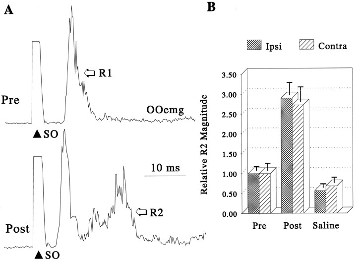
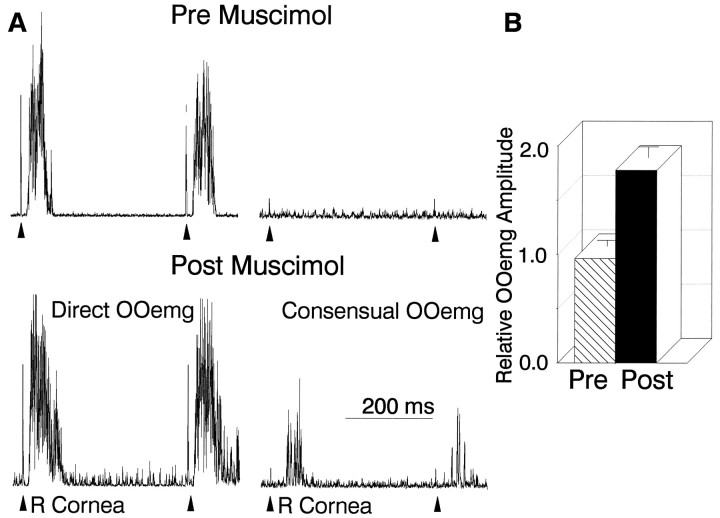
As suggested by the effects of modulating SNr activity, muscimol inactivation of the SC increased the amplitude of reflex blinks bilaterally. In two alert rats, muscimol microinjections only enhanced the amplitude of the long-latency, R2 component of reflex blinks (Fig. 4) but shortened the latency of both components. Microinjections of saline vehicle failed to alter R2 amplitude significantly (Fig. 4B). Similarly, microinjections as small as 0.5 μl of 0.01 μg of muscimol, a GABA agonist, into the SC of eight urethane-anesthetized rats markedly increased the amplitude (Wilcoxon T+ = 7, n = 9, p = 0.03; Fig. 5B) and decreased the latency of corneally evoked blinks. The increase in the amplitude of reflex blinks after unilateral SC microinjection of muscimol was also seen in response to stimulation of either cornea in acute rat preparations. Microinjection of muscimol into the SC also turned the normally unilateral corneally evoked blink into a bilateral response (Fig. 5A, Consensual OOemg). Similarly, increases in reflex blink excitability, evidenced by a reduction in suppression in the paired-stimulus paradigm, were evident in anesthetized rats after muscimol microinjection in the SC (Fig.5A). Injecting muscimol into the SC of anesthetized rats mimicked the effect of 6-OHDA lesions on reflex blinking and, therefore, mimicked the behavioral pathology of Parkinsonism on trigeminally evoked reflex blinks.
Fig. 4.
Microinjection of muscimol into the superior colliculus increases R2 blink component amplitude in alert rats.A, Each record is the average of 15 rectified, direct OOemg responses to stimulation of the supraorbital branch of the trigeminal nerve (SO) before (top trace, Pre) and after (bottom trace, Post) a 0.1 μg muscimol microinjection into the superior colliculus contralateral to the SO stimulus. B, Relative R2 OOemg amplitude evoked by SO stimuli presented ipsilateral (hatched bars) or contralateral (striped bars) to the injected superior colliculus. The R2 component increased bilaterally (Post) relative to stimuli delivered before the microinjection (Pre) or after control microinjections of saline (Saline). All integrated OOemg data were normalized to the mean preinjection OOemg amplitude, and error bars are 1 SEM.
Fig. 5.
Microinjection of muscimol into the superior colliculus of urethane-anesthetized rats mimics the effects of 6-OHDA lesions. A, Each record is the average of 10 rectified OOemg responses to two stimuli delivered to the right cornea (R Cornea) with an interstimulus interval of 400 msec. Theleft traces are from the right OOemg (Direct OOemg), and right traces are from the left OOemg (Consensual OOemg). Top traces are direct and consensual blinks evoked before muscimol microinjection (Pre Muscimol), and the bottom traces are blinks evoked after a 0.01 μg microinjection of muscimol into the left superior colliculus (Post Muscimol). B, Relative direct OOemg amplitude evoked by SO stimulation before (Pre, striped bars) and after (Post, solid bars) muscimol microinjection into the SC contralateral to the corneal stimulus. The bars are means of nine microinjections normalized to the mean preinjection OOemg amplitude of all animals, and the error bars are 1 SEM.
Microstimulation in the acute preparation
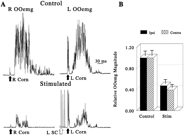
The increase in reflex blink amplitude and excitability after a reduction in SC activity suggested that increasing SC activity wouldreduce the amplitude of reflex blinks. Presenting a 70 msec train of stimuli before the corneal stimulus dramatically suppressed the amplitude of the subsequent corneally evoked reflex blinks bilaterally in anesthetized rats (Fig. 6; ANOVAF(1,60) = 392.34, p < 0.0001). Regardless of which cornea was used to elicit blinks, the amount of suppression caused by unilateral SC microstimulation did not differ significantly between the two sides (Fig. 6B, ANOVAF(1,60) = 1.63, p not significant; Fig. 6B).
Fig. 6.
Microstimulation of the superior colliculus suppresses cornea reflex blinks in urethane-anesthetized rats.A, Each record is the average of 10 rectified OOemg responses to stimulation of the right (R Corn) or left (L Corn) cornea. Right traces are from the left OOemg (L OOemg), and left tracesare from the right lid OOemg (R OOemg). Top traces show normal, control responses, and bottom traces illustrate responses preceded by a 70 msec train of microstimulation to the left superior colliculus (L SC) that terminated 5 msec before the corneal stimulation.B, Reduction in the amplitude of the direct OOemg response caused by microstimulation of the superior colliculus ipsilateral (Ipsi, solid bars) or contralateral (Contra, hatched bars) to the stimulated cornea. All integrated OOemg data were normalized to the mean preinjection OOemg amplitude for the group, and error bars are 1 SEM.
Superior colliculus microstimulation and microinjection sites
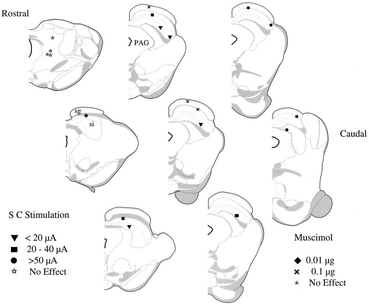
The effective sites for microinjection of muscimol and microstimulation were restricted to specific SC locations (Fig.7). Muscimol-induced increases in blink reflex excitability occurred with concentrations as low as 0.01 μg when the microinjection was in the deep layers of the rostral SC. Microinjections at more caudal sites in the SC had smaller effects or required higher concentrations of muscimol to produce effects equivalent to those obtained from microinjections into the rostral SC. Figure 7 also shows the locations and the required stimulus intensities that produced a 50% reduction in reflex blink amplitude. The sites with the lowest stimulus intensity were in the deep layers of the SC and tended to be lateral and rostral. Two microinjection sites and two microstimulation sites that were outside of the SC had no effect on reflex blinking. Consistent with the modulation of reflex blinking via the nigro-collicular pathway, the effective microstimulation and microinjection sites coincided with nigro-collicular termination zones (Bickford and Hall, 1992; May and Hall, 1993; May et al., 1993).
Fig. 7.
Reconstruction of the sites of muscimol microinjections and microstimulation sites in anesthetized rats. Microinjection and microstimulation sites from all animals mapped on schematic representation of coronal brain sections spaced 200 μm apart (Swanson, 1992). Filled diamonds are sites where 0.01 μg of muscimol increased blink amplitude; × are sites where 0.1 μg of muscimol increased blink amplitude. Inverted filled triangles show sites where currents <20 μA caused a 50% reduction in blink amplitude. Filled squares show sites where currents between 20 and 40 μA caused a 50% reduction in blink amplitude. Filled circles show sites requiring currents > 50 μA to cause a 50% reduction in blink amplitude.Filled stars are sites where 0.1 μg of muscimol did not alter reflex blinks. Open stars show sites where currents > 100 μA failed to alter reflex blinks.PAG, Periaqueductal gray; sg, stratum griseum superficiale; si, stratum griseum intermediale.
DISCUSSION
It is clear from experimental (Shallert et al., 1989; Basso et al., 1993) as well as clinical (Pearce et al., 1968; Penders and Delwaide, 1971; Messina et al., 1972; Kimura, 1973; Esteban et al., 1981; Caliguiri et al., 1987; Masumoto et al., 1992) studies that dopamine depletion in the basal ganglia increases reflex blink excitability. In humans and alert rats, this excitability increase occurs primarily in the long-latency R2 component of SO-evoked reflex blinks. As with other reflexes in Parkinsonian patients, the short-latency R1 component shows only modest increases in excitability (Kimura, 1973). This enhanced excitability of long-latency reflexes develops because of the BG’s increased inhibition of its target structures (Albin et al., 1989; DeLong, 1990; DeLong and Wichmann, 1993). The present study demonstrates that the increased excitability of reflex blinks seen in the rat model of Parkinson’s disease (Basso et al., 1993) also results from increased inhibition by BG output structures. In this respect, our results support current models of Parkinsonian symptomatology. Moreover, our results extend these models of Parkinsonism to include a critical role for the SNr–SC inhibitory projection in the expression of reflex blink hyperexcitability.
Three lines of evidence support the role of the superior colliculus in Parkinsonian reflex blink hyperexcitability. First, blocking the excessive GABAergic inhibition of the superior colliculus of 6-OHDA-lesioned animals with a GABA antagonist, bicuculline, virtually eliminated reflex blink hyperexcitability. The GABA blockade of the superior colliculus exerted its strongest effect on the long-latency R2 component of the blink, the component most profoundly altered in experimental animals with dopamine depletion (Basso et al., 1993) and in humans with Parkinson’s disease (Pearce et al., 1968; Penders and Delwaide, 1971; Messina et al., 1972; Kimura, 1973; Esteban et al., 1981; Caliguiri et al., 1987; Masumoto et al., 1992). Second, reducing the inhibitory output of the nigro-collicular pathway by microinjecting the GABA agonist muscimol into the SNr decreased reflex blink amplitude of the long-latency R2 component in normal rats. Third, simulating the increased activity of the inhibitory nigro-collicular pathway associated with 6-OHDA lesions by injecting muscimol into the superior colliculus caused normal rats to exhibit reflex blink hyperexcitability normally associated with dopamine depletion. The sites where muscimol microinjections effectively increased reflex blink excitability were restricted to the superior colliculus and overlapped the termination zones of the nigro-collicular input (May and Hall, 1984, 1986; Bickford and Hall, 1992; May et al., 1993). Because the SC microinjection procedure did not affect basal ganglia inhibitory inputs to thalamus, the nigro-collicular pathway was sufficient to control trigeminal reflex blink excitability. Moreover, because cortical or thalamic damage in humans decreased or did not alter reflex blink amplitude and excitability (Ongerboer de Visser and Moffie, 1979; Ongerboer de Visser, 1981; Berardelli et al., 1983), the increased inhibition of the pallido-thalamic pathway in Parkinson’s disease failed to explain increased reflex blink excitability. In addition to demonstrating the critical role of the superior colliculus in basal ganglia modulation in Parkinsonian reflex blink hyperexcitability, the current study revealed a mechanism through which the basal ganglia can modulate reflex blinks.
Basal ganglia disorders can both increase and decrease reflex blink excitability. For instance, patients suffering from Huntington’s disease have a normal level of striatal dopamine but, because of striatal cell loss, have a decreased inhibitory drive from the GPi and, presumably, SNr to target structures (Albin et al., 1990). This reduction in nigro-collicular inhibition should increase collicular activity. As predicted from the superior colliculus microstimulation experiment and the SNr muscimol experiment, patients with Huntington’s disease typically exhibit hypoexcitable reflex blinks (Esteban and Giminez-Roldan, 1975; Caraceni et al., 1976). Thus, increasing inhibition of the superior colliculus enhances reflex blink excitability, whereas decreasing inhibition of the superior colliculus reduces reflex blink excitability. The simplest explanation for these data is that the BG inhibit a neural pathway, mediated through the superior colliculus, that tonically inhibits the trigeminal reflex blink circuit.
The well established role of the superior colliculus in orienting behaviors such as saccadic gaze shifts (for review, see Grantyn, 1988) suggests that collicular modulation of reflex blinking relates to the interactions between blinking and saccadic gaze shifts (Evinger et al., 1994). It is probably more advantageous, however, to view SC modulation as part of the more general relationship between orienting and defensive behaviors. Microstimulation of the rodent SC activates both orienting movements and defensive movements (Sahibzada et al., 1986;Dean et al., 1988a,b, 1989; Northmore et al., 1988; Westby et al., 1990). Microstimulation of the medial SC evokes ipsilateral cringing behavior, whereas microstimulation of the lateral SC elicits contralateral orienting. Consistent with the medial SC organizing defensive behaviors, neurons in the rostral, medial SC respond to both nociceptive and non-nociceptive cutaneous stimulation (McHaffie et al., 1989) and project to the intralaminar nucleus of the thalamus (Yamasaki and Krauthamer, 1990). Neurons in the lateral SC are active with rapid head (Guitton et al., 1980), eye (Wurtz and Goldberg, 1972; Stein et al., 1976), and pinna (Stein and Clamann, 1981) orienting movements. McHaffie et al. (1989) proposed a circuit in which the activation of the medial SC inhibited neurons in the lateral SC by activating the intralaminar nucleus of the thalamus, which led to an increase in SNr inhibition of lateral SC. Suppression of the lateral SC would permit a withdrawal response to occur unopposed by a competing orienting response. The present data are consistent with this prediction of the model because inhibiting lateral SC inhibition dramatically enhances reflex blinks, an important element in a defensive withdrawal response. Although we did not quantify sites in the medial SC, we never evoked blinks from microstimulation of more medial sites. Rather, our qualitative impression was that the microstimulation became less effective in suppressing blinks as we moved more medially. In this regard, our data are not consistent with the model of McHaffie et al. (1989). Our observation suggests that SC effects on reflex blinking share elements with the orienting system, rather than being associated with defensive behaviors. This hypothesis is consistent with very recent findings that tonically active neurons in the lateralSC of the rat respond to both nociceptive and wide dynamic range cutaneous stimuli (Redgrave et al., 1996).
Activation of the lateral SC suppresses reflex blinks. One possibility is that the BG influence brainstem reflex blink circuits by altering the amount of inhibition on these cells in the lateral SC output neurons with cutaneous response profiles. The cells with tonic activity could continuously regulate the level of excitability of brainstem structures involved in reflex blinking. In Parkinson’s disease, the increased inhibition of these cells leads to an increase in reflex excitability through a tonic disinhibition of trigeminal reflex blink circuits. Another possibility is that dopamine depletion in the basal ganglia alters the phasic response of lateral SC neurons to cutaneous stimulation. For example, these neurons may respond phasically to cutaneous stimulation of the face, which would modulate a brainstem inhibitory interneuron to ensure an appropriate amplitude blink given the stimulus parameters. The level of BG inhibition of SC output could regulate the excitability of reflex blinks during different movement programs and perhaps regulate the sensory input the movements themselves create. Thus, the hyperexcitable reflexes of Parkinson’s disease may reflect a motor system that inadequately regulates reflexes with other types of movements.
Footnotes
This work was supported by National Eye Institute Grant EY07391 (C.E.) and summer fellowships from the Parkinson’s Disease Foundation (M.A.B.). We thank Donna Schmidt for her expert technical assistance.
Correspondence should be addressed to Craig Evinger, Department of Neurobiology and Behavior, SUNY Stony Brook, Stony Brook, NY 11794-5230.
Dr. Basso’s current address: Laboratory of Sensorimotor Research, National Eye Institute, Building 49, Room 2A50, Bethesda, MD 20892-4435.
REFERENCES
- 1.Albin RL, Young AB, Penney JB. The functional anatomy of basal ganglia disorders. Trends Neurosci. 1989;12:366–375. doi: 10.1016/0166-2236(89)90074-x. [DOI] [PubMed] [Google Scholar]
- 2.Basso MA, Evinger C. An explanation for blink reflex hyperexcitability in Parkinson’s disease. II. Nucleus raphe magnus. J Neurosci. 1996;16:7318–7330. doi: 10.1523/JNEUROSCI.16-22-07318.1996. [DOI] [PMC free article] [PubMed] [Google Scholar]
- 3.Basso MA, Strecker RE, Evinger C. Midbrain 6-hydroxydopamine lesions modulate blink reflex excitability. Exp Brain Res. 1993;94:88–96. doi: 10.1007/BF00230472. [DOI] [PubMed] [Google Scholar]
- 4.Berardelli A, Accornero N, Cruccu G, Fabiano F, Guerrisi V, Manfredi M. The orbicularis oculi response after hemispheral damage. J Neurol Neurosurg Psychiatry. 1983;46:837–843. doi: 10.1136/jnnp.46.9.837. [DOI] [PMC free article] [PubMed] [Google Scholar]
- 5.Bickford ME, Hall WC. The nigral projection to predorsal bundle cells in the superior colliculus of the rat. J Comp Neurol. 1992;319:11–33. doi: 10.1002/cne.903190105. [DOI] [PubMed] [Google Scholar]
- 6.Caliguiri MP, Abbs JH. Response properties of the perioral reflex in Parkinson’s disease. Exp Neurol. 1987;98:563–572. doi: 10.1016/0014-4886(87)90265-2. [DOI] [PubMed] [Google Scholar]
- 7.Caraceni T, Avanzini G, Spreafico R, Negri S, Broggi G, Girotti F. Study of the excitability cycle of the blink reflex in Huntington’s in Parkinsonian signs. Clin Neurosci. 1976;1:18–26. doi: 10.1159/000114774. [DOI] [PubMed] [Google Scholar]
- 8.Dean P, Mitchell IJ, Redgrave P. Contralateral head movements produced by microinjection of glutamate into superior colliculus of rats: evidence for mediation by multiple output pathways. Neuroscience. 1988a;24:491–500. doi: 10.1016/0306-4522(88)90344-2. [DOI] [PubMed] [Google Scholar]
- 9.Dean P, Mitchell IJ, Redgrave P. Responses resembling avoidance behaviour produced by microinjection of glutamate into superior colliculus of rats. Neuroscience. 1988b;24:501–510. doi: 10.1016/0306-4522(88)90345-4. [DOI] [PubMed] [Google Scholar]
- 10.DeLong MR. Primate models of movement disorders of basal ganglia origin. Trends Neurosci. 1990;13:281–285. doi: 10.1016/0166-2236(90)90110-v. [DOI] [PubMed] [Google Scholar]
- 11.DeLong MR, Wichmann T. Basal ganglia-thalamocortical circuits in Parkinsonian signs. Clin Neurosci. 1993;1:18–26. [Google Scholar]
- 12.Esteban A, Mateao D, Gimenez-Roldan S. Early detection of Huntington’s disease: blink reflex and levodopa load in presymptomatic and incipient subjects. J Neurol Neurosurg Psychiatry. 1981;44:43–48. doi: 10.1136/jnnp.44.1.43. [DOI] [PMC free article] [PubMed] [Google Scholar]
- 13.Evinger C, Basso MA, Manning KA, Sibony PA, Pellegrini JJ, Horn AKE. A role for the basal ganglia in nicotinic modulation of the blink reflex. Exp Brain Res. 1993;92:507–515. doi: 10.1007/BF00229040. [DOI] [PubMed] [Google Scholar]
- 14.Evinger C, Manning KA, Pellegrini JJ, Basso MA, Powers AS, Sibony PA. Not looking while leaping: the linkage of blinking and saccadic gaze shifts. Exp Brain Res. 1994;100:337–344. doi: 10.1007/BF00227203. [DOI] [PubMed] [Google Scholar]
- 15.Filion M, Tremblay L. Abnormal spontaneous activity of globus pallidus neurons in monkeys with MPTP induced Parkinsonism. Brain Res. 1991;547:142–151. [PubMed] [Google Scholar]
- 16.Filion M, Tremblay L, BÄdard PJ. Abnormal influences of passive limb movement on the activity of globus pallidus neurons in Parkinsonian monkeys. Brain Res. 1988;444:165–176. doi: 10.1016/0006-8993(88)90924-9. [DOI] [PubMed] [Google Scholar]
- 17.Filion M, Tremblay L, BÄdard PJ. Effects of dopamine agonists on spontaneous activity of globus pallidus neurons in monkeys with MPTP-induced Parkinsonism. Brain Res. 1991;547:152–161. [PubMed] [Google Scholar]
- 18.Grantyn R. Gaze control through the superior colliculus. In: Büttner-Ennever JA, editor. Neuroanatomy of the oculomotor system. Elsevier; Amsterdam: 1988. pp. 273–333. [PubMed] [Google Scholar]
- 19.Guitton D, Crommelinck M, Roucoux A. Stimulation of the superior colliculus in the alter cat. I. Eye movements and neck EMG activity evoked when the head is restrained. Exp Brain Res. 1980;39:63–73. doi: 10.1007/BF00237070. [DOI] [PubMed] [Google Scholar]
- 20.Kimura J. Disorders of interneurons in Parkinson’s disease. Brain. 1973a;96:87–96. doi: 10.1093/brain/96.1.87. [DOI] [PubMed] [Google Scholar]
- 21.Leigh RJ, Newman SA, Folstein SE, Lasher AG, Jensen BA. Abnormal ocular motor control in Huntington’s disease. Exp Neurol. 1983;33:1268–1275. doi: 10.1212/wnl.33.10.1268. [DOI] [PubMed] [Google Scholar]
- 22.Matsumoto H, Noro H, Kaneshige T, Chiba S, Miyano N, Motoi Y, Yanada Y. A correlation study between blink reflex habituation and clinical state in patients with Parkinson’s disease. J Neurol Sci. 1992;107:155–159. doi: 10.1016/0022-510x(92)90283-q. [DOI] [PubMed] [Google Scholar]
- 23.May PJ, Hall WC. Relationship between the nigrotectal pathway and the cells of origin of the predorsal bundle. J Comp Neurol. 1984;226:357–376. doi: 10.1002/cne.902260306. [DOI] [PubMed] [Google Scholar]
- 24.May PJ, Hall WC. The sources of the nigrotectal pathway. Neuroscience. 1986;19:159–180. doi: 10.1016/0306-4522(86)90013-8. [DOI] [PubMed] [Google Scholar]
- 25.May PJ, Hall WC, Porter JD, Sakai ST. The comparative anatomy of nigral and cerebellar control over tectally initiated orienting movements. In: Mano N, Hamada I, DeLong MR, editors. Role of the cerebellum and basal ganglia in voluntary movement. Elsevier; Amsterdam: 1993. pp. 221–231. [Google Scholar]
- 26.McHaffie JG, Stein BE. Eye movements evoked by electrical stimulation of the superior colliculus of rats and hamsters. Brain Res. 1982;247:243–253. doi: 10.1016/0006-8993(82)91249-5. [DOI] [PubMed] [Google Scholar]
- 27.Messina C, DiRosa AE, Tomasello F. Habituation of blink reflexes in Parkinson’s patients under levodopa and amantadine treatment. J Neurol Sci. 1972;17:141–148. doi: 10.1016/0022-510x(72)90136-0. [DOI] [PubMed] [Google Scholar]
- 28.Northmore DPM, Levine ES, Schneider GE. Behavior evoked by electrical stimulation of the hamster superior colliculus. Exp Brain Res. 1988;73:595–605. doi: 10.1007/BF00406619. [DOI] [PubMed] [Google Scholar]
- 29.Ongerboer de Visser BW. Corneal reflex latency in lesions of the lower postcentral region. Neurol. 1981;31:701–707. doi: 10.1212/wnl.31.6.701. [DOI] [PubMed] [Google Scholar]
- 30.Ongerboer de Visser BW, Moffie D. Effects of brain-stem and thalamic lesions on the corneal reflex: an electrophysiological and anatomical study. Brain. 1979;102:595–608. doi: 10.1093/brain/102.3.595. [DOI] [PubMed] [Google Scholar]
- 31.Pan HS, Walters JR. Unilateral lesion of the nigrostriatal pathway decreases the firing rate and alters the firing pattern of globus pallidus neurons in the rat. Synapse. 1988;2:650–656. doi: 10.1002/syn.890020612. [DOI] [PubMed] [Google Scholar]
- 32.Pearce J, Aziz H, Gallagher JC. Primitive reflex activity in primary and symptomatic Parkinsonism. J Neurol Neurosurg Psychiatry. 1968;31:501–508. doi: 10.1136/jnnp.31.5.501. [DOI] [PMC free article] [PubMed] [Google Scholar]
- 33.Penders CA, Delawaide PJ. Blink reflex studies in patients with Parkinsonism before and during therapy. J Neurol Neurosurg Psychiatry. 1971;34:674–678. doi: 10.1136/jnnp.34.6.674. [DOI] [PMC free article] [PubMed] [Google Scholar]
- 34.Redgrave P, McHaffie JG, Stein BE. Nocioceptive neurons in the rat superior colliculus. I. Antidromic activation from the contralateral predorsal bundle. Exp Brain Res. 1996;109:185–196. doi: 10.1007/BF00231780. [DOI] [PubMed] [Google Scholar]
- 35.Sahibzada N, Dean P, Redgrave P. Movements resembling orientation or avoidance elicited by electrical stimulation of the superior colliculus in rats. J Neurosci. 1986;6:723–733. doi: 10.1523/JNEUROSCI.06-03-00723.1986. [DOI] [PMC free article] [PubMed] [Google Scholar]
- 36.Shallert T, Petrie BF, Whishaw IQ. Neonatal dopamine depletion: spared and unspared sensorimotor and attentional disorders and effects of further depletion in adulthood. J Psychobiol. 1989;17:386–396. [Google Scholar]
- 37.Stein BE, Clamann HP. Control of pinna movements and eye movements are in sensorimotor register in cat superior colliculus. Brain Behav Evol. 1981;19:180–192. doi: 10.1159/000121641. [DOI] [PubMed] [Google Scholar]
- 38.Stein BE, Magalhïes B, Kriger L. Relationship between visual and tactile representation in cat superior colliculus. J Neurophysiol. 1976;30:401–419. doi: 10.1152/jn.1976.39.2.401. [DOI] [PubMed] [Google Scholar]
- 39.Swanson LW. Brain maps: structure of the rat brain. Elsevier; Amsterdam: 1992. [Google Scholar]
- 40.Thomas RC, Wilson VJ. Precise localization of Renshaw cells with a new marking technique. Nature. 1965;206:211–213. doi: 10.1038/206211b0. [DOI] [PubMed] [Google Scholar]
- 41.Westby GWM, Keay KA, Redgrave P, Dean P, Bannister M. Output pathways from the rat superior colliculus mediating approach and avoidance have different sensory properties. Exp Brain Res. 1990;81:626–638. doi: 10.1007/BF02423513. [DOI] [PubMed] [Google Scholar]
- 42.Yamasaki DSG, Krauthamer GM. Somatosensory neurons projecting from the superior colliculus to the intralaminar thalamus in the rat. Brain Res. 1990;523:188–194. doi: 10.1016/0006-8993(90)91486-z. [DOI] [PubMed] [Google Scholar]
- 43.Zee DS, Chu FC, Leigh RJ, Savino PJ, Schatz NJ, Reingold DB, Cogan D. Blink-saccade synkinesis. Neurol. 1983;33:12330–12336. doi: 10.1212/wnl.33.9.1233. [DOI] [PubMed] [Google Scholar]


