Abstract
The physiological and pharmacological properties of the α1E calcium (Ca) channel subtype do not exactly match any of the established categories described for native neuronal Ca currents. Many of the key diagnostic features used to assign cloned Ca channels to their native counterparts, however, are dependent on a number of factors, including cellular environment, β subunit coexpression, and modulation by second messengers and G-proteins. Here, by examining the intrinsic pore characteristics of a family of transiently expressed neuronal Ca channels, we demonstrate that the permeation properties of α1E closely resemble those described for a subset of low-threshold Ca channels. The α1A (P-/Q-type), α1B(N-type), and α1C (L-type) high-threshold Ca channels all exhibit larger whole-cell currents with barium (Ba) as the charge carrier as compared with Ca or strontium (Sr). In contrast, macroscopic α1E currents are largest in Sr, followed by Ca and then Ba. The unique permeation properties of α1E are maintained at the single-channel level, are independent of the nature of the expression system, and are not affected by coexpression of α2 and β subunits. Overall, the permeation characteristics of α1E are distinct from those described for R-type currents and share some similarities with native low-threshold Ca channels.
Keywords: calcium channel, permeation, barium, strontium, transient expression, conductance, pore properties
Calcium (Ca) influx through voltage-dependent Ca channels mediates a wide range of neurophysiological functions, including gene expression, neurotransmitter release, and firing patterns (Tsien et al., 1988). On the basis of their electrophysiological and pharmacological properties, Ca channels have been classified into T-, L-, N-, P-, and Q-types (for review, see Stea et al., 1995b). T-type channels transiently activate at relatively negative membrane potentials, whereas the other channel types first activate at more positive potentials and display diverse kinetic characteristics. The high threshold channels can also be distinguished pharmacologically: L-type channels are sensitive to dihydropyridine agonists and antagonists, N-type channels are blocked by ω-conotoxin GVIA, and P-type channels are sensitive to nanomolar concentrations of ω-agatoxin IVA. Q-type channels are sensitive to both ω-conotoxin MVII-C and ω-agatoxin IVA and may be related structurally to P-type channels (Stea et al., 1994; Dunlap et al., 1995). To date, there are no known specific blockers of native low voltage-activated channels.
In addition to their differential kinetic and pharmacological profiles, the major types of Ca channels display distinct permeation characteristics (Bean, 1985; Nilius et al., 1986; Carbone and Lux, 1987; Fox et al., 1987; Akaike et al., 1989; Takahashi et al., 1991). Of particular note, the relative permeabilities for Ca, barium (Ba), and strontium (Sr) are dependent on the Ca channel subtype. In general, Ba permeates high voltage-activated Ca channels more effectively than Ca does, whereas low voltage-activated (T-type) Ca channels are permeated by Ca as well as or better than by Ba (Hille, 1992). Most neurons express multiple subtypes of Ca channels, however, making it difficult to determine the exact permeation properties of individual subtypes of neuronal Ca channels. Furthermore, to date, there has been no side-by-side comparison of the permeation characteristics of the major Ca channel subtypes under identical experimental conditions.
The expression of cloned Ca channels in exogenous systems has allowed the study of individual Ca channel subtypes in isolation. Five distinct Ca channel α1 subunit genes are expressed in the mammalian CNS (α1A, α1B, α1C, α1D, and α1E). Functional expression and immunoprecipitation studies have demonstrated that α1C and α1D encode dihydropyridine-sensitive L-type channels (Williams et al., 1992a; Hell et al., 1993; Tomlinson et al., 1993), whereas α1B encodes an ω-conotoxin-sensitive N-type channel (Dubel et al., 1992; Williams et al., 1992b; Fujita et al., 1993; Stea et al., 1993). The α1A channel displays properties similar to P- and Q-type currents (Mori et al., 1991, Sather et al., 1993; Stea et al., 1994), whereas α1E does not fit exactly into any of the Ca channel subtypes described in native cells. Although α1E channels differ from T-type channels expressed in endocrine and cardiac cells, they display several properties consistent with a subset of mid- to low-threshold Ca channels expressed in the brain, including relatively more negative potentials for half-activation and inactivation and a high sensitivity to blockade by nickel (Soong et al., 1993). On the basis of these properties, together with cellular and subcellular distribution, Soong and coworkers (1993) have suggested that α1Econstitutes a novel member of the heterogeneous family of low voltage-activated Ca channels, although this notion is controversial (Ellinor et al., 1993; Zhang et al., 1993; Williams et al., 1994).
In this study, we show that consistent with native P-/Q-, N-, and L-type channels, peak whole-cell currents recorded from α1A, α1B, and α1C were consistently smaller with Ca or Sr as the carrier ion compared with Ba. A similar behavior was observed at the single-channel level. In contrast, Ca or Sr substitution for Ba resulted in consistently larger peak whole-cell currents for α1E. At the single-channel level, Ba, Ca, and Sr permeated α1E equally, suggesting that this channel exhibits pronounced differences in its inner pore properties compared with the other Ca channel subtypes. Overall, the permeation properties observed with α1E resemble those described for T-type Ca channels and support the notion that α1E constitutes a novel member of a diverse family of low voltage-activated Ca channels.
MATERIALS AND METHODS
Isolation of Xenopus oocytes and nuclear injection of cloned Ca channel subunits. Stage V and VI oocytes were surgically removed from anesthetized adult Xenopus laevis and treated for 2–3 hr with 2 mg/ml collagenase (Type 1A; Sigma, St. Louis, MO) in a Ca-free medium. After a recovery period of 3–10 hr, nuclear injection was performed using 10 nl of a 1:1:1 mix of cDNAs encoding rat brain Ca channel α1, α2, and β subunits inserted into the pMT2 expression vector (2.5 ng of each cDNA). The cDNA constructs carrying the α1A, α1B, α1C, α1E,α2, and β1b have been described previously (Soong et al., 1993; Tomlinson et al., 1993;Bourinet et al., 1994; Stea et al., 1994, 1995a). Before electrophysiological recording, oocytes were incubated at 19°C under gentle shaking on a rotating platform for 3–5 d in standard oocyte saline [(in mm): 100 NaCl, 2 KCl, 1.8 CaCl2, 1 MgCl2, 5 HEPES, at pH 7.5] containing 2.5 mm sodium pyruvate and 10 μg/ml gentamycin sulfate.
Electrophysiological recording. For oocytes, macroscopic currents were recorded using the two-electrode voltage–clamp technique with either a GeneClamp 500 amplifier or an Axoclamp 2A amplifier (Axon Instruments, Burlingame, CA). Acquisition and data analysis were performed using pCLAMP (v6.0) software (Axon Instruments). Leak currents and capacitive transients were subtracted using a P/5 procedure. Oocytes were placed in a 150 μl recording chamber and superfused continuously with a solution containing (in mm): either 5 Ba(OH)2, 5 Ca(OH)2, or 5 Sr(OH)2, 60 TEA-OH, 25 NaOH, 2 CsOH, 5 HEPES (titrated to pH 7.3 with methane sulfonic acid). KCl-Agar bridges were used as ground electrodes to minimize any junction potential attributable to the change in ionic composition of the bath solution. Pipettes of typical resistance ranging from 0.5 to 1.5 MΩ were filled with 2.8 m CsCl, 0.2 m CsOH, 10 mm HEPES, and 10 mmBAPTA-free acid. To record Ca channel activity accurately, the endogenous oocyte Ca-activated Cl current was suppressed by injection of 10–30 nl of a solution containing 100 mmBAPTA-free acid and 10 mm HEPES (pH titrated to 7.2 with CsOH) using a third pipette connected to an electric microinjector. The estimated final intraoocyte BAPTA concentration was 2–5 mm. An effective exchange of the chamber solution was achieved within 1–2 sec, as judged by superperfusion of a solution containing 100 μm Cd and by monitoring the development of block. For each oocyte, solutions were switched from Ba to Ca to Sr and then again to Ba to eliminate possible errors arising from rundown during the time course of the experiment.
For concentration–conductance experiments, a different set of solutions was used because the hydroxide salts of Ca, Ba, and Sr are not soluble at physiological pH at concentrations greater than ∼50 mm. In these cases we used 40 mm BaCl2 or 40 mm CaCl2, 2 mm CsCl, 36 mm TEA, 0.4 mm niflumic acid, 20 μm5-nitro-2-(3-phenylpropylamino) benzoic acid (NPPB), 5 mm HEPES, pH 7.6. In lower ionic strength solutions, sucrose was substituted for Ca or Ba; in 100 mm solutions, TEA and CsCl were omitted. In these experiments current–voltage (I–V) curves were acquired using a ramp stimulation from −100 to 100 mV (dV/dt = 1 mV/msec). This protocol permitted the acquisition of a completeI–V relation in 2 sec and allowed the study of a range of different ionic conditions on the same oocyte. There were no detectable differences between I–V curves recorded using either the more typical increased steps to various potentials or the ramp protocol.
Macroscopic currents in HEK 293 cells were obtained by the whole-cell patch-clamp technique using an Axopatch 200A amplifier. The internal solution contained (in mm): 140 N-methyl-d-glucamine (NMG)-MeSO3, 5 EGTA, 1 MgCl2, 4 MgATP, and 10 HEPES, pH 7.3, adjusted with NMG. The external solution contained (in mm): 130 NMG-aspartate, 1 MgCl2, 10 glucose, 10 4-aminopyridine, 10 HEPES, and 10 BaCl2 or CaCl2, pH 7.3, adjusted with NMG.
Single-channel recordings were performed on oocytes in the cell-attached mode using a Gene Clamp 500 amplifier. The sampling frequency for acquisition was 5–10 kHz, and records were filtered at 1 kHz. After a short incubation (1–3 min) in a hyperosmotic medium to shrink the cell membrane, the vitelline envelope was removed manually from the oocyte. The membrane potential was reduced essentially to zero by placing the oocytes in a high potassium solution (100 mm KCl, 10 mm EGTA, 2 mm MgCl2, and 10 mm HEPES, pH 7.3, with NaOH). Sylgard-coated pipettes with resistances between 15 and 25 mΩ were filled with a solution containing either 100 mmBaCl2, 100 mmCaCl2, or 100 mmSrCl2, and 10 mm HEPES, pH 7.5, with NaOH. For recordings of α1C, 5 mm Bay K8644 was included in the pipette.
Transient expression in HEK cells. HEK 293 cells were transiently transfected as described previously (Dhallan et al., 1990), using 10 μg each of α1E and β2a cDNAs subcloned into the vector pGW1H (British Biotechnology, Abingdon, UK).
Data analysis and curve fitting. MacroscopicI–V relations were fitted using the equation:
where I is the current at the potential V,G is the maximum slope conductance, andErev, V0.5, andK are the reversal potential, the potential of half activation, and the slope factor of the activation curve, respectively. Single-channel conductances were obtained from linear regression through open single-channel I–V relationships. Unless stated otherwise, error bars represent SE.
Materials. Drugs were purchased from Sigma, except for BayK 8644 and NPPB, which were purchased from RBI (Natick, MA), and for Sr(OH)2, which was a kind gift from Dr. Joël Nargeot.
RESULTS
α1E channels exhibit permeation properties distinct from other Ca channel subtypes
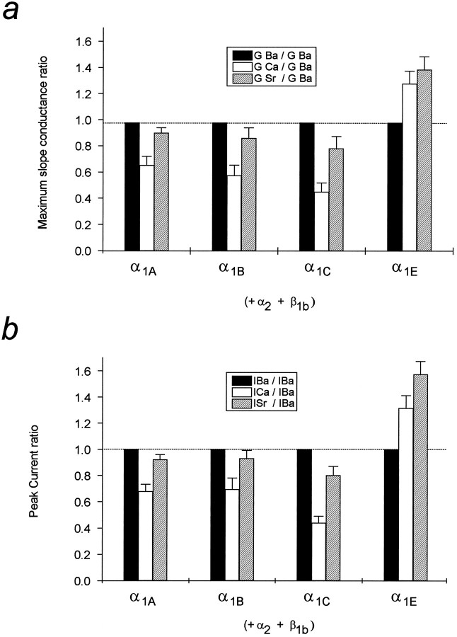
Figure 1 illustrates the whole-cell properties of four major rat brain Ca channel subtypes, α1A, α1B, α1C, and α1E (coexpressed with α2 and β1b subunits), using Ba, Sr, or Ca as the charge carriers. For each channel type, switching the external solution from Ba to Sr to Ca induced a depolarizing shift of the I–V relation toward more depolarized potentials. Furthermore, at least for α1C, the steepness of the activation curve appeared reduced, consistent with that observed for native L-type Ca channels (Byerly et al., 1985). Of particular note, both the peak currents and the maximum slope conductances were differentially affected among various neuronal Ca channel subtypes. Although the peak currents of α1A, α1B, and α1C were all significantly larger in Ba than in Sr or Ca (IBa > ISr > ICa), α1E showed larger currents in Sr compared with Ca or Ba (ISr > ICa > IBa). Although switching the type of permeant ion also appeared to result in changes in the reversal potential, producing a more positive reversal potential with Ca as the charge carrier compared with Sr and Ba (Erev,Ca > Erev,Sr > Erev,Ba), we did not attempt to estimate permeability ratios from the measured reversal potential values, because the intracellular divalent cation concentration could not be controlled precisely.
Fig. 1.
Comparison of macroscopic currents carried by 5 mm Ba, Ca, or Sr. Current traces obtained at the peak of the I–V relations for α1A, α1B, α1C, and α1E(coexpressed with α2 and β1b) are presented with their respectiveI–V relations. I–Vrelations were fitted as described in Materials and Methods. Note that Ba produces the largest currents through α1A, α1B, and α1C, but the smallest currents through α1E channels. Also note the pronounced Ca-dependent inactivation for α1C.
If changes in reversal potential and half-activation potential differed in their absolute magnitudes, the peak current ratios would be a poor indication of the true conductance ratio. To circumvent this issue, we used the slope of the I–V relation on the plateau of the activation curve (i.e., the maximum slope conductance,G) as an indication of the ability of the carrier ion to permeate the channel. As shown in Figure 2a, Ca and Sr produced a larger conductance compared to Ba for α1E, whereas the order was reversed for the other channel isoforms (for α1A, α1B, and α1C:GBa > GSr > GCa). A comparison with the peak ratios (Fig. 2b) revealed a similar profile, consistent with the observation that the shifts in reversal potential and the shifts in half-activation voltage that occur when switching from Ba to Ca (or Sr) were of similar magnitude.
Fig. 2.
Summary of the conductance properties of the different channels. Apparent maximum slope conductances obtained from fits to individual I–V relations are compared ina. Values are presented in the form of conductance ratios (Gion/GBa).b, The peak current values from the same experiments, normalized to those seen in Ba (Imax(ion)/Imax(Ba)). Note the different permeation profile of the α1E channel. Error bars are SE based on 5–14 determinations.
Ca channel permeation characteristics are determined by the α1 subunit
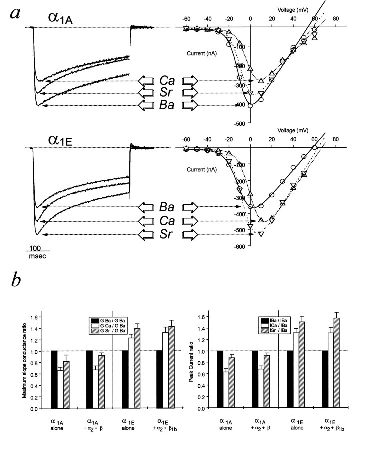
The coexpression of ancillary Ca channel subunits modulates several biophysical properties of cloned Ca channel α1 subunits (for review, see Stea et al., 1995b). To determine whether α2 and β subunits affected permeation, we examined the conductance profiles of α1A and α1E alone (both of these channel types result in high levels of expression in oocytes in the absence of ancillary subunits) (Soong et al., 1993; Stea et al., 1994). Figure 3 shows that no significant differences in the response to switching the type of permeant ion were observed when α2 and β subunits were omitted. All of the changes induced by the Ba–Ca–Sr substitutions described in Figures 1and 2 were maintained, including the unique profile of the α1E channel. Taken together, these data suggest that the basic permeation properties are intrinsic to the α1 subunit and are not significantly affected by coexpression with α2 or β subunits. Furthermore, the permeation differences between α1E and the other channel types is indicated to be an inherent property of the α1E protein.
Fig. 3.
Comparison of macroscopic Ba, Ca, and Sr currents through α1A and α1E in the absence of ancillary subunits. a, Peak current traces with their respective current–voltage relationships as described in Figure 1 (○, Ba; △, Ca; ▿, Sr). b, Comparison of the maximum slope conductance ratios (Gion/GBa) and peak current ratios (Imax(ion)/Imax(Ba)) in the presence and absence of α2 and β1b subunits. Note that the ancillary subunits do not significantly affect the permeation properties of the channels. Error bars are SE on 5–12 determinations.
Dependence of the different α1 channels on [Ba2+]o and [Ca2+]o
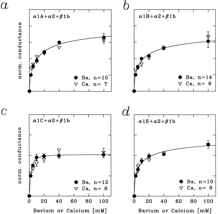
To characterize further the permeation characteristics of the Ca channel isoforms, we recorded concentration–conductance relations for the four Ca channel subtypes in both Ba and Ca (Fig. 4). Increasing the external concentration of either cation progressively increased the whole-cell conductance. The conductance subsequently leveled off at higher concentrations and was consistent with the existence of one or more saturable binding sites for both Ca and Ba within the pore. In Figure 4, the data obtained in Ba and Ca are scaled and superimposed, revealing that the binding sites along the permeation pathway become similarly saturated with either Ba or Ca as the carrier ion for each of the channel subtypes. A pronounced difference, however, becomes apparent on comparison of α1C with the other channel subtypes. The concentration–conductance relation rose more steeply for α1C and saturated at a lower concentration (Fig. 4), suggesting that the divalent binding sites in the pore of α1C exhibit a higher affinity for permeating ions relative to the other channel types. Because the saturation for α1E channels was similar to that for α1A and α1B, a difference in affinity for a putative divalent binding site as the reason for the permeation differences in Ba and Ca can be excluded.
Fig. 4.
Concentration–conductance relations obtained for the four α1 subunits coexpressed with β1b and α2. The data were normalized to 1 at an ion concentration of 2 mm and superimposed. Note that the conductance depends similarly on both Ba and Ca concentration for each of the Ca channel subtypes. The α1C channels seem to differ from the other subtypes in that the current saturates at lower concentrations. The data were obtained from fits to macroscopicI–V relations. Error bars indicate SE; thesolid lines are a smooth approximation of the Ba data based on the Hill equation.
Unitary current as a function of the permeant ion
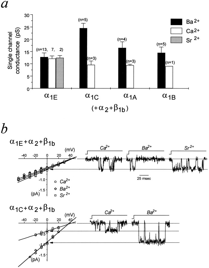
The maximum slope conductance G is a product of the single-channel conductance (g), the number of channels (n) and their open probability (Po). To determine whether the dependence of the whole-cell conductance was attributable to a change in g, we examined the single-channel conductances of the four Ca channel subtypes in both Ba and Ca (and Sr in the case of α1E). Figure5 shows that the single-channel conductances of α1C, α1A, and α1B all were reduced notably when Ca replaced Ba. In contrast, the single-channel conductance of α1E was similar in Ba, Ca, or Sr. The two most extreme cases are illustrated in Figure 5b, with a side-by-side comparison of the unitary conductance–voltage relations for α1E and α1C and single-channel records at a test potential of 0 mV. Consistent with the whole-cell data, the single-channel amplitude of currents carried by α1C channels was about half of that seen with Ba. For α1E, the unitary currents were nearly identical with either Ba, Ca, or Sr. These data indicate that the increases in slope conductance associated with Sr or Ca substitution for Ba were more likely attributable to an increase in plateau open probability than to a change in single-channel conductance. This notion is supported by a preliminary analysis of steady-state activation at the single-channel level (not shown) and is consistent with previous studies (Shuba et al., 1991). Overall, the single-channel data support the uniqueness of the permeation properties of α1E as compared with the other neuronal Ca channel subtypes.
Fig. 5.
Dependence of the single-channel conductance on the type of external permeant ion. The histogram in asummarizes the results obtained with the four α1 subunits coexpressed α2 and β1b. The conductance of α1E is not significantly different with 100 mm Ba, Ca, or Sr as permeant ion, whereas α1C, α1A, and α1B exhibit larger conductances in Ba than in Ca. Error bars indicate SD. b, α1E and α1CI–V relations and current traces evoked by a step depolarization from −100 to 0 mV. Solid lines in the current–voltage relations are linear regressions through the data.
The distinct properties of α1E are not dependent on the expression system
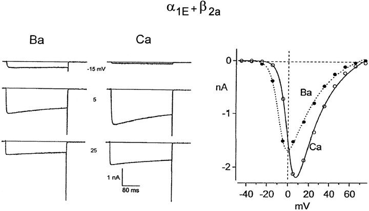
To exclude contributions of the amphibian cellular environment to the distinct properties of α1E, we investigated the whole-cell properties of α1E transiently expressed in human embryonic kidney cells (HEK 293). In this series of experiments, the α1E subunit was coexpressed with β2a to slow inactivation, thereby maximizing resolution of peak currents. As expected from the Ca-induced shift inV0.5, macroscopic Ba currents were initially larger than Ca currents at modest depolarizations (−15 mV), but Ca currents exceed Ba currents at stronger depolarization (to +5 and +25 mV). A comparison of Figure 6 and Figure 1 shows a similar α1E dependence on permeant ion type in both oocytes and mammalian cells. These results indicate that the distinctive properties of α1E are attributable to the intrinsic permeation and gating properties of the channel and are independent of the expression system.
Fig. 6.
Comparison of Ba and Ca whole-cell currents of α1E as expressed in HEK 293 cells.Left, Whole-cell Ba and Ca currents elicited by the indicated test depolarizations from a holding potential of −90 mV. The tail potential is −80 mV. Records have been leak-subtracted by a P/8 algorithm, filtered at 2 kHz (4-pole Bessel filter), and sampled at 10 kHz. Right, Peak current versus test voltage relation. Smooth curves are drawn by eye. Series resistance of 5 MΩ, compensated by 70%; cell capacitance of 34 pF. All recordings at room temperature.
DISCUSSION
α1E permeation properties resemble those of low-threshold Ca channels
In contrast with the higher threshold α1A, α1B, and α1C Ca channels, the biophysical and pharmacological properties of α1E do not exactly match any of the established classes of native Ca channels. That α1E represents a novel type of neuronal mid- to low-threshold Ca channel that is distinct from the commonly accepted T-type channel has been a subject of controversy (Soong et al., 1993; Zhang et al., 1993; Williams et al., 1994). It has been suggested that the α1E isoform from human brain encodes a high voltage-threshold channel (Williams et al., 1994). Similarly, an α1E homolog from the marine ray nervous system (Ellinor et al., 1993) exhibitsI–V characteristics more similar to high voltage-threshold Ca channels and has been correlated with a rat granular cell Ca current resistant to dihydropyridines, ω-conotoxin GVIA, and ω-agatoxin IVA (termed “R-type”; Zhang et al., 1993). It has been argued that the time course of inactivation seen with transiently expressed α1E channels is slow compared with that of native T-type channels. When RNA fractions presumably encoding for T-type channels from thalamic neurons are injected into Xenopus oocytes, however, the resulting waveform is also slowed compared with the channels in their native environment (Dzhura et al., 1994), suggesting that gating kinetics may in some instances be a poor diagnostic for identifying native counterparts to cloned Ca channels. The rat brain α1E channel shows two pronounced differences compared with the residual R-type current (Zhang et al., 1993). First, unlike α1E (de Leon et al., 1995), the R-type current described by Zhang and coworkers (1993) exhibits a faster rate of inactivation with Ca as the charge carrier. We could not detect a similar effect on α1E channels in either oocytes or HEK cells, although this property is readily observed for α1C channels expressed in these two systems (Charnet et al., 1994; de Leon et al., 1995). Second, the permeation profile of the residual R-type current indicates a higher permeation for Ba over Ca (Zhang et al., 1993) and is significantly different from that reported here for α1E.
As suggested by Soong and coworkers (1993), our results provide further support for the notion that α1E possesses properties consistent with a subset of neuronal low-threshold Ca channels. As summarized in Table 1, most low-threshold (T-type) Ca channels have been shown to carry larger whole-cell currents with Ca compared with Ba. This effect is observed in various cell types, including atrial and ventricular myocytes (Bean, 1985,Nilius et al., 1986), skeletal and smooth muscle cells (Beam and Knudson, 1988, Neveu et al., 1994), pancreatic B cells (Hiriat and Matteson, 1988), and both peripheral and central neurons (Carbone and Lux, 1987; Fox et al., 1987; Akaike et al., 1989; Takahashi et al., 1991). An exception to this general trend is an unusual T-type channel from the reticular nucleus of the thalamus, which exhibits larger currents in Ba than in Ca (Huguenard and Prince, 1992). Overall, at both the whole-cell and single-channel levels, our results with α1E are consistent with those reported for the majority of native low voltage-threshold channels.
Table 1.
Relative Ca2+, Ba2+, and Sr2+ conduction through native and cloned Ca2+ channel subtypes
| Native type | T | L | N | P/Q | R |
|---|---|---|---|---|---|
| ICa/IBa | 1–1.8 | 0.2–0.6 | 0.6–0.8 | 0.5 | 0.8 |
| (1, 2, 3, 4, 5, 6, 7, 8)1_a | (1, 5, 6, 7, 9, 10, 11)1_a | (1, 13)1_a | (14)1_a | (15)1_a | |
| ISr/IBa | 1.5–1.9 | 0.6–0.8 | 0.7 | ? | ? |
| (2, 4, 8)1_a | (12, 13)1_a | (13)1_a |
| Expressed cDNA | α1E | α1C | α1B | α1A | |
|---|---|---|---|---|---|
| ICa/IBa | 1.3 | 0.4 | 0.7 | 0.7 | |
| (16)1_a | (16)1_a | (16)1_a | (16)1_a | ||
| ISr/IBa | 1.5 | 0.8 | 0.9 | 0.9 | |
| (16)1_a | (16)1_a | (16)1_a | (16)1_a |
Numbers refer to some of the studies showing the permeation properties of the different channels: 1, Fox et al., 1987; 2, Akaike et al., 1989; 3, Nilus et al., 1984; 4, Carbone et al., 1986; 5, Bean, 1986; 6, Beam and Knudson, 1988; 7, Hiriat and Matteson, 1988; 8, Takahashi et al., 1992; 9, Hess and Tsien, 1984; 10, Almers and McCleskey, 1984; 11, Neveu et al., 1994; 12, Hagiwara and Ohmori, 1982; 13, Kasai and Neher, 1992; 14, Regan, 1991; 15, Zhang et al., 1993; 16, this study.
At the macroscopic level, Williams et al. (1994) reported that the human α1E isoform (>95% identical to the rat brain subtype) displayed a more prominent activity in Ba rather than in Ca. The difference may be attributable to the small differences in amino acid sequence between the species-specific isoforms. Alternatively, as a significant degree of rundown of α1E currents occurs, the magnitude of the whole currents may seem to decrease in some cells if the external solutions are not applied cyclically (e.g., Ba to Ca and then back to Ba; not shown). In our experiments, we minimized the possibility of induced errors arising from rundown by performing a cyclic application of divalent ions (see Materials and Methods).
For α1E, the whole-cell current magnitude varied with the relative ratio Sr > Ca > Ba. In contrast, the single-channel conductances were virtually identical with either Ba, Ca, or Sr. On the basis of a preliminary analysis of the open probability at the plateau of the activation curve, our results suggest that the higher maximum slope conductances obtained in Sr and Ca are attributable to an increase in the open probability of the channel (see α1E single-channel traces in Fig.5b) and a reduction in the number of blank sweeps (not shown). A similar dependence of open probability on the type of permeant ion has been described previously for native low-threshold channels (McDonald et al., 1994).
Ca substitution for Ba differentially affects neuronal Ca channels
A major goal of this study was to determine permeation characteristics of the four major classes of cloned neuronal Ca channels under identical conditions. A number of previous studies indicate that native high voltage-threshold Ca channels carry larger currents with Ba as the permeant ion as compared with Ca or Sr (see Table 1). The most detailed descriptions of divalent cation permeation has been carried out for native L-type Ca channels from both heart and skeletal muscle (Almers and McCleskey, 1984; Hess et al., 1986; Yue and Marban, 1990; McDonald et al., 1994). Current permeation models assume double occupancy of the pore and electrostatic repulsion between both ions (Almers and McCleskey, 1984; Hess and Tsien, 1984) and suggest that Ca ions bind more tightly within the pore than Ba ions, which results in a smaller current amplitude. This feature seems to be common among all L-type Ca channels; however, the absolute value of the conductance ratio in Ba and Ca shows some variability among channels from different tissues (e.g., cardiac vs skeletal muscle) (Beam and Knudson, 1988) and between distinct populations of L-type channels present in a same tissue (e.g., in aortic myocytes) (Neveu et al., 1994). There have also been reports demonstrating that both N-type (Fox et al., 1987; Kasai and Neher, 1992) and P-type (Regan, 1991) channels carry larger currents in Ba compared with Ca. Our results showing increased whole-cell Ba currents for α1A, α1B, and α1C support these studies. Similar to that observed with native cardiac and neuronal L-type channels, Ca substitution for Ba also increased the rate of inactivation and seemed to decrease the steepness of the activation curve of the α1C L-type channel (Byerly et al., 1985; Charnet et al., 1994; Galli et al., 1994; Imredy and Yue, 1994; Neely et al., 1994; Perez-Reyes et al., 1994; de Leon et al., 1995). Although it is possible that some of the effects observed at the whole-cell level are attributable to altered channel kinetics (McDonald et al., 1994), the single-channel records support the notion that the dependence of the whole-cell conductance on the type of permeant ion is an intrinsic property of the permeation pathway (all of the high voltage-activated subtypes studied here exhibited smaller single-channel conductances when the permeant ion was switched from Ba to Ca). The α1E channels display a unique dependence of nickel pore block on the type of permeant ion compared with α1A and α1Cchannels, consistent with the distinct permeation properties observed with α1E (Zamponi et al., 1996).
Implications for models of Ca channel permeation
Concentration–conductance curves obtained for the four different types of Ca channels revealed two additional qualitative features of Ca channels. First, with the concentration–conductance curves scaled, the data obtained in Ba and Ca superimposed for each of the Ca channel isoforms. Second, the concentration–conductance curves obtained for α1C appeared steeper than those of the other channel subtypes (at 40 mm, a concentration that produces only a 70% saturation of each α1A, α1B, and α1E, currents through α1C are essentially saturated). Although the multi-ion nature of the pore precludes us from estimating a precise Kd value for binding of permeant ions to proposed intrapore sites, these qualitative observations suggest a stronger affinity of both Ca and Ba ions for the pore of α1C than with the remaining channel subtypes. Second, the results also suggest that each of the channel types becomes similarly saturated with either ion.
The half-saturation concentration is mainly dependent on the depth of the energy wells in the pore, whereas the maximal conductance at saturating concentrations depends predominantly on the height of the exit barrier toward the cytoplasmic side (Hille, 1992). We observed an effect of the type of permeant ion only on maximal conductance, and the data suggest that the unique properties observed for α1E may be attributable to the distinct properties of its exit barrier. Amino acids in the pore-forming regions of the Ca channel isoforms are highly conserved across the Ca channel isoforms (for review, see Stea et al., 1995b). It has been proposed recently that four glutamate residues in the pore are the most crucial determinant of permeation (Tang et al., 1993; Yang et al., 1993). The glutamates are thought to cooperate in forming a high-affinity interaction between the channel and permeating ions (Ellinor et al., 1995). Electrostatic repulsion between two Ca ions ultimately drives ions across the pore, as proposed previously for native L-type channels (Almers and McCleskey, 1984; Hess and Tsien, 1984). Because all Ca channels cloned to date, including α1E, possess the four glutamate residues in similar positions, the data presented in this study suggest that additional structural determinants within the channel pore are likely to play a role in ion permeation. The existence of additional determinants for permeation is also supported by the recent report that an aspartate residue in the pore of the cardiac α1C isoform is critical for Ca binding and Cd block (Parent et al., 1995). One conspicuous difference between α1E and the remaining Ca channel subtypes occurs in domain I, where α1E lacks a conserved aspartate residue at position 264. The absence of a negatively charged residue may affect the shape of the barrier profile within the pore. Future studies involving site-directed mutagenesis of each of the Ca channel subtypes may identify additional amino acid residues involved in permeation. In this regard, the unique properties of the α1E channel may prove particularly useful.
In conclusion, our data are consistent with the previous proposal (Soong et al., 1993) that α1E channels reflect a subset of native neuronal mid- to low-threshold Ca channels, and it is likely that additional gene products encode other types of T-type Ca channels. Ultimately, antisense and/or gene knockout experiments are required to establish conclusively the exact nature of the native counterpart of α1E channels.
Footnotes
This work was supported by postdoctoral fellowships from European Molecular Biology and Institut National de la Santé et de la Recherche Médicale to E.B. and by postdoctoral fellowships from the Medical Research Council (MRC) of Canada to G.W.Z. and A.S.; G.W.Z. also holds a postdoctoral fellowship from the Alberta Heritage Foundation for Medical Research. T.W.S. is supported by a postdoctoral fellowship and is on leave from the National Institute of Molecular and Cell Biology, National University of Singapore. B.A.L. and L.P.J. are supported by the National Institutes of Health Medical Scientist Training Program. D.T.Y. is supported by a Presidential Faculty Fellowship of the National Science Foundation. T.P.S. is an MRC Scientist and is supported by grants from the MRC and the Howard Hughes Medical Institute International Research Scholars Program. Travel support was provided by NATO Grant CRG 890374 to Joël Nargeot. We thank Drs. John Hanrahan and Mary Gilbert for comments on this manuscript and Dr. Andy Randall for providing unpublished information concerning R-type currents.
Correspondence should be addressed to Dr. Terry P. Snutch, Biotechnology Laboratory, Room 237, 6174 University Boulevard, University of British Columbia, Vancouver, British Columbia, Canada V6T 1Z3.
REFERENCES
- 1.Akaike N, Kostyuk PG, Osipchuk YV. Dihydropyridine-sensitive low-threshold calcium channels in isolated rat hypothalamic neurones. J Physiol (Lond) 1989;412:181–195. doi: 10.1113/jphysiol.1989.sp017610. [DOI] [PMC free article] [PubMed] [Google Scholar]
- 2.Almers W, McCleskey EW. Non-selective conductance in calcium channels of frog muscle: calcium selectivity in a single-file pore. J Physiol (Lond) 1984;353:585–608. doi: 10.1113/jphysiol.1984.sp015352. [DOI] [PMC free article] [PubMed] [Google Scholar]
- 3.Beam KG, Knudson CM. Calcium current in embryonic and neonatal mammalian skeletal muscle. J Gen Physiol. 1988;91:781–798. doi: 10.1085/jgp.91.6.781. [DOI] [PMC free article] [PubMed] [Google Scholar]
- 4.Bean BP. Two kinds of calcium channels in canine atrial cells. J Gen Physiol. 1985;86:1–30. doi: 10.1085/jgp.86.1.1. [DOI] [PMC free article] [PubMed] [Google Scholar]
- 5.Bourinet E, Charnet P, Stea A, Tomlinson JW, Snutch TP, Nargeot J. Voltage dependent facilitation of a neuronal α1C L-type calcium channel. EMBO J. 1994;13:5032–5039. doi: 10.1002/j.1460-2075.1994.tb06832.x. [DOI] [PMC free article] [PubMed] [Google Scholar]
- 6.Byerly L, Chase PB, Stimers J. Permeation and interaction of divalent cations in calcium channels of snail neurons. J Gen Physiol. 1985;85:491–518. doi: 10.1085/jgp.85.4.491. [DOI] [PMC free article] [PubMed] [Google Scholar]
- 7.Carbone E, Lux HD. Kinetics and selectivity of a low-voltage-activated calcium current in chick and rat sensory neurones. J Physiol (Lond) 1987;386:547–570. doi: 10.1113/jphysiol.1987.sp016551. [DOI] [PMC free article] [PubMed] [Google Scholar]
- 8.Charnet P, Bourinet E, Dubel SJ, Snutch TP, Nargeot J. Calcium currents recorded from a neuronal α1C L-type calcium channel in Xenopus oocytes. FEBS Lett. 1994;344:87–90. doi: 10.1016/0014-5793(94)00357-2. [DOI] [PubMed] [Google Scholar]
- 9.de Leon M, Wang Y, Jones L, Perez-Reyes E, Wei X, Soong TW, Snutch TP, Yue DT. Essential Ca2+-binding motif for Ca2+-sensitive inactivation of L-type Ca2+ channels. Science. 1995;270:1502–1506. doi: 10.1126/science.270.5241.1502. [DOI] [PubMed] [Google Scholar]
- 10.Dhallan RS, Yau KW, Schrader KA, Reed RR. Primary structure and functional expression of a cyclic nucleotide-activated channel from olfactory neurons. Nature. 1990;347:184–187. doi: 10.1038/347184a0. [DOI] [PubMed] [Google Scholar]
- 11.Dubel SJ, Starr TV, Hell J, Ahlijannian MK, Enyeart JJ, Catterall WA, Snutch TP. Molecular cloning of the α1 subunit of an ω-conotoxin-sensitive calcium channel. Proc Natl Acad Sci USA. 1992;89:5058–5062. doi: 10.1073/pnas.89.11.5058. [DOI] [PMC free article] [PubMed] [Google Scholar]
- 12.Dunlap K, Luebke JI, Turner TJ. Exocytotic Ca2+ channels in the mammalian central nervous system. Trends Neurosci. 1995;18:89–98. [PubMed] [Google Scholar]
- 13.Dzhura I, Kostyuk P, Lyubanova O, Naidenov V, Shuba Y. Expression of low-voltage activated Ca2+-channels from rat brain neurons in Xenopus oocytes. NeuroReport. 1994;5:1960–1962. doi: 10.1097/00001756-199410000-00030. [DOI] [PubMed] [Google Scholar]
- 14.Ellinor PT, Zhang JF, Randall AD, Zhou M, Schwarz TL, Tsien RW, Horne WA. Functional expression of a novel neuronal voltage-dependent calcium channel. Nature. 1993;363:455–458. doi: 10.1038/363455a0. [DOI] [PubMed] [Google Scholar]
- 15.Ellinor PT, Yang J, Sather WA, Zhang JF, Tsien RW. Ca2+ channel selectivity at a single locus for high affinity Ca2+ interactions. Neuron. 1995;15:1121–1132. doi: 10.1016/0896-6273(95)90100-0. [DOI] [PubMed] [Google Scholar]
- 16.Fox AP, Nowycky MC, Tsien RW. Kinetic and pharmacological properties distinguishing three types of calcium currents in chick sensory neurones. J Physiol (Lond) 1987;394:149–172. doi: 10.1113/jphysiol.1987.sp016864. [DOI] [PMC free article] [PubMed] [Google Scholar]
- 17.Fujita Y, Mynlieff M, Dirksen RT, Kim MS, Niidome T, Nakai J, Friedrich T, Iwabe N, Miyata T, Furuchi T, Furutama D, Mikoshiba K, Mori Y, Beam KG. Primary structure and functional expression of the ω-conotoxin-sensitive N-type calcium channel from rabbit brain. Neuron. 1993;10:585–598. doi: 10.1016/0896-6273(93)90162-k. [DOI] [PubMed] [Google Scholar]
- 18.Galli A, Ferroni A, Bertollini L, Mazzanti M. Inactivation of single Ca2+channels in rat sensory neurons by extracellular Ca2+. J Physiol (Lond) 1994;477:15–26. doi: 10.1113/jphysiol.1994.sp020167. [DOI] [PMC free article] [PubMed] [Google Scholar]
- 19.Hagiwara S, Ohmori H. Studies of calcium channels in rat clonal pituitary cells with patch electrode voltage clamp. J Physiol (Lond) 1982;49:807–818. doi: 10.1113/jphysiol.1982.sp014371. [DOI] [PMC free article] [PubMed] [Google Scholar]
- 20.Hell JW, Westenbroek RE, Warner C, Ahlijanian MA, Prystay W, Gilbert MM, Snutch TP, Catterall WA. Identification and differential subcellular localization of the neuronal class C and class D L-type calcium channel α1 subunits. J Cell Biol. 1993;123:949–962. doi: 10.1083/jcb.123.4.949. [DOI] [PMC free article] [PubMed] [Google Scholar]
- 21.Hess P, Tsien RW. Mechanism of ion permeation through calcium channels. Nature. 1984;309:453–456. doi: 10.1038/309453a0. [DOI] [PubMed] [Google Scholar]
- 22.Hess P, Lansman JB, Tsien RW. Calcium channel selectivity for divalent and monovalent cations. J Gen Physiol. 1986;88:293–319. doi: 10.1085/jgp.88.3.293. [DOI] [PMC free article] [PubMed] [Google Scholar]
- 23.Hille B. Sinauer; Sunderland, MA: 1992. Ionic channels of excitable membrane. [Google Scholar]
- 24.Hiriat M, Matteson DR. Na channels and two types of Ca channels in rat pancreatic B cells identified with the reverse hemolytic plaque assay. J Gen Physiol. 1988;91:617–639. doi: 10.1085/jgp.91.5.617. [DOI] [PMC free article] [PubMed] [Google Scholar]
- 25.Huguenard JR, Prince DA. A novel T-type current underlies prolonged Ca2+-dependent burst firing in GABAergic neurons of rat thalamic reticular nucleus. J Neurosci. 1992;12:3804–3817. doi: 10.1523/JNEUROSCI.12-10-03804.1992. [DOI] [PMC free article] [PubMed] [Google Scholar]
- 26.Imredy JP, Yue DT. Mechanism of Ca2+-sensitive inactivation of L-type Ca2+ channels. Neuron. 1994;12:1301–1318. doi: 10.1016/0896-6273(94)90446-4. [DOI] [PubMed] [Google Scholar]
- 27.Kasai H, Neher E. Dihydropyridine-sensitive and ω-conotoxin-sensitive calcium channels in a mammalian neuroblastoma-glioma cell line. J Physiol (Lond) 1992;448:161–188. doi: 10.1113/jphysiol.1992.sp019035. [DOI] [PMC free article] [PubMed] [Google Scholar]
- 28.McDonald TF, Pelzer S, Trautwein W, Pelzer DJ. Regulation and modulation of calcium channels in cardiac, skeletal, and smooth muscle cells. Physiol Rev. 1994;74:365–507. doi: 10.1152/physrev.1994.74.2.365. [DOI] [PubMed] [Google Scholar]
- 29.Mori Y, Friedrich T, Kim MS, Mikami A, Naaki J, Ruth P, Bosse E, Hofmann F, Flockerzi V, Furuichi T, Mikoshiba K, Imoto K, Tanabe T, Numa S (1991) Primary structure and functional expression from complementary DNA of a brain calcium channel. Nature 350: 398–402. [DOI] [PubMed]
- 30.Neely A, Olcese R, Wei X, Birnbaumer L, Stefani E. Ca2+-dependent inactivation of a cloned cardiac Ca2+ channel α1 subunit (α1C) expressed in Xenopus oocytes. Biophys J. 1994;66:1895–1903. doi: 10.1016/S0006-3495(94)80983-X. [DOI] [PMC free article] [PubMed] [Google Scholar]
- 31.Neveu D, Quignard JF, Fernandez A, Richard S, Nargeot J. Differential β-adrenergic regulation and phenotypic modulation of voltage-activated calcium currents in rat aortic myocytes. J Physiol (Lond) 1994;479:171–182. doi: 10.1113/jphysiol.1994.sp020286. [DOI] [PMC free article] [PubMed] [Google Scholar]
- 32.Nilius B, Hess P, Lansman JB, Tsien RW. A novel type of cardiac calcium channel in ventricular cells. Nature. 1986;316:443–446. doi: 10.1038/316443a0. [DOI] [PubMed] [Google Scholar]
- 33.Parent L, Davila DD, Gopalakrishnan M, Brown AM. Aspartate residue D737 in pore II of the cardiac calcium channel α1C subunit contributes to high-affinity calcium and cadmium binding. Biophys J. 1995;68:A264. [Google Scholar]
- 34.Perez-Reyes E, Yuan W, Wei X, Bers DM. Regulation of the cloned L-type cardiac calcium channel by cyclic-AMP-dependent protein kinase. FEBS Lett. 1994;342:119–123. doi: 10.1016/0014-5793(94)80484-2. [DOI] [PubMed] [Google Scholar]
- 35.Regan LJ. Voltage-dependent calcium currents in Purkinje cells from rat cerebellar vermis. J Neurosci. 1991;11:2259–2269. doi: 10.1523/JNEUROSCI.11-07-02259.1991. [DOI] [PMC free article] [PubMed] [Google Scholar]
- 36.Sather WA, Tanabe T, Zhang JF, Mori Y, Adams ME, Tsien RW. Distinctive biophysical and pharmacological properties of Class A (BI) calcium channel α1subunits. Neuron. 1993;11:291–303. doi: 10.1016/0896-6273(93)90185-t. [DOI] [PubMed] [Google Scholar]
- 37.Shuba YM, Teslenko VI, Savchenko AN, Pogorelaya NH. The effect of permeant ions on single calcium channel activation in mouse neuroblastoma cells: ion-channel interaction. J Physiol (Lond) 1991;443:25–44. doi: 10.1113/jphysiol.1991.sp018820. [DOI] [PMC free article] [PubMed] [Google Scholar]
- 38.Soong TW, Stea A, Hodson CD, Dubel SJ, Vincent SR, Snutch TP. Structure and functional expression of a member of the low voltage-activated calcium channel family. Science. 1993;260:1133–1136. doi: 10.1126/science.8388125. [DOI] [PubMed] [Google Scholar]
- 39.Stea A, Dubel SJ, Pragnell M, Leonard JP, Campbell KP, Snutch TP. A β-subunit normalizes the electrophysiological properties of a cloned N-type Ca2+ channel α1-subunit. Neuropharmacology. 1993;32:1103–1116. doi: 10.1016/0028-3908(93)90005-n. [DOI] [PubMed] [Google Scholar]
- 40.Stea A, Tomlinson JW, Soong TW, Bourinet E, Dubel SJ, Vincent SR, Snutch TP. The localization and functional properties of a rat brain α1A calcium channel reflect similarities to neuronal Q- and P-type channels. Proc Natl Acad Sci USA. 1994;91:10576–10580. doi: 10.1073/pnas.91.22.10576. [DOI] [PMC free article] [PubMed] [Google Scholar]
- 41.Stea A, Soong TW, Snutch TP. Determinants of PKC-dependent modulation of a family of neuronal calcium channels. Neuron. 1995a;15:929–940. doi: 10.1016/0896-6273(95)90183-3. [DOI] [PubMed] [Google Scholar]
- 42.Stea A, Soong TW, Snutch TP. Voltage-gated calcium channels. Ligand- and voltage-gated ion channels. In: North RA, editor. Handbook of receptors and channels. CRC; Boca Raton, FL: 1995b. pp. 113–152. [Google Scholar]
- 43.Takahashi K, Ueno S, Akaike N. Kinetic properties of T-type Ca2+ currents in isolated rat hippocampal CA1 pyramidal neurons. J Neurophysiol. 1991;65:148–155. doi: 10.1152/jn.1991.65.1.148. [DOI] [PubMed] [Google Scholar]
- 44.Tang S, Mikala G, Bahinski A, Yatani A, Varadi G, Schwartz A. Molecular localization of ion selectivity sites within the pore of a human L-type cardiac calcium channel. J Biol Chem. 1993;268:13026–13029. [PubMed] [Google Scholar]
- 45.Tomlinson JW, Stea A, Bourinet E, Charnet P, Nargeot J, Snutch TP (1993) Functional properties of a neuronal class C L-type calcium channel. Neuropharmacology 32: 1117–1126. [DOI] [PubMed]
- 46.Tsien RW, Lipscombe D, Madison DV, Bley KR, Fox AP. Multiple types of neuronal calcium channels and their selective modulation. Trends Neurosci. 1988;11:431–438. doi: 10.1016/0166-2236(88)90194-4. [DOI] [PubMed] [Google Scholar]
- 47.Williams ME, Feldman DH, McCue AF, Brenner R, Velicelebi G, Ellis SB, Harpold MM. Structure and functional expression of α1, α2, and β subunits of a novel human neuronal calcium channel subtype. Neuron. 1992a;8:71–84. doi: 10.1016/0896-6273(92)90109-q. [DOI] [PubMed] [Google Scholar]
- 48.Williams ME, Brust PF, Feldman DH, Patthi S, Simerson S, Maroufi A, McCue AF, Velicelebi G, Ellis SB, Harpold MM. Structure and functional expression of an ω-conotoxin-sensitive human N-type calcium channel. Science. 1992b;257:389–395. doi: 10.1126/science.1321501. [DOI] [PubMed] [Google Scholar]
- 49.Williams ME, Marubio LM, Deal CR, Hans M, Brust PF, Philipson LH, Miller RJ, Johnson EC, Harpold MM, Ellis SB. Structure and functional characterization of neuronal α1ECa2+ channel subtype. J Biol Chem. 1994;269:22347–22357. [PubMed] [Google Scholar]
- 50.Yang J, Ellinor PT, Sather WA, Zhang JF, Tsien RW. Molecular determinants of Ca2+ selectivity and ion permeation in L-type Ca2+ channels. Nature. 1993;366:158–161. doi: 10.1038/366158a0. [DOI] [PubMed] [Google Scholar]
- 51.Yue DT, Marban E. Permeation in the dihydropyridine-sensitive calcium channel: multi-ion occupancy but no anomalous mole fraction effect between Ba2+ and Ca2+. J Gen Physiol. 1990;95:911–939. doi: 10.1085/jgp.95.5.911. [DOI] [PMC free article] [PubMed] [Google Scholar]
- 52.Zamponi GW, Bourinet E, Snutch TP. Nickel block of a family of neuronal calcium channels. Subtype- and subunit-dependent action at multiple sites. J Membr Biol. 1996;151:77–90. doi: 10.1007/s002329900059. [DOI] [PubMed] [Google Scholar]
- 53.Zhang JF, Randall AD, Ellinor PT, Horne WA, Sather WA, Tanabe T, Schwarz TL, Tsien RW. Distinctive pharmacology and kinetics of cloned neuronal Ca2+ channels and their possible counterparts in mammalian CNS neurons. Neuropharmacology. 1993;32:1075–1080. doi: 10.1016/0028-3908(93)90003-l. [DOI] [PubMed] [Google Scholar]


