. 2019 May 22;8:e44904. doi: 10.7554/eLife.44904

Figure 7. Mechanism of completion of PTC assembly and its quality control.

Insets show the sequential interactions of base A2971 with Nmd3, uL16 and SBDS during PTC completion. Models for 60S-bound EFL1 and SBDS are based on EMD-3145, EMD-3146 and EMD-3147 (Weis et al., 2015). Proteins targeted by mutations in Shwachman-Diamond syndrome (yellow) and paediatric T-ALL (blue) are highlighted.

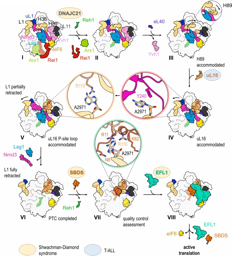
Figure 7.

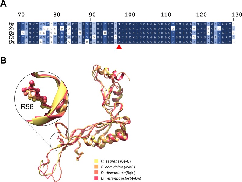
Figure 7—figure supplement 1. The uL16 sequence and structure are highly conserved.

Figure 7—figure supplement 1.

(A) Protein sequence alignment of uL16 from Homo sapiens, Saccharomyces cerevisiae, Dictyostelium discoideum, Caenorhabditis elegans and Drosophila melanogaster. Residue R98 (mutated in T-ALL) is highlighted in bold and marked with a red arrow (▲). The alignment was performed using Clustal Omega (Sievers et al., 2011). (B) Superposition of corresponding atomic models for uL16 (pdb entries 6ek0 [Natchiar et al., 2017], 4v88 [Ben-Shem et al., 2011], 6qkl [Weis et al., 2015] and 4v6w [Anger et al., 2013]).