Abstract
We present a patient who suffered an unstable intertrochanteric hip fracture and underwent osteosynthesis with a trochanteric nail. During the postoperative period, he presented a pseudoaneurysm of the lateral circumflex branch of the deep femoral artery secondary to a displaced fracture of the lesser trochanter. With the suspected diagnosis due to indirect clinical and radiological signs and confirmation by Doppler ultrasound and computed tomography angiography, a transverse arterial embolization with resolution of the symptoms was carried out. The pseudoaneurysm of the deep femoral artery or its branches is a very rare complication after intertrochanteric hip fractures, which must be taken into account in the late appearance of edema and hematoma in the thigh and evidence of medial and superior displacement of the lesser trochanter. The diagnosis is confirmed by CT angiography and the treatment by percutaneous arterial embolization has good results without the need of excising the lesser trochanter.
INTRODUCTION
Hip fractures in the elderly are very frequent events while vascular injuries related to trauma during the surgical procedure are exceptional. The treatment of choice in intertrochanteric hip fractures is the trochanteric nail through minimally invasive procedure, so in those cases in which the lesser trochanter (LT) is displaced, most of the time, it is not necessary to reduce or to fix it.
In this type of fracture the deep femoral artery or its branches are the vessels most at risk of injury during surgery [1]. However, the displaced fracture of the LT and its possible secondary mobilization in the cranial-medial direction by traction of the iliopsoas may cause late lesion of the deep femoral artery or its branches in contact with its sharp edges [1].
We present a case of a deep femoral artery pseudoaneurysm of late presentation secondary to the displacement of the LT after an intertrochanteric hip fracture, satisfactorily treated with a minimally invasive endovascular technique.
CASE REPORT
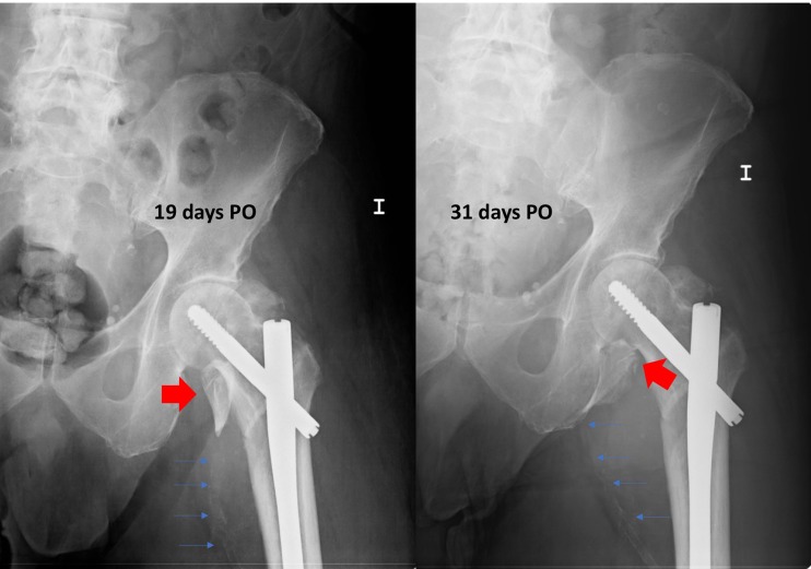
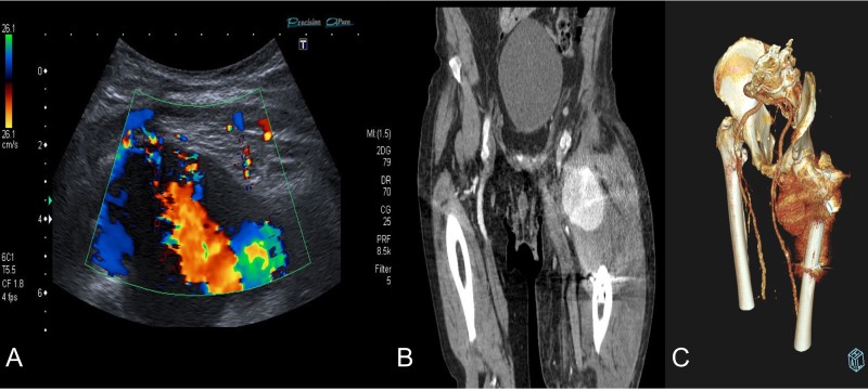
We present a case of an 80-year-old institutionalized man, who suffered an unstable intertrochanteric fracture of the left hip, type 31.A2.1 (AO-OTA) due to a low energy accident after falling from his own height. The patient previous medical history included bipolar disorder, hypertension, DM, and right inguinal herniorrhaphy surgery. The patient underwent closed reduction and internal fixation with an intramedullary nail type Gamma3 (Stryker Trauma GmbH Prof. Küntscher-Str. 1-5 24 232 Schönkirchen, Germany) of the fracture during the first 24 hours after injury. The surgical procedure was performed without any intraoperative complications. He was discharged from the hospital on the sixth postoperative day, asymptomatic, with a postoperative hematoma in the left thigh, walking with a walking frame, with a postoperative radiographic control showing a correct nail placement without alterations and with an hemoglobin value (HGB) of 10.7 g/dl and a hematocrit value (HCT) of 30.8%. On the 18th postoperative day, he suffered a fall after having a syncopal episode, so he went to the emergency room and was admitted to the hospital for study. The hematoma in his thigh was evolving correctly, and the radiographs remained unchanged with respect to the previous ones. He was discharged two days later. One month after surgery, he returned to the hospital due to persistent pain, volume increase and progressive hematoma in the left thigh. The blood analysis showed an anemia (HGB 9.5 g/dL, HCT 29%). The pedia pulse was present and the posterior tibial pulse was weaker than the contralateral but also present. An X-ray showed a superior and medial displacement of the lesser trochanter compared to previous radiographs and a medialization of the femoral artery, visible because it was calcified (Fig. 1). A Doppler ultrasound was performed showing a hypoechoic lesion with turbulent flow inside compatible with pseudoaneurysm at the level of the deep femoral artery or one of its branches. The study was completed with a CT angiography that confirmed the presence of a pseudoaneurysm of 7.3 × 6.7 × 6 cm at the level of the deep femoral artery at the beginning of the lateral circumflex branch (Fig. 2). At that time, the patient was referred to the Interventional Radiology Service. The vascular lesion was immediately treated by femoral transcatheter embolization with two distal coils in the lateral circumflex artery measuring 3 and 4 mm (Axium 3D Medtronic 9775 Toledo Way Irvine, CA 92 618 USA) and proximal embolization by a liquid embolic agent, Onyx 34 (Covidien 106-108 Rue la Boetie 75 008 Paris, France).
Figure 1:
Postoperative radiographs after intertrochanteric fracture synthesis with trochanteric nail 19 and 31 days after surgery. Secondary displacement of LT can be observed (wide arrows) and of the calcified femoral vessels (thin arrows), indirect signs of the presence of a pseudoaneurysm.
Figure 2:
Diagnosis confirmation by Doppler ultrasound (A), shows hypoechoic lesion with turbulent flow inside with 5–6 cm of maximum diameter; and CT angiography (B) and three-dimensional reconstruction (C) that confirm a lateral circumflex artery pseudoaneurym branch of the deep femoral artery of 7,3 × 6,7 × 6 cm size.
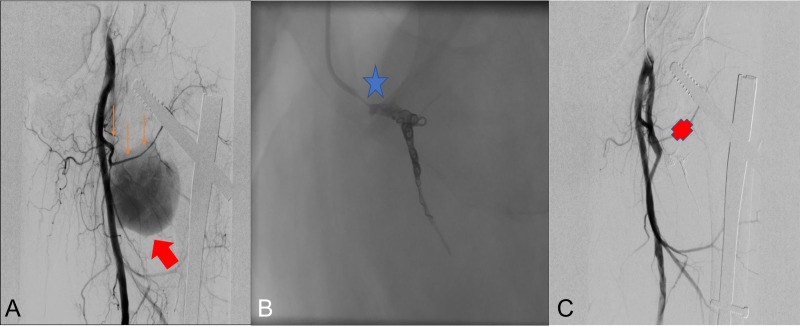
The procedure achieved complete closure of the pseudoaneurysm and the vascular leakage was completely resolved stopping the active arterial bleeding, with permeability of the deep femoral artery (Fig. 3). The patient left the hospital 24 hours after procedure.
Figure 3:
Radioescopic images sequence of the interventional procedure of embolization by retrograde right femoral puncture. A: Lateral femoral circumflex artery (thin arrows) branch of the deep femoral artery, pseudoanerysm (wide arrow). B: Embolization using two Axium 3D distal coils (star). C: Final result with embolized lateral circumflex artery (Cross).
DISCUSSION
Hip fractures are very frequent in elderly patients and their incidence is progressively increasing. About half of them are extracapsular and approximately one in four is unstable with involvement of LT [2]. The treatment of choice in these cases is closed reduction and internal fixation with trochanteric nails although other devices are also used, such as Dinamic Hip Screw—DHS. Vascular lesions during these procedures are very rare, around 0.21%, those that have a higher risk of injury are the deep femoral artery and its branches [1] due to their anatomical location posterior and close to the femoral bone, representing 80% of cases of vascular injury [3]. Deep femoral artery is a branch of the femoral artery and supplies the anterior muscles of the thigh, adductors and hamstrings. It divides into the medial and lateral circumflexes and in 2-6 perforating branches. Circumflex arteries present anastomosis with the gluteal artery and with the first perforanting branch, wich means that one or the other can be ligated or embolizated if necessary. The injury can result from trauma, iatrogenic causes such as surgical procedures, or during the postoperative period, frequently by acute edges of the LT [1]. The cause is frequently traumatic, either due to contusions at the time of fracture, injuries during surgery, with surgical retractors, needles or screws; or during the postoperative rehabilitation period, due to the displacement of the LT by the contraction of the iliopsoas. The pseudoaneurysm is frequently located at the level of the third or fourth hole of the DHS system or at the level of the distal locking screw on the intramedullary nail [4].
Among the underlying factors are the decrease in the elasticity of the vessels due to arteriosclerosis or calcification and the position of the patient during surgery with the limb in adduction, gentle traction and internal rotation using a traction table and the pressure of the perineal pole, putting the deep femoral artery closer to the femur, increasing the risk of injury [5]. Most of the cases found in the literature in which the lesion is suspected to be caused by the displacement of the LT, the fractures are type 31.A2 of the AO-OTA classification in wich the LT fragment is free and can be tractioned and displaced by the iliopsoas. Diagnosis is usually delayed due to the unspecific symptoms. Cases have been described to be diagnosed from the third post-operative day to 14 years after the fracture [6], although the diagnosis at the moment of lesion by the displacement of the LT usually occurs during the first 18-36 days [7], 30 days in our case. In the pseudoaneurysm of the superficial femoral artery the appearance of a pulsatile mass is frequent [8], but in the deep femoral artery cases, due to its anatomical location, it presents with more unspecific signs, such as swelling, edema, persistent pain and anemia, being able to be camouflaged by the typical hip surgery symptoms [9]. There are indirect radiographic signs that, accompanied by compatible clinical symptoms, should make us suspect the possible existence of a pseudoaneurysm, such as the medial and proximal displacement of the lesser trochanter or the medial displacement of the calcified femoral vessels, which is very representative in our case. The confirmation diagnosis can be made by Doppler ultrasound, showing a hypoechoic lesion with turbulent flow in it, and confirmed by a CT angiography. Treatment options include open surgery or endovascular procedure. We think that the technique of choice in these cases is an arterial embolization independent of the size of the pseudoaneurysm. However, other authors defend that in cases of large pseudoaneurysms open surgery would be the treatment of choice, reserving endovascular techniques for smaller cases [10]. In cases in which the aneurysm is produced by the displacement of the LT, the definitive treatment would be achieved with the extraction of the bone fragment after the arterial ligature [10], although in these cases, endovascular treatment has been shown to be effective and without risk of recurrence, as in our case [7]. Endovascular treatments include Coils, Stents, coagulant agents, or combinations of them, without evidence recommending one technique over another in this pathology. In our case, we performed a ‘sandwich’ embolization technique to close the pseudoaneurysm, technique that has shown good long-term results. The particularity of the treatment performed is that we did not remove the shifted LT fragment. Other authors also performed endovascular treatments in cases of pseudoaneurysm due to displacement of the LT with satisfactory results without evidence of recurrence [1, 7, 8, 10]. The late onset of a pseudoaneurysm of the deep femoral artery or its branches is a rare complication after hip fractures, but it should be suspected in a clinical setting of persistent pain after nailing, together with the late onset of edema and hematoma in the thigh and indirect signs in radiographs, such as medial and superior displacement of the lesser trochanter or the calcified femoral vessels. Confirmation must be made using Doppler ultrasound and CT angiography, and percutaneous treatment has good results without the need of excising the lesser trochanter.
CONFLICT OF INTEREST STATEMENT
None declared.
REFERENCES
- 1. Luria S, Eylon S, Peyser A. Vascular aneurysm secondary to a femoral intertrochanteric fracture concealed by anticoagulation therapy. Isr Med Assoc J 2015;17:777–9. [PubMed] [Google Scholar]
- 2. Lindskog DM, Baumgaertner MR. Unstable intertrochanteric hip fractures in the elderly. J Am Acad Orthop Surg 2004;12:179–90. [DOI] [PubMed] [Google Scholar]
- 3. Whitehill R, Wang GJ, Edwards JR, Stamp WG. Late injuries to femoral vessels after fracture of the hip. Case report. J Bone Joint Surg Am 1978;60:541–2. [PubMed] [Google Scholar]
- 4. Wolfgang GL, Barnes WT, Hendricks GL. False aneurysm of the profundal femoris artery resulting from anil-plate fixation of intertrochanteric fractures. Clin Orthop 1974;100:143–50. [PubMed] [Google Scholar]
- 5. Yang KH, Yoon CS, Park HW, Won JH, Park SJ. Position of the superficial femoral artery in closed hip nailing. Arch Orthop Trauma Surg 2004;124:169–72. [DOI] [PubMed] [Google Scholar]
- 6. Molfetta L, Chiapale D, Caldo D, Leonardi F False. Aneurysm of the superficial femoral artery after total hip arthroplasty: a case report. Hip Int 2009;17:234–6. [DOI] [PubMed] [Google Scholar]
- 7. Piolanti N, Giuntoli M, Nucci AM, Battistini P, Lisanti M, Andreani L. Profunda femoris artery pseudoaneurysm after intramedullary fixation for a pertrochanteric hip fracture. J Orthop Case Rep 2017;7:74. [DOI] [PMC free article] [PubMed] [Google Scholar]
- 8. Craxford S, Gale M, Lammin K. Arterial injury to the profunda femoris artery following internal fixation of a neck of femur fracture with a compression hip screw. Case Rep Orthop 2013;2013:181293. [DOI] [PMC free article] [PubMed] [Google Scholar]
- 9. de Raaff CA, van Nieuwenhuizen RC, van Dorp TA. Pseudoaneurysm after pertrochanteric femur fracture: a case report. Skeletal Radiol 2016;45:575–8. [DOI] [PubMed] [Google Scholar]
- 10. Kizilates U, Nagesser SK, Krebbers YM, Sonneveld DJA. False aneurysm of the deep femoral artery as a complication of intertrochanteric fracture of the hip: options of open and endovascular repairs. Perspect Vasc Surg Endovasc Ther 2009;21:245–8. [DOI] [PubMed] [Google Scholar]


