Abstract
Objectives: To study the long‐term outcomes of inactivated bone reimplantation with preservation of the epiphysis in children with distal femoral osteosarcomas.
Methods: Over 10 years, five children (mean age 9.2 years, one boy and four girls) with distal femoral osteosarcomas underwent inactivated bone reimplantation with preservation of the epiphysis following chemotherapy in our hospital. Three patients were type I on MRI classification (one with pathological fracture), and two type II. The therapeutic regime was two cycles of preoperative chemotherapy, surgery and six cycles of postoperative chemotherapy.
Results: Five patients were followed up for 60–126 months (mean 82 months). No local tumor recurrences or metastases occurred. Three patients with fractures of inactivated bone were treated by open reduction, bone grafting and internal fixation; their fractures had united by 6 months after reoperation. The mean functional score of the affected limbs was 25.6 points (13–30 points).
Conclusions: Inactivated bone reimplantation with preservation of the epiphysis for distal femoral osteosarcomas in children optimizes recovery of limb function and preservation of limb length. The main measures for improving clinical outcomes include preoperative analysis of the lesion's boundaries and extent of tumor invasion, bone grafting between inactivated and host bone, and timely treatment of complications.
Keywords: Epiphyses, Femur, Osteosarcoma
Introduction
The treatment of osteosarcoma has evolved remarkably during the past 3 decades, with corresponding dramatic improvements in prognosis and reconstructive surgery. The current standard treatment for osteosarcoma includes neoadjuvant chemotherapy and limb salvage with surgical resection and reconstruction. However, in children many reconstruction methods, such as prostheses, can lead to discrepancies in length between the normal and affected limbs and poor limb function. Recently, surgeons have performed limb salvage surgery with preservation of the epiphysis (LSOPE) in order to overcome the above‐mentioned problems 1 . Since 1999, we have treated 11 patients with osteosarcoma of the distal femur without epiphyseal invasion by preoperative chemotherapy followed by en bloc resection of the tumor and reconstruction with inactivated tumor‐bone shell (treated with 95% alcohol) and bone cement. In the present study, our aim was to assess the long‐term outcomes, especially in regard to limb length discrepancy and limb function. We retrospectively studied five patients who had undergone LSOPE and been followed‐up for over 5 years to evaluate the long‐term outcomes and complications.
Materials and methods
Patient selection criteria
-
1
Diagnosis of distal femoral osteosarcoma by puncture biopsy and histopathology. No open biopsy performed or radiotherapy administered before commencing chemotherapy.
-
2
No epiphyseal involvement evident on MRI (Type I or II epiphysis on the MRI classification system 2 ).
-
3
No distal metastatic lesions found on chest X‐ray films and CT scanning.
-
4
Administration of two cycles of chemotherapy with high‐dose methotrexate, doxorubicin and ifosfamide followed by surgical removal of tumor and inactivated bone reimplantation with preservation of the epiphysis.
-
5
All operations performed by the first author and attendance for regular follow‐up for at least 5 years.
Clinical characteristics
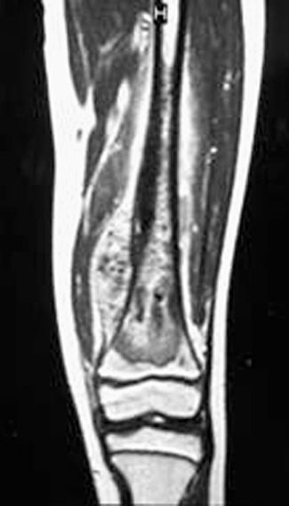
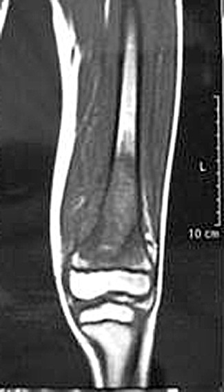
Between January 1999 and January 2005, five patients conformed to the above‐described criteria: one boy and four girls, with a mean age of 9.2 years (6–11 years). Their disease history ranged from one to six months. Before chemotherapy, three patients were type I (Fig. 1) and two type II (Fig. 2) on the MRI classification system 2 , and one of the type I patients had a pathological fracture.
Figure 1.

MRI of an osteosarcoma in the distal femur showing that the epiphyseal plate and physis are not invaded by tumor tissue.
Figure 2.

MRI of an osteosarcoma in the distal femur showing that the tumor tissue is in contact with the epiphyseal plate.
Surgical technique
The patient was placed on the operating table in the supine position under epidural or general anesthesia. A long midline incision was made, beginning in the mid thigh, and a medial parapatellar arthrotomy performed to enable wide exposure of the distal part of the femur and the knee joint. The biopsy track was preserved in continuity with the specimen. The distal part of the femur was approached through the gap between the rectus femoris and vastus medialis. If there was an extra‐osseous tumor component, a cuff of normal muscle was excised. Proximal femoral osteotomy was performed at the location determined by preoperative imaging studies, this location being 1–2 cm from the distal edge of tumor growth, which was defined as the point at which the marrow signal intensity changed from abnormal to normal. Measurements on preoperative MRI were correlated with anatomical landmarks to ensure the correct location of the osteotomy. After the posterior and medial structures had been protected and retracted, the osteotomy was performed perpendicular to the long axis of the femur. All remaining soft tissues at the level of the transection were then cleared as follows. The distal part of the femur was pulled forward in order to expose the soft‐tissue attachments of the popliteal space. The popliteal artery was mobilized and the geniculate vessels ligated and transected. Both heads of the gastrocnemius were released and the posterior capsule opened. The cruciate and collateral ligaments were identified, left intact and attached to the epiphysis, which was preserved. Following the osteotomy, the distal part of the femur, including both condyles, was removed. After cytological confirmation there were no tumor cells at the osteotomy site, reconstruction of the bone defect was performed with inactivated tumor‐bone shell that had been treated with 95% alcohol for 30 min and bone cement containing doxorubicin (20 g bone cement:10 mg doxorubicin). Next, the osteotomy site was stabilized by means of internal fixation with cancellous screws compressing the metaphyseal bone. An intramedullary nail was inserted to fix the diaphyseal osteotomy site. A suction drain was inserted and, after lavage of the wound with saline solution, meticulous suture repair of the quadriceps was performed, followed by layered closure of the subcutaneous tissues and skin. Finally, the knee was splinted externally in full extension with a brace.
Postoperative management
Antibiotics were given intravenously according to the usual prophylactic protocol; routine anticoagulation therapy was not used. The drainage tube was removed when the drainage amount was <50 ml/24 h. Progressive passive exercises were initiated with the affected limb protected by a plaster cast for 8 weeks. Sutures were removed 12–14 days postoperatively, after which postoperative chemotherapy was initiated, the drugs and dosage being based on the response to the preoperative chemotherapy. Eight weeks after surgery, the patients were permitted to walk with the aid of two crutches for the next 4 months.
Follow‐up
All patients were followed‐up regularly, including periodic evaluation of the length and function of the affected limb and to investigations to detect any local recurrence or distal metastases. Dynamic imaging was used to assess bone healing and osteosynthesis. The affected limb function was assessed according to the Musculoskeletal Tumor Society 93 system 3 .
Results
There were no surgical complications, and the absence of tumor cells at the osteotomy site determined by cytology intraoperatively was confirmed by post‐operative histopathology. No blood vessel or nerve injuries occurred and all incisions healed well.
All patients were followed‐up for 60–126 months (mean 82 months). Three patients could flex the affected knee joint ≥110°, one to 90°, and one to 70°. The length of the lower extremities was equal in one patient, the affected limb was 2 cm shorter than that of the control in three cases, and 8 cm shorter in one case. Four patients could walk without orthopedic footwear. Limb function scores were 13–30 points (mean 25.6). No patient had developed metastases or local recurrence by the final follow‐up. Table 1 shows general patient information including the follow‐up results and Table 2 gives the results of assessment of limb function.
Table 1.
General characteristics of five children with osteosarcoma
| NO. | Sex | Age (years) | MRI type | Pathological fracture | Follow‐up duration (months) | Comparison of limb length | Flexion of affected knee (0) | Relapse | Metastasis | Death | Other |
|---|---|---|---|---|---|---|---|---|---|---|---|
| 1 | Female | 9 | I | No | 126 | 2 cm shorter | 110 | No | No | No | Fracture |
| 2 | Female | 14 | II | No | 89 | Equal length | 135 | No | No | No | No |
| 3 | Female | 6 | II | No | 66 | 2 cm shorter | 90 | No | No | No | No |
| 4 | Male | 9 | I | No | 60 | 1 cm shorter | 135 | No | No | No | Fracture |
| 5 | Female | 8 | I | Yes | 69 | 8 cm shorter | 70 | No | No | No | Fracture |
Table 2.
Musculoskeletal Tumor Society scores of patients
| NO. | Pain | Function | Emotional | Supports | Walking | Gait | Total |
|---|---|---|---|---|---|---|---|
| 1 | None | Slightly restricted | Happy | None | Normal | Claudication | 28 |
| 2 | None | Normal | Happy | None | Normal | Normal | 30 |
| 3 | None | Slightly restricted | Happy | None | Normal | Claudication | 27 |
| 4 | None | Normal | Happy | None | Normal | Normal | 30 |
| 5 | None | Some apraxia | Satisfactory | Crutches | Restricted | Severe abnormalities | 13 |
Using dynamic imaging inspection to assess bone healing between the inactivated bone and epiphysis, we found callus formation between the inactivated bone and diaphysis at 2 months after surgery; increased callus at 4 months and complete bone union between the inactivated bone and diaphysis at 6 months after surgery.
Complications
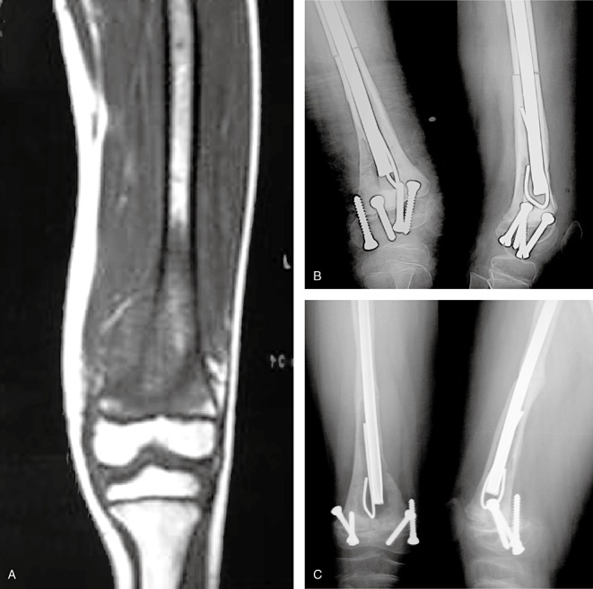
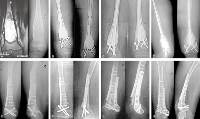
During follow‐up, four patients underwent reoperation, the exception being No. 3 (Fig. 3). No. 1 patient underwent open reduction, bone grafting and internal fixation because of inactivated bone fracture at 26 months after initial surgery; the bone had healed well by 4 months after reoperation. This patient can now walk without orthopedic footwear, the affected limb being 2 cm shorter than the unaffected limb, and flexion of the affected knee joint is ≥110° with a limb function score of 28 at 126 months after the first operation. Like No.1, No. 4 patient required reoperation 18 months after operation; this patient can now walk normally, the affected limb being shorter 1 cm than the unaffected limb and flexion of the affected knee joint ≥135°with a limb function score of 30 at 42 months after reoperation. No. 5 patient (Fig. 4) underwent open reduction and allografting combined with autografting for inactivated bone fracture 12 months after the initial surgery. We performed autografting and fixing with plate again at 20 months after reoperation because of resorption of allografted bone. Although the grafted bone had healed well at 36 months after the third operation, her affected limb was much shorter than her unaffected limb and her knee could flex only 70°. No. 2 patient underwent arthroscopic release, quadricepsplasty and continuous passive motion for knee stiffness at 3 years after the initial operation. At the last follow‐up 89 months postoperatively, the length of the lower extremities was equal and the affected knee flexed to normal.
Figure 3.

A 6 year‐old girl with an osteosarcoma in the left distal femur. (A) An MRI image showing no involvement of the epiphysis. (B) An X‐ray image one week after surgery showing internal fixation in good position. (C) An X‐ray image 40 months after surgery showing bone healing with internal fixation in good position.
Figure 4.

An 8‐year‐old girl with an osteosarcoma in the right distal femur (A) MRI and X‐ray images show no involvement of the epiphysis (Type I). (B) Four days after surgery, the X‐ray image shows a visible gap between the host and inactivated bone with internal fixation in good position. (C) Three months after surgery, the gap has decreased with internal fixation in good position. (D) Two years after surgery, the inactivated bone was found to have fragmented with internal fixation still in good position. (E) One week after reoperation and implantation of allograft showing internal fixation in good position. (F) Twenty months after reoperation, the allograft has fragmented. (G) Three months after the third operation, the internal fixation and autograft are in good position. (H) Twenty‐eight months after the third operation, bone healing is visible in the X‐ray images with internal fixation in good position.
Discussion
The aims of LSOPE, an accepted innovative operation for osteosarcoma in children, are to avoid postoperative discrepancies in limb length and improve limb function without increasing the risk of local recurrence or jeopardizing the patient's life. Nowadays, many techniques for performing LSOPE have been described in published reports. Cañadell et al. applied physeal distraction that literally pulled the tumor away from the epiphysis, leaving behind a widened boundary of newly formed bone between the tumor and the epiphysis 4 . En bloc resection of the tumor was then performed without sacrificing the adjacent joint and reconstruction of the bone defect with allograft undertaken as soon as absence of tumor at the edges of the resected segment had been confirmed pathologically. Manfrini et al. described a surgical technique in which resection is performed with fluoroscopic guidance to confirm the absence of tumor cells in the osteotomy regions while preserving the epiphysis, following which reconstruction of the bone defect with allograft and contralateral fibular graft is undertaken, the residual epiphysis being fixed to the allograft bone by screws 5 . The procedure described by Tsuchiya et al. involves en bloc tumor resection with preservation of the epiphysis and temporary shortening of the affected limb, following which distraction osteogenesis is performed to prevent limb length discrepancy 6 . Wang et al. reported limb salvage surgery with epiphyseal preservation in children and adolescents and bone defect reconstruction with massive allograft bone transplantation 7 . Since 1999, we have used inactivated bone shell reimplantation with preservation of the epiphysis to treat osteosarcoma in children without impinging on the epiphysis, and have previously reported the short term outcomes and complications of this operation 8 . All aspects of the short‐term outcomes were satisfactory.
The probability of postoperative complications is much higher with LSOPE than with any other surgery for osteosarcoma, however it has the advantages of avoiding limb length discrepancy and improving affected limb function. The main complications that have been reported are infection, graft bone resorption, fracture and internal fixation loosening. Among 20 patients treated by Cañadell et al., two had infection, three absorption of graft bone, one fibular nerve palsy and one graft bone fracture 4 . Of the patients reported by Tsuchiya et al., four had superficial infection, seven deep infection, three fractures, five fibular nerve palsy, and seven delayed bone healing 6 . In 11 of the 13 cases reported by Muscolo et al. who were followed up, complications occurred in seven, including grafting bone fractures in three, metaphyseal non‐union in two, deep infection in one and soft tissue recurrence in one 9 . The main complications in the patients treated by Wang et al. (mean follow‐up 37.6 months, range 12–72 months) were bone non‐union, graft bone fracture and nerve injury 7 .
In our group, in five patients who were followed up for 5–10 years the long‐term complications were inactivated bone fractures and flexion limitation of the affected knee. Three patients developed inactivated bone fractures 1–2 years after surgery, the fracture locations being at the junction between inactivated and host bone. We therefore think it is important to perform bone grafting around the osteotomy region during primary surgery to enhance bone healing. One of the three patients who had been type I on preoperative MRI imaging and had presented with a pathological fracture developed an inactivated bone fracture. An inappropriate choice of surgical procedure was the reason for this result. We now realize that our procedure is not suitable for patients with associated pathological fracture. Although our patient underwent allograft bone reimplantation and open reduction with autograft bone reimplantation, her affected limb was much shorter (8 cm) than the normal limb and she had limited knee flexion. Our experience indicates that patients with type I on MRI combined with pathological fractures can undergo LSOPE after effective preoperative chemotherapy, but en bloc resection of the tumor should be performed, preserving the epiphysis, and the bone defect reconstructed with massive allograft bone transplantation; intramedullary nail is the best choice for fixation.
Knee stiffness, a common complication of surgery on the distal femur, is an important factor influencing clinical outcomes. No. 2 patient, who we had found to be tumor‐free on many follow‐up examinations, underwent arthroscopic knee release and quadricepsplasty 3 years after the primary operation because of her affected knee stiffness. At her last follow‐up, she could move her affected knee normally and her lower extremities were of equal length.
One of purposes of performing LSOPE is to avoid postoperative limb length discrepancy. Manfrini et al. followed up six patients with proximal tibial tumors 5 . They documented that even an epiphyseal segment that is only 5 mm thick can survive and grow to full skeletal maturity; none of their patients had a limb length discrepancy of greater than 3.5 cm, their functional results averaged 95% of those for the normal lower limbs, and they reported no cases of knee instability or anterior cruciate ligament laxity. They also found the presence of screws did not stop growth but could be a factor in producing smaller proximal tibias. Wang et al. reported an excellent or good rate for the affected limb of 82.8%, the mean limb length discrepancy was 3.2 cm (2–6 cm) 7 . They found no joint instability or osteoarthritis during follow‐up. In our group, all patients were followed up for 60–126 months (mean 82 months). Five patients could flex the affected knee joint 70°–135°. The length of the lower extremities was equal in one patient, the affected limb was 2 cm shorter than the control in three cases, and 8 cm in one (this large discrepancy resulted from an inappropriate choice of surgical procedure). Four patients could walk without orthopedic footwear (the exception being No.5 patient). Limb function scores averaged 25.6 points (range, 13–30 points). We also found the presence of screws did not influence the function of the affected limb. It is our experience that the long‐term outcomes of inactivated bone reimplantation with preservation of the epiphysis are satisfactory provided we conform strictly to the indications for LSOPE and choose the appropriate surgical procedure.
Disclosure
No benefits in any form have been, or will be, received from a commercial party related directly or indirectly to the subject of this manuscript.
References
- 1. Yu XC, Liu XP, Li KH. Inactivated bone replantation with preservation of the epiphysis: clinical report of two cases and literature review (Chin). Zhonghua Xiao Er Wai Ke Za Zhi, 2002, 23: 565–566. [Google Scholar]
- 2. Yu XC, Liu XP, Zhou Y, et al Value of MRI for detecting epiphyseal extension of osteosarcoma (Chin). Zhongguo Jiao Xing Wai Ke Za Zhi, 2003, 11: 874–877. [Google Scholar]
- 3. Enneking WF, Dunham W, Gebhardt MC, et al A system for the functional evaluation of reconstructive procedures after surgical treatment of tumors of the musculoskeletal system. Clin Orthop Relat Res, 1993, 286: 241–246. [PubMed] [Google Scholar]
- 4. Cañadell J, Forriol F, Cara JA. Removal of metaphyseal bone tumours with preservation of the epiphysis. Physeal distraction before excision. J Bone Joint Surg Br, 1994, 76: 127–132. [PubMed] [Google Scholar]
- 5. Manfrini M, Gasbarrini A, Malaguti C, et al Intraepiphyseal resection of the proximal tibia and its impact on lower limb growth. Clin Orthop Relat Res, 1999, 358: 111–119. [PubMed] [Google Scholar]
- 6. Tsuchiya H, Abdel‐Wanis ME, Sakurakichi K, et al Osteosarcoma around the knee. Intraepiphyseal excision and biological reconstruction with distraction osteogenesis. J Bone Joint Surg Br, 2002, 84: 1162–1166. [DOI] [PubMed] [Google Scholar]
- 7. Wang Z, Guo Z, Liu JZ, et al Limb salvage surgery for malignant bone tumor of the extremities (Chin). Zhonghua Gu Ke Za Zhi, 2006, 26: 813–818. [Google Scholar]
- 8. Yu XC, Liu XP, Zhou Y, et al Epiphyseal preservation and reconstruction with inactivated bone in distal femur for metaphyseal osteosarcoma in children (Chin). Zhongguo Jiao Xing Wai Ke Za Zhi, 2007, 15: 811–813. [Google Scholar]
- 9. Muscolo DL, Ayerza MA, Aponte‐Tinao LA, et al Partial epiphyseal preservation and intercalary allograft reconstruction in high‐grade metaphyseal osteosarcoma of the knee. J Bone Joint Surg Am, 2005, 87 (Suppl 1, Pt 2): 226–236. [DOI] [PubMed] [Google Scholar]


