

Abstract
The authors describe a hybrid technique that involves a combination of open decompression and posterior lumbar interbody fusion (PLIF) and percutaneously inserted pedicle screws. This technique allows performance of PLIF and decompression via a midline incision and approach without compromising operative time and visualization. Furthermore, compared to standard open decompression, this approach reduces post‐operative wound pain because the small midline incision significantly reduces muscle trauma by obviating the need to dissect the paraspinal muscles off the facet joint complex and by avoiding posterolateral fusion, thus requiring limited lateral muscle dissection off the transverse processes. A series of patients with Grade I‐II spondylolisthesis at L4–5 and moderate–severe canal/foraminal stenosis underwent midline PLIF at L4–5, with closure of the midline incision. Percutaneous pedicle screws were inserted, thereby minimizing local muscle trauma, reduction of the spondylolisthesis being performed by using a pedicle screw construct. Rods were inserted percutaneously to link the L4 and L5 pedicle screws. Image intensification was used to confirmed satisfactory screw placement and reduction of spondylolisthesis. The results of a prospective study comparing a standard open decompression and fusion technique for spondylolisthesis versus the minimally invasive hybrid technique are discussed. The minimally invasive technique resulted in shorter hospital stay, earlier mobilization and reduced postoperative narcotic usage. The long‐term clinical outcomes were equivalent in the two groups.
Keywords: Percutaneous Lumbar Pedicle Screws, Posterior Lumbar Interbody Fusion, Technique, 80/20, Technique, Hybrid, Spondylolisthesis
Introduction
Degenerative lumbar spondylolisthesis is a challenging clinical entity. When associated with lumbar canal and/or foraminal stenosis, patients present with claudicant and/or radicular symptoms, respectively. Surgical intervention provides more positive outcomes than conservative management. The spondylolisthesis arm of the spine patient outcomes research trial (SPORT) concluded that in a non‐randomized as‐treated environment (with control of potentially confounding baseline factors), after 2 years outcomes were significantly better in regards to pain and function for patients with degenerative spondylolisthesis and spinal stenosis than for those treated non‐surgically (SPORT Trial)1. The correlation with mechanical low back pain is less clear and will not be discussed here.
One of the treatment methods for degenerative spondylolisthesis with claudicant and/or radicular symptoms is lumbar laminectomy with instrumented fusion. This can be performed via a posterior (pedicle screw fixation ± posterolateral graft ± posterior or transforaminal lumbar interbody fusion, PLIF/TLIF), or anterior approach (anterior lumbar interbody fusion, ALIF), or a combination of the above. PLIF is reportedly equivalent to TLIF in regards to post‐operative outcomes2. In one prospective study, PLIF was found to have a higher fusion rate than posterolateral fusion alone, but at the cost of a higher rate of complications related to hardware biomechanics3. Kim et al. directly compared PLIF, posterolateral fusion and PLIF with posterolateral fusion and found no difference in regards to clinical results and union rates between the three. However, they noted that PLIF alone resulted in less donor site pain, shorter operating time and less blood loss (it has been noted that these benefits are, at least in part, attributable to not taking any iliac crest bone graft in the PLIF group)4. A review of PLIF versus posterolateral fusion for management of isthmic spondylolisthesis reported a higher fusion rate for PLIF (93%) versus 68% for posterolateral fusion. However, the only statistically significant improvement in outcome was for high grade slipping managed with PLIF; there was no difference in outcomes for low grade slips5. An earlier review noted that, for isthmic spondylolisthesis, PLIF conferred some statistically, but not clinically, significant improvements in some outcome indicators compared to posterolateral fusion6.
Disadvantages of the open posterior over other approaches include the morbidity of the increased muscle dissection this approach entails, increased post‐operative wound pain (both short and long term), slower post‐operative mobilization (and subsequent longer hospital stays) and increased intra‐operative blood loss. An alternative to the “traditional” open approach is to a minimally invasive method using percutaneous pedicle screws in combination with a minimally invasive bilateral laminotomy and PLIF. However, disadvantages of this method include longer operating times and higher complication rates7.
The alternative method presented here involves a combination of open and minimally invasive approaches and has the aim of maximizing the benefits while avoiding the disadvantages of both the open and minimally invasive approaches. Open laminectomy and PLIF is performed at the level of the affected disc to minimize operative time and provide adequate exposure. This allows minimization of intra‐operative adverse events while providing adequate access in the event of occurrence of adverse events (e.g. incidental durotomy). A PLIF alone is performed (without a posterolateral graft) to minimize muscle dissection and retraction laterally without compromising rates of fusion. Finally, percutaneous pedicle screws are placed in all pedicles to minimize muscle dissection off the facet joints, thus minimizing postoperative wound pain (Fig. 1).
Figure 1.
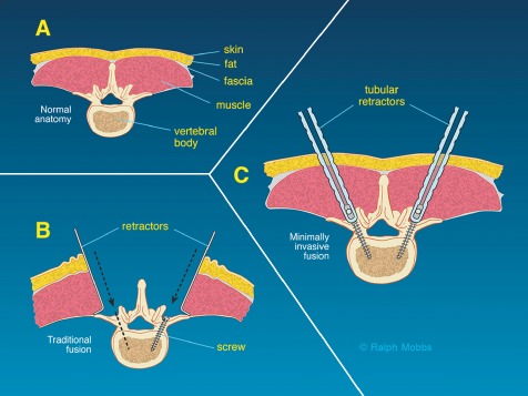
Diagrams showing rationale for MIS pedicle screw fixation: minimization of muscle trauma with a percutaneous pedicle screw insertion technique (A) Normal anatomy of the lumbar vertebra. (B) Traditional fusion. (C) Minimally invasive fusion.
Technique
The name of the hybrid (“80/20”) technique described here (Fig. 2) was coined by the senior author (RJM) to describe the relative importance of each step in the procedure. The initial “80%” is the primary goal of the technique: decompression of the neurological elements, preparation of the vertebral endplates and insertion of an interbody cage on either side of the thecal sac (Fig. 3). The final “20%” is the percutaneous insertion of the pedicle screws and reduction of the spondylolisthesis. RJM has also previously described the “50/50 technique” (Fig. 4). In this procedure, the caudal pedicle screws are inserted via an open approach. This technique may be required if the caudal pedicle anatomy is difficult to determine on anterior‐posterior (AP) X‐ray films and the surgeon is not comfortable with inserting percutaneous pedicle screws at the affected level.
Figure 2.
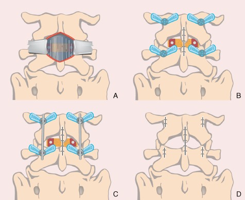
Workflow with hybrid (80/20) technique: (A) Performance of midline incision and PLIF. (B) Closure of the midline incision. (C) Percutaneous screw insertion via four incisions with reduction using a pedicle screw construct. (D) Closure of the percutaneous incisions.
Figure 3.
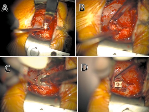
Midline PLIF technique via mini‐open approach: (A) Midline incision, decompression and preparation of interbody (disc) space. (B) Endplate preparation. (C) Insertion of a rotatable cage packed with graft. (D) Interbody cage in position.
Figure 4.
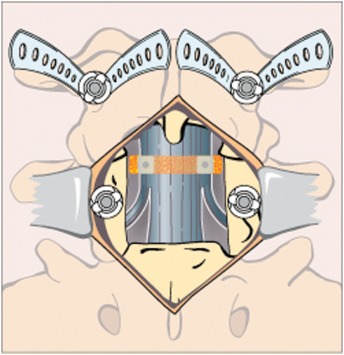
Workflow with 50/50 technique: Midline incision and PLIF are performed and a pedicle screw inserted into the caudal pedicle. Insertion of a percutaneous screw into the cranial pedicle avoids damage to the cranial/mobile facet joint.
Description of the hybrid 80/20 technique
Step 1
Under general anesthesia, the patient is positioned prone on a Jackson or similar operating table. Radiolucency of the operating table at the level of the surgery is essential to allow use of AP image intensifier X‐rays.
Step 2
A midline incision is performed directly over the L4–5 disc space with radiological confirmation of the level of the spondylolisthesis. Because lateral retraction is not necessary, only a short incision is required. Most incisions are between 3.5–5 cms long. Retraction using a Versa‐Trac (Medtronic Sofamor Danek, Memphis, TN, USA) or Trim‐Line (Medtronic Sofamor Danek) is followed by performance of a laminectomy at L4–5. A bilateral medial facetectomy at L4–5 with rhizolysis of both L5 nerve roots is then performed ½. The inferior ½–½ of the L4 spinous process is removed to assist with neural retraction. The disc at L4–5 is then removed and the endplates prepared (Fig. 3). The bone from the L4 spinous process, laminae and L4–5 facets is cleaned of residual ligament/soft tissue and prepared using a bone mill. It is then combined with osteobiologic material (Kasios TCP granules; Kasios Biomaterials, L'Union, France) and bone marrow aspirate before being packed into two PLIF cages (Vigor rotatable PLIF cages; A‐spine Asia, Taipei, Taiwan) and inserted into the L4–5 disc space. After achieving homeostasis, the midline wound is closed in layers (Fig. 2).
Step 3
The X‐ray/II machine is moved into a position that targets the L4 and L5 pedicles. A Jamshidi (CareFusion, San Diego, CA, USA) needle is introduced via a stab incision along the lateral aspect of the pedicle on the AP view. The Jamshidi is introduced into the pedicle to a depth of 20–25 mms making sure not to breach the medial border of the pedicle wall by observing the AP view on the image intensifier. A lateral X‐ray film is taken to confirm the position of the Jamshidi in the vertebral body. After confirmation that the pedicles have been penetrated by the needle, the trochar is removed and Kirschner (K)‐wires introduced through the barrel of the Jamshidi needle. Their position is confirmed prior to advancement of the K‐wires through the pedicles under lateral fluoroscopy. Once satisfactory penetration of the pedicles with the K‐wire has been achieved, the Jamshidi needle is removed whilst taking care to keep the K‐wires in the same position. Appropriate skin incisions are then made. A pedicle tap is introduced down the K‐wire through the pedicle into the trabecular bone of the vertebral body, this then being confirmed by image intensifier. The tap is then removed and appropriate pedicle screws (measurements based on pre‐operative CT scans) sited. Confirmation of pedicle screw placement is obtained with the image intensifier. Reduction of the spondylolisthesis is then performed using the standard reduction instrumentation that is provided with both systems used in the study (Serengeti, K2M, Leesburg, VA, USA and MANTIS, Stryker, Allendale, NJ, USA). At the completion of the procedure, the four stab incisions are closed (Fig. 2) with a single suture for the deep fascial and a single suture for the skin incision.
Step 4
Following reversal of anesthesia, the patient is extubated and transferred to the ward. Mobilization can be attempted from the first postoperative day. Post‐operative CT of the lumbar spine allows confirmation of reduction of the spondylolisthesis, as well as satisfactory positioning of the interbody devices, bone graft and all four pedicle screws. Follow up is routinely performed at 6 weeks and 3 months with flexion/extension X‐ray films to confirm solid fusion and reduction of the spondylolisthesis at L4–5 (Fig. 5).
Figure 5.
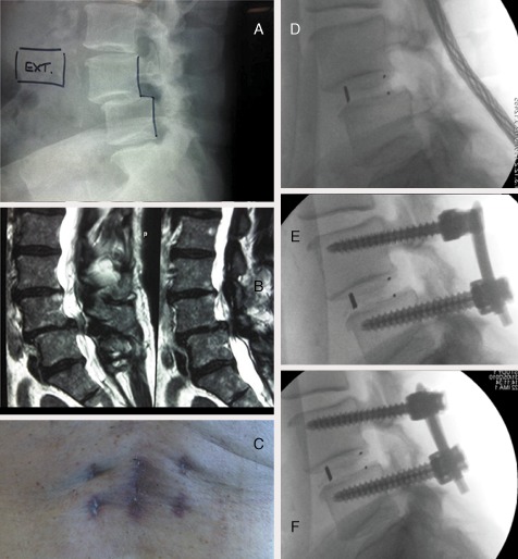
A case example: pre‐ and post‐ operative: (A) L 4–5 spondylolisthesis. (B) Severe canal stenosis. (C) Appearance of incisions 4 weeks postoperatively. (D) Performance of initial posterior lumbar interbody fusion. (E) Insertion of percutaneous screws. (F) Reduction maneuver and correction of spondylolisthesis.
Prospective Clinical Study
From 2007–2011, a prospective clinical study was conducted to compare the results of open versus minimally invasive fusion (MIS, the hybrid 80/20 technique) for degenerative lumbar spine pathologies. Eighty‐two patients were studied prospectively following appropriate ethics approval and consent (41 MIS spinal fusion, 41 open surgical equivalents) under a single surgeon (RJM). Data collected on all patients included Oswestry disability index (ODI) scores, Short Form 12 (SF‐12) v1, visual analogue scale (VAS), patient satisfaction index (PSI), length of hospital stay, time to mobilization, postoperative medication and complications. Inclusion criteria were patients aged 35–75 years with degenerative spondylolisthesis grade 1–2. All patients presented with either back pain, radiculopathy, claudication or a combination of these three symptoms. All patients had pain resistant to prolonged (at least six months) conservative therapy.
Clinical Results
Relevant patient variables, pre and post‐operative ODI and VAS responses are shown in Table 1. The indication for surgery was degenerative spondylolisthesis grade 1 or 2. All diagnoses were confirmed by dynamic X‐rays, CT scans and MRI. The average follow‐up time was 18.7 months (range, 8–40 months). The ODI and SF‐12 were utilized to assess the impact of these surgical techniques on patient disability and quality of life and the VAS to assess pain.
Table 1.
Relevant patient variables and outcome data in Hybrid technique and open groups
| Index | Hybrid technique (41 cases) | Open (41 cases) | P value |
|---|---|---|---|
| Age (years) | 68 ± 12 | 67 ± 13 | N/A |
| Sex (Male/Female) | 19/22 | 17/24 | N/A |
| Follow‐up time (months) | 17 | 19 | N/A |
| Median length of hospital stay (days) | 5.4 (37 cases) | 9.8 (32 cases) | 0.0019 |
| Average time to mobilization (mean ± SD, hours ) | 19 ± 8 (37 cases) | 29 ± 16 (37 cases) | 0.0025 |
| ODI (mean ± SD, %) | |||
| Preoperative | 54 ± 19 | 52 ± 17 | 0.81 |
| Postoperative | 25 ± 16 | 28 ± 16 | 0.23 |
| Change | 29 ± 19 | 24 ± 17 | 0.12 |
| SF‐12 (mean ± SD) | |||
| PCS | 41.0 ± 11.4 | 36.9 ± 10.6 | 0.1568 |
| MCS | 50.0 ± 8.8 | 50.9 ± 10.6 | 0.7634 |
| VAS (mean ± SD) | |||
| Preoperative | 7.9 ± 1.5 | 8.3 ± 1.5 | 0.2113 |
| Postoperative | 2.4 ± 2.2 | 3.3 ± 1.5 | 0.0144 |
| Change | 5.5 ± 2.4 | 5.0 ± 2.0 | 0.3199 |
Unpaired Student's t‐tests were utilized to compare normally distributed continuous data (age, follow‐up time, post‐operative ODI scores, SF‐12 mental and physical component scores [SF‐12 MCS and PCS], change in VAS). ODI (preoperative, change), VAS (pre‐ and postoperative), length of stay, time to mobilization, opioid and non‐opioid use were analyzed using the Mann Whitney test. Dichotomous variables (sex) were analyzed using the X2 test, whilst preoperative diagnosis and PSI and complications using the Fisher exact test. A P value of < 0.05 was considered to be statistically significant.
Both groups had significant improvements in quality of life and reduction in disability following their operations, ODI scores falling from 54% to 25% for the MIS technique (P < 0.0001) within the mean 16 month follow‐up time period, and from 52% to 28% for the open technique (P < 0.0001). Significant reductions in postoperative pain were observed after both procedures, VAS scores falling from 7.9 to 2.4 for the MIS technique (P < 0.0001) and from 8.2 to 3.3 for the open technique (P < 0.0001). Postoperative pain was significantly less following the MIS technique (2.4 vs 3.3). Despite this, the amount of pain relief (VAS change) provided by both procedures was not significantly different.
A similar proportion of MIS (83%) and open (78%) patients were satisfied with the benefits they experienced from their procedures. However, surgery met the expectations of a significantly greater proportion of MIS patients than of open patients (P = 0.023, Fig. 6). The MIS technique resulted in significantly shorter hospital stays (P = 0.0016) and time to mobilization (P = 0.002) than did the open technique.
Figure 6.
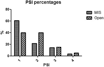
Patient Satisfaction Index. 1, surgery met my expectations; 2, I did not improve as much as I had hoped but I would undergo the same operation for the same results; 3, surgery helped but I would not undergo the same operation for the same outcome; 4, I am the same or worse as compared to before surgery.
The minimally invasive cohort had significantly less postoperative pain and met the expectations of a significantly greater proportion of patients than did conventional open surgery. The minimally invasive approach also had a significantly shorter length of stay and time to mobilization, less opioid use and a reduced total complication rate. In our study, minimally invasive and conventional open techniques were of similar efficacy; however, the former was superior in regards to patient satisfaction, length of hospital stay, time to mobilization and complication rates.
The minimally invasive technique had a significantly lower complication rate than the conventional open technique. There were two cases of infection with the open technique in comparison to one with the MIS technique (UTIs, no wound infections). One member of the minimally invasive group developed a painful hematoma postoperatively and presented with sacral and bilateral leg numbness. Motor function remained intact. One open patient also experienced postoperative radiculopathy. These patients were observed without treatment and their radiculopathy improved with time, however they did experience long‐term sensory impairment. One member of the open group had a dural tear and experienced headaches and vomiting following the procedure, which prolonged the length of stay. There were two cases of non‐union in the open group. They were identified following complaints of worsening mechanical lower back pain at the operation site over 9–12 months and finding of significant motion and subsidence on flexion‐extension lateral radiographs and CT. Both cases required subsequent revision surgery via an anterior approach (ALIF). There were also three cases of postoperative paralytic ileus, identified after complaints of nausea and vomiting, in the open group. A single case of deep vein thrombosis occurred in the open cohort.
Discussion
Posterior lumbar decompression and fusion is an established technique for providing symptomatic and functional relief from a complex degenerative process. The multiple alternatives currently available for approaching degenerative spondylolisthesis potentially create a decision and management dilemma. The SPORT trial, spondylolisthesis arm, concluded that surgical management provides better results than non‐surgical management1.
Researchers have previously shown that PLIF provides high fusion rates that are at least as good as, if not superior to, posterolateral fusion in regard to correction of spondylolisthesis and improvements in coronal and sagittal balance8. However, the traditional open approach has significant morbidity with regards to intra‐operative blood loss and postoperative wound pain and time to mobilization. Minimally invasive pedicle screw fixation and minimally invasive TLIF using the METRx (Medtronic, Memphis, TN, USA) system have reportedly been combined, the authors citing the advantages of decreased blood loss, wound pain and average length of post‐operative hospital stay9 , 10. The authors did acknowledge, however, that the limited exposure potentially provides an environment for an increased chance of intra‐operative adverse events and a reduced operative field for correcting any such events (such as unintended durotomies). Previously published reports indicate that intra‐operative durotomy rates are significantly increased, as is the length of the procedure7 , 9 , 10. Combining an open laminectomy with the PLIF procedure avoids the disadvantages of minimally invasive TLIF/PLIF but retains the benefits of percutaneous pedicle screws.
One source of post‐operative wound pain is muscle dissection off the facet joints and transverse process. With traditional open fusion, this is necessary to provide exposure for the pedicle screw entry points, especially the most rostral screw. Use of percutaneous pedicle screws requires minimal muscle dissection and thus avoids this morbidity. Another source of post‐operative wound pain is far lateral muscle dissection off the transverse processes to allow for a posterolateral graft. Because the procedure described here involves PLIF alone, no lateral dissection of muscle off the transverse processes is necessary.
Finally, in combination with the above two means of reducing postoperative pain, the smaller exposure required both laterally and craniocaudally allows for a more minimalistic incision involving less soft tissue dissection, without compromising access11. Wound size is reportedly independent of post‐operative pain12, but at the very least it is logical that a smaller wound facilitates reduced muscular exposure and greater patient satisfaction.
The senior author (RJM) has performed 72 hybrid “80/20” type procedures for degenerative spondylolisthesis. To date, no patient has required a blood transfusion and their average length of stay has been approximately 4 days. In addition, over 50% of patients have not required postoperative morphine/narcotic based analgesia.
We conclude that the hybrid technique for decompression and fusion is a safe and reproducible approach to management of degenerative spinal pathologies such as spondylolisthesis.
The “80/20” approach proposed by the authors has been successfully employed at our institution with encouraging results. The method of open PLIF and percutaneous pedicle screw fixation allows for minimization of muscular dissection, thus reducing morbidity and requirements for postoperative pain medication and allow earlier mobilization, whilst providing effective decompression and stabilization of the degenerative motion segment.
Disclosure: No financial support was obtained for this work.
References
- 1. Weinstein JN, Lurie JD, Tosteson TD, et al Surgical versus nonsurgical treatment for lumbar degenerative spondylolisthesis. N Engl J Med, 2007, 356: 2257–2270. [DOI] [PMC free article] [PubMed] [Google Scholar]
- 2. Yan DL, Pei FX, Li J, Soo CL. Comparative study of PILF and TLIF treatment in adult degenerative spondylolisthesis. Eur Spine J, 2008, 17: 1311–1316. [DOI] [PMC free article] [PubMed] [Google Scholar]
- 3. Cheng L, Nie L, Zhang L. Posterior lumbar interbody fusion versus posterolateral fusion in spondylolisthesis: a prospective controlled study in the Han nationality. Int Orthop, 2009, 33: 1043–1047. [DOI] [PMC free article] [PubMed] [Google Scholar]
- 4. Kim KT, Lee SH, Lee YH, Bae SC, Suk KS. Clinical outcomes of 3 fusion methods through the posterior approach in the lumbar spine. Spine (Phila Pa 1976), 2006, 31: 1351–1357. [DOI] [PubMed] [Google Scholar]
- 5. Dehoux E, Fourati E, Madi K, Reddy B, Segal P. Posterolateral versus interbody fusion in isthmic spondylolisthesis: functional results in 52 cases with a minimum follow‐up of 6 years. Acta Orthop Belg, 2004, 70: 578–582. [PubMed] [Google Scholar]
- 6. La Rosa G, Conti A, Cacciola F, et al Pedicle screw fixation for isthmic spondylolisthesis: does posterior lumbar interbody fusion improve outcome over posterolateral fusion? J Neurosurg, 2003, 99 (2 Suppl): 143–150. [DOI] [PubMed] [Google Scholar]
- 7. Park Y, Ha JW. Comparison of one‐level posterior lumbar interbody fusion performed with a minimally invasive approach or a traditional open approach. Spine (Phila Pa 1976), 2007, 32: 537–543. [DOI] [PubMed] [Google Scholar]
- 8. Wang JC, Mummaneni PV, Haid RW. Current treatment strategies for the painful lumbar motion segment: posterolateral fusion versus interbody fusion. Spine (Phila Pa 1976), 2005, 30 (16 Suppl): S33–S43. [DOI] [PubMed] [Google Scholar]
- 9. Foley KT, Holly LT, Schwender JD. Minimally invasive lumbar fusion. Spine (Phila Pa 1976), 2003, 28 (15 Suppl): S26–S35. [DOI] [PubMed] [Google Scholar]
- 10. Khoo LT, Palmer S, Laich DT, Fessler RG. Minimally invasive percutaneous posterior lumbar interbody fusion. Neurosurgery, 2002, 51 (5 Suppl): S166–S181. [PubMed] [Google Scholar]
- 11. Mobbs RJ, Sivabalan P, Li J. Technique, challenges and indications for percutaneous pedicle screw fixation. J Clin Neurosci, 2011, 18: 741–749. [DOI] [PubMed] [Google Scholar]
- 12. Datta G, Gnanalingham KK, Peterson D, et al Back pain and disability after lumbar laminectomy: is there a relationship to muscle retraction? Neurosurgery, 2004, 54: 1413–1420. [DOI] [PubMed] [Google Scholar]