Abstract
Diabetes can affect the skin structure as well as the cutaneous vascular permeability. However, effective methods to quantitatively evaluate diabetes-induced skin disorders in vivo are still lacking. Here, we visualized the skin by using in vivo two-photon imaging and quantitatively evaluated the collagen morphology. The results indicated that diabetes could cause a significant reduction in the number of collagen fibers and lead to the disorder of skin collage fibers. And, the classic histological analysis also showed diabetes did lead to the change of skin filamentous structure. Additionally, the Evans Blue dye was used as an indicator to evaluate vascular permeability. We in vivo monitored cutaneous microvascular permeability by combining spectral imaging with the skin optical clearing method. This work is very useful for quantitative evaluation of skin disorders based on in vivo optical imaging, which has a great reference value in the clinical diagnosis.
1. Introduction
Diabetes mellitus ranks among the most common diseases, and approximately thirty percent of diabetic patients suffer skin lesions during their lifetime [1,2]. Diabetes-induced dermatological complications mainly includes xerosis (dry skin) and foot ulcer, besides, the diabetics’ skin is more vulnerable to infection [3–5]. In diabetic patients, the skin structure in foot ulcer changed by assessment of ex vivo skin in hematoxylin-eosin–stained sections [1]. Diabetes not only leads to changes in skin structure, but also in skin components, such as variation of epidermal and dermal features [2], as well as nerve fiber loss [1,6]. Some studies show that diabetes induces skin dysfunctions like reduced skin barrier functions [7], changes in mechanical properties of dermal collagen fibril [8], mechanical hyposensitivity and abnormal temperature of skin [9,10]. In addition, the abnormal changes in motor behavior of skin immune cells or skin microvascular function occur with the development of diabetes [11,12]. Regardless of these diabetic-induced effects, most research studies mainly focus on the wounded diabetic skin. The study of non-wounded skin to evaluate skin disorders as a result of diabetes should also be performed, since it provides valuable diagnostic information.
Skin optical properties also changed due to diabetes-induced variation of structure or component of skin, which provides the direction for developing the optical method for quantitative evaluation of skin disorders. Tissue glycation is an important aspect for diabetes related biochemical process, in which advanced glycation end products (AGE) significantly accumulates. Increase of AGEs in various tissues has been usually performed in skin biopsies [13]. Besides, Accumulation of AGEs, as a normal physiological processes, is related to intrinsic aging and some age-related chronic diseases [14,15]. However, the methods for evaluating AGEs have some limitations, since it can’t be used for measuring non-fluorescent AGEs, furthermore the excitation spectrums of fluorophores (such as amino acids, e.g. tryptophan) overlapped [15,16]. To above mentioned restrictions, two-photon microscopic technique based on second harmonic generation (SHG) and autofluorescence in skin is used as a great tool for label-free in vivo 3D visualization and analysis of the skin [17–19]. The study showed SHG intensity of skin decreased due to diet-induced obesity by in vivo two-photon imaging of skin [15]. Some researchers reported that the SHG intensity decreased and autofluorescence intensity of skin increased during ex vivo skin glycation by using two-photon imaging [20]. These studies only focused on analyzing the intensity of SHG autofluorescence in skin, while largely neglecting the assessment of skin structural changes under some pathological conditions.
Additionally, diabetes affects not only the skin structure and components, but also the cutaneous permeability. Skin microvascular permeability generally determined based on assessment of Evans blue dye (EBd) from ex vivo skin sample [21]. However, the evaluation of extravasated EBd usually needs complicated biochemical methods to extract it from the ex vivo samples, furthermore the residual dyes in blood vessels may affect the measurement. The EBd leakage process cannot also be dynamically visualized. Spectral imaging, as a low-cost, simply-operated method, has been proposed for evaluation of microvascular permeability [22]. But the high scattering characteristic of skin limits the imaging properties of optical techniques. Fortunately, in vivo skin optical clearing method can be used for improving the performance in spectral imaging [12,23].
In this work, we used two-photon imaging to realize label-free in vivo 3D visualization of skin, and quantitatively evaluated the changes in skin collagen structure by texture analysis based on analysis of SHG images of type 1 diabetic (T1D) mice. In addition, we combined spectral imaging and skin optical clearing method to in vivo monitor skin microvascular permeability by quantitative evaluation of EBd leakage process.
2. Materials and methods
2.1 T1D model
Male Balb/c mice (8 weeks old) were intraperitoneally injected with 150 mg/kg alloxan (30 mg/ml) for a four-day administering to establish the T1D mice model. The blood glucose in mice were measured for validating the T1D mice model (fasting blood glucose >7 mmol/L). The experiment was performed after the mice kept hyperglycemia for 4 weeks. All the experimental procedures were performed according to animal experiment guidelines of the Experimental Animal Management Ordinance of Hubei Province, P. R. China.
2.2 Visualization of skin structure using two-photon imaging
In the experimental process, the mice were anesthetized by intraperitoneal injection of the mixture of 2% α-chloralose and 10% urethane (8 mL/kg). The dorsal hair was removed by depilatory cream (Veet, China).
The TPLSM (A1R MP, Nikon, Japan) is employed to image the structure of skin. Ti-sapphire laser (Mai Tai DeepSee, Spectra-Physics, Santa Clara, USA), offering over 2.4 W average power and the tunable wavelength ranging from 690 nm to 1040 nm, was employed as the excitation light source. The 16 × water-immersion objective was used for collecting the emitted fluorescence with a numerical aperture of 0.8 and a working distance of 3 mm. The size of the images in each stack was 1024 × 1024 pixels and the z-step was 1 μm. The dwell time was 2.4 μs/pixel, and the zoom factor was 2. In this experiment, the excitation wavelength was set at 770 nm, and the detection wavelength of skin two-photon autofluorescence (TPAF) was 525 ± 25 nm. Additionally, the filter of detection channel for SHG was 492 nm SP.
2.3 Quantitative evaluation of the disorder of skin collagen
To evaluate the relative collagen orientation, the ImageJ/FIJIs directionality plugin was employed to obtain the distribution of structure orientation. In addition, texture analysis method was used for quantitative evaluation of the disorder of skin collagen in each dermal depths with the MATLAB. We created a gray-level co-occurrence matrix () from the SHG collagen image for texture analysis, which was named as gray-level spatial dependence matrix. The in four directions (horizontal: 0°, vertical: 45°, left diagonal: 90°, right diagonal: 135°) was expressed as follow [24–26]:
was a matrix, and was the number of gray levels in the SHG collagen image. The element of this matrix () was generated by counting the number of adjacent pixels under the combination of different gray levels for each direction (For example, g(1,1,0°) represents the number of pixel groups with gray level of 1 and 1 for horizontally adjacent pixels (d = 0°)).
The gray level of co-occurrence matrix from the image can reflect the changes of gray levels between pixels of the image. It was the basis for analysis of local pattern and the arrangement of textures from the image. In order to visually evaluate the textures information of the images, the described two statistics were calculated based on the co-occurrence matrix [25,26]. Energy reflects the uniformity of the grayscale distribution and the regular pattern of texture. The Homogeneity can be used for evaluating the degree of the local change of image texture. The larger value of Homogeneity, the less change of image texture in different regions, and the more homogeneous the local image is. The mean values of energy and homogeneity in four directions can be calculated as follow:
2.4 Histopathologic examination of skin
The hair of dorsal skin in mice was removed. Then we cleaned the surface of skin and cut the skin samples down with small sharp scissors. The skin samples were fixed in 4% neutral paraformaldehyde, dehydrated and embedded in paraffin. After cutting into 4-μm sections, the samples were stained with Masson.
2.5 Spectral imaging combing with the skin optical clearing method for monitoring cutaneous permeability
In order to in vivo monitor skin microvascular permeability, we used the EBd as the indicator and employed the spectral imaging method that developed in previous work [22] to acquire the spectral images under the help of skin optical clearing method. The spectral imaging setup consists of a charge coupled device (CCD) camera (Pixelfly USB, PCO Company, Germany), a liquid crystal tunable filter (LCTF, CRi Varispec VIS, Perkin Elmer, USA; Bandwidth: 7 nm), a stereo microscopy (SZX7, Olympus, Japan) and a ring-like LED light with polarizer.
To image cutaneous microvessels, we established an optical clearing skin window. The usage about in vivo skin optical clearing method is detailed in reference [27]. After establishing optical clearing skin window, the anaesthetic mice were placed on a temperature stabilized heating pad at 37 °C. Then, we acquired images before intravenous injection of EBd for obtaining the spectral data of skin tissue and cutaneous vascular hemoglobin. After intravenous injection of 125 μL EBd (2% mass volume ratio, Sigma) and 10 min circulation, the spectral images were continuously recorded for 55 min with an interval of 5 min. Finally, a certified reflectance standard (SRS-99-020, Labsphere, USA) was used to acquire the illuminated calibration spectral images, and the dark noise of camera was measured by closing the camera shutter.
2.6 Quantitative evaluation of skin microvascular permeability
All spectral raw images were normalized to obtain the reflectance value () for each pixel as follows:
Where for each pixel, the is the radiance values of camera acquisition from spectral images, and represent the radiance values of the certified reflectance standard and the dark current at each wavelength λ, respectively. The absorption of each pixel can be calculated as follows:
The EBd concentration was measured according to the linear fitting equation of absorption at 620 nm. The skin EBd absorption of each pixel can be expressed as:
The represents the normalized reflectance value of each pixel at 620 nm, is the normalized reflectance value of each pixel which contains tissue and hemoglobin information from the acquired raw images before injection of EBd. Then we can evaluate the EBd concentration (C) based on the linear fitting equation that was proposed in our previous work as follows:
Where, is the skin EBd absorption of each pixel at 620 nm.
2.7 Statistical analysis
In this work, significant differences between the T1D group and Normal group were analyzed using Student’s t test. Significant differences among different time points were analyzed using one way analysis of variance with a Tukey’s multiple comparison test with MATLAB.
3. Results
3.1 The T1D-induced changes of skin using TPLSM
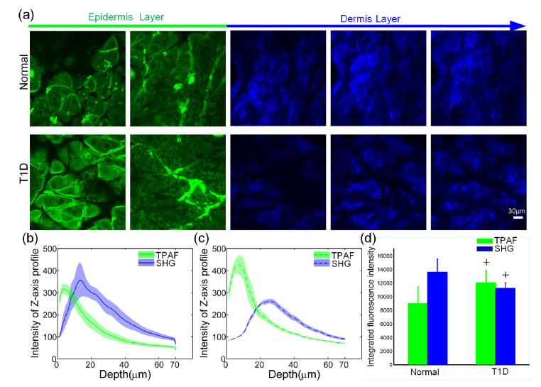
Here, the TPLSM was employed for imaging the TPAF and SHG signals of skin from epidermis layer to dermis layer in the normal and T1D mice as shown in Fig. 1(a). Further, the intensity of SHG and TPAF were quantitatively evaluated with z-axis. Under the same conditions, there were significant differences in TPAF and SHG intensity between the T1D (Fig. 1(c)) and normal mice (Fig. 1(b)). We obtained the integrated TPAF and SHG intensity by calculating the area under the profiles. It can be noticed that comparing to the normal mice, the integrated TPAF intensity of the T1D mice was significantly increased (p < 0.05) but the integrated SHG intensity was significantly decreased (p < 0.05) as shown in Fig. 1(d). It indicated that the T1D may cause the decrease of skin collagen. The increase of TPAF intensity for T1D could be related to the oxidative stress-induced increase of NAD(P)H due to the T1D [28–30].
Fig. 1.
Two-photon imaging of skin in normal and T1D mice. (a) The image of each layer for the dorsal skin in normal and T1D mice from the skin surface to deep layer of the dermis. (b) and (c) were the fluorescence intensity profiles of normal and T1D mice, respectively (The green and blue curves are mean values, and the shadows represent standard error, n = 6). (d) The integrated fluorescence intensity profiles of normal and T1D mice (Error bars represent the standard error, n = 6). + p < 0.05 compared with the normal mice.
3.2 Quantitative evaluation of T1D-induced changes in SHG collagen
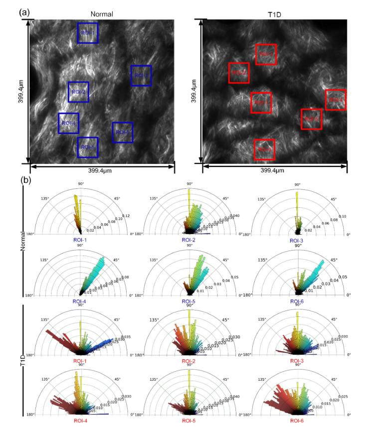
It can be found that there is a great difference in the structure of SHG collagen between the normal and T1D mice in the dermis as indicated in Fig. 2(a). The dermal collagen structure of the normal mice was relatively ordered and fiber-rich, while it obviously changed in T1D mice. Here, we analyzed the proportion of collagen fibers in MIP maps at 0°-180° direction via directionality analysis to assess the disorder degree of collagen fibers. Figure 2(b) shows that in the normal group, the collagen fibers are almost distributed in a certain direction, but they are distributed in all directions in T1D group. It indicated that T1D could lead to changes in the degree of disorder in fibril orientation.
Fig. 2.
The disorder degree of longitudinal orientated collagen of skin. (a) Typical maximum intensity projection maps (from 20 µm to 60 µm) of dermal SHG collagen in normal and T1D mice. (b) Quantitative evaluation of the skin collagen orientation from 0° to 180°corresponding to the blue and red regions of interest as indicated in (a) for normal mice and T1D mice. (The polar axis represents the proportion of collagen in each direction.)
Further, in order to quantitatively evaluate the disorder degree of skin collagen fibers, here, the texture analysis method was employed, and we selected the regions in Figure 2(a) for analysis. The energy and homogeneity of SHG images at different dermal depths were calculated respectively as shown in Fig. 3(a) and (b). The energy values of T1D group were lower than that of the normal group in Fig. 3(a), which means that there were some uniformities of gray distribution in SHG images caused by the T1D. The homogeneity values for T1D group were almost lower than that of the normal group as shown in Fig. 3(b), which suggested that the texture of SHG images in T1D mice changed more in different locations, and the local distribution of SHG collagen fibers was uneven. Further, the four-direction average values of energy and homogeneity of SHG maximum intensity projection maps from the normal and T1D mice were calculated respectively as shown in Fig. 3(c) and 3(d). Similarly, the energy and homogeneity values of T1D group were significantly lower than that of the normal group, these results suggest that T1D can cause the changes in textural features of dermal collagen, and the texture analysis method based on collagen SHG images could effectively evaluate the T1D-induced changes in skin filamentous structure.
Fig. 3.
The texture analysis of skin SHG collagen images. The energy (a) and homogeneity (b) of SHG images at different dermal depths in normal and T1D mice. (The dotted lines represent the average values of gray-level co-occurrence matrix of SHG images in four directions. The red and blue shadows represent standard deviations.). The four-direction average values of energy (c) and homogeneity (d) of SHG maximum intensity projection maps from the normal and T1D mice. (Error bars represent the standard error, n = 6, + p < 0.05 compared with the normal mice.)
3.3 Histological analysis of T1D-induced changes in skin with Masson stained sections
The T1D-induced changes in collagen fibers of skin were verified by classical Masson staining. The collagen in the skin of the normal mice were dense and their distribution was uniform. For the T1D mice, the color of collagen was shallow, the amount of collagen decreased obviously, and the collagen was arranged disorderly as shown in Fig. 4(a). The area ratio of collagen fibers was calculated and showed in Fig. 4(b). This result indicated that T1D could cause a significant reduction in the number of collagen fibers (p < 0.01) and also lead to their random orientation. This is consistent with the above results of texture analysis, suggesting that diabetes does lead to the changes in collagen fibers in the T1D mice model.
Fig. 4.
The Masson staining of skin. (a) The images of Masson stained skin of normal and T1D mice. (b) The area ratio of collagen for normal and T1D mice. + p <0.01 compared with the normal mice (n = 9, mean ± standard deviations).
3.4 Visualization of T1D-induced changes in skin microvascular permeability
We also monitored skin microvascular permeability in vivo by quantitatively evaluating the EBd concentration by combing spectral imaging and skin optical clearing method. With the help of optical clearing skin window, the cutaneous microvessels can be monitored clearly by the spectral imaging. The flow chart of monitoring the changes in vascular EBd was showed in Fig. 5(a). Figure 5(b) shows the cutaneous microvascular EBd concentration maps of the normal and T1D mice. For the normal mice, distribution of the EBd concentration changed little over time, while for the T1D mice, the EBd concentration of skin tissue increased as time went by. Further, the time-dependent changes in EBd concentration at vascular and extravascular positions were quantitatively analyzed as shown in Fig. 5(c) and (d), respectively. For the normal mice, there was no significant difference in the vascular and extravascular EBd concentration at different time points. However, for T1D mice, the vascular and extravascular EBd concentration started to significantly increase at 10 min. It indicated that T1D caused the increase of cutaneous microvascular permeability, and the combination of spectral imaging and skin optical clearing technique provided an efficient and low-cost method for in vivo visualization of skin microvascular permeability.
Fig. 5.
The permeability of cutaneous microvessels. (a) The flow chart of monitoring the vascular EBd. (b) The skin microvascular EBd concentration maps obtained by spectral imaging with the help of in vivo skin optical clearing method (The colorbar represents the EBd concentration). The time-dependent changes of EBd concentration at vascular positions (c) and extravascular positions (d) (The vascular positions and extravascular positions were white and red rectangular areas as indicated in (b), respectively. Mean ± standard deviations). + + p <0.01 compared with EBd concentration at 5 min.
4. Discussion
The elastin, NAD(P)H, collagen and melanin in skin have autofluorescence, in which the NAD(P)H in cells is present in the stratum corneum, stratum spinosum and basal layer [28]. The SHG signal is due to collagen in skin. Thus, we can in vivo visualize skin structure based on collagen SHG and epidermal autofluorescence by using TPLSM. The obtained distribution of SHG and the autofluorescence by this approach can well reflect the skin structure, which is consistent with that known from histology [28]. We found the TPAF intensity of T1D mice was significantly increased compared to the normal mice. The TPAF mainly origins from epidermal autofluorescence, due to NAD(P)H, tryptophan, keratin and melanin [31]. And some studies showed that diabetes could induce upregulation of the expression of NAD(P)H oxidase [32,33], which may lead to the increase of NAD(P)H in skin cells of T1D mice. Jo-Ya Tseng et al. also demonstrated that skin glycation caused the increase of TPAF signals based on ex vivo skin experiments [20]. These results are consistent with ours.
It is well known that SHG signal origins from collagen in biological tissue. Therefore, the collagen SHG have been widely used as a diagnostic parameter [34,35]. We found that for the T1D mice, the collagen SHG intensity decreased. Abd A. Tahrani et al. assess the collagen fibril by using ex vivo skin samples in hematoxylin-eosin–stained sections, and show that the diabetes-induced change in collagen fibril can lead to the abnormal changes in skin structure in foot ulcer [1]. Further, Dóra Haluszka et al. employ the two-photon imaging to in vivo monitor the diabetes-induced obesity skin changes, which illustrate that diabetes-induced obesity could lead to the decrease of SHG intensity [15]. And some study showed that skin glycation can lead to the decrease of SHG intensity by two-photon imaging ex vivo skin [20]. These results is consistent with ours.
Presently most of the researches focus on the analysis of SHG or autofluorescence intensity of skin in pathological state, but quantitative assessment of skin structural changes based on SHG receives relatively few attentions. Wenyan Hu et al. proposed the orientation-dependent gray level co-occurrence matrix method to quantitatively evaluate the SHG images from biological samples in pathological conditions [35]. It provides a useful approach for evaluation of collagen morphology. Thus, in this work, we used the texture analysis method based on gray-level co-occurrence matrix to quantitatively evaluate T1D-induced structural changes on collagen SHG. We found that T1D could lead to changes in the distribution of collagen, such as orientation and clutter, furthermore, the results about histological analysis also showed that T1D could induce the changes of collagen. It indicated that this analysis method based on SHG images could effectively evaluate diabetes-induced abnormal collagen morphology.
Additionally, diabetes can induce the increase of skin vascular permeability [36]. The in vitro studies on living cells show that the exudate concentration of the bovine serum protein is positively proportional to the concentration of glucose in the culture medium [37,38]. C. Schaefer et al. found that the vascular permeability increases due to diabetes by using intravital microscopic imaging with the skin-fold chamber, it leads to the massive exudation of the vascular fluorescent dye [36]. However, the skin-fold chamber established with surgery still has limitations, which will inevitably lead to a local or even systemic pro-inflammatory stimulus [39]. Besides, diabetes can facilitate inflammation of wound caused by the surgery, which may influence the vascular permeability [11]. Tissue optical clearing technique has great potential in improving the performance of optical imaging [27,40–43] and optical therapy [44,45]. Thereinto, in vivo skin optical clearing method provides an effective imaging window to fundamentally overcome the high scattering of skin. Therefore, in this work, we realized in vivo monitoring the dynamic changing process of EBd leakage due to the increase of cutaneous microvascular permeability by combining spectral imaging with the optical clearing skin window.
Evans Blue (68 kDa when combined with plasma albumin) has been widely used as an indicator for the evaluation of plasma volume in human and the study of vascular permeability in animal models [46]. Evaluation of EBd extravasation based on the extraction of EBd from ex vivo samples has been a classical method to reflect vascular barrier function [47]. However, extraction of EBd from the samples is a complicated biochemical process, which is very difficult for accurate quantitation of EBd [46]. Spectral imaging provides a useful tool for noninvasive evaluation of the characteristics of biological tissue [48]. In this work, we combined the spectral imaging with in vivo skin optical clearing method to visualize the dynamic changes in cutaneous microvascular permeability by quantitative evaluation of EBd. To accurately evaluate the EBd concentrations in the skin, the absorption from skin tissue and cutaneous vascular hemoglobin needs to be considered and subtracted. Therefore, before injection of EBd, we acquired the spectral image that was regarded as the background. Additionally, in this work, we performed the study on mice, but in the future study we hope this method based on in vivo skin optical imaging for quantitative evaluation of skin disorders can also be applied in human.
5. Conclusion
We quantitatively evaluated the skin disorders based on in vivo skin optical imaging in T1D mice. Visualization of skin structure was realized by using in vivo label-free two-photon imaging. T1D-induced changes in skin structure were quantitatively evaluated by texture analysis method based on collagen SHG images. The histological analysis also showed that diabetes did lead to the changes of collagen in this T1D mice model, indicating that texture analysis method based on collagen SHG images could effectively evaluate the T1D-induced changes in skin filamentous structure. Additionally, EBd, as an indicator, was used for quantitative evaluation of cutaneous microvascular permeability. We in vivo monitored T1D-induced increase of cutaneous microvascular permeability by combining spectral imaging with the in vivo skin optical clearing method, which provides a low-cost, simply-operated tool for in vivo monitoring skin microvascular permeability. This research shows a very useful tool for evaluation of collagen morphology and microvascular permeability in skin, which has a great reference value in the clinical diagnosis.
Acknowledgments
We thank to the Optical Bioimaging Core Facility of WNLO-HUST (Wuhan National Laboratory for Optoelectronics-Huazhong University of Science and Technology) for support in data acquisition.
Funding
National Natural Science Foundation of China (NSFC) (Grant No. 61860206009, 81870934, 31571002, 61721092); the National Key Research and Development Program of China (2017YFA0700501); the Fundamental Research Funds for the Central Universities; HUST (2019KFYXMBZ069; 2019KFYXJJS052); Innovation Fund of WNLO.
Disclosures
The authors declare that there are no conflicts of interest related to this article.
References
- 1.Tahrani A. A., Zeng W., Shakher J., Piya M. K., Hughes S., Dubb K., Stevens M. J., “Cutaneous structural and biochemical correlates of foot complications in high-risk diabetes,” Diabetes Care 35(9), 1913–1918 (2012). 10.2337/dc11-2076 [DOI] [PMC free article] [PubMed] [Google Scholar]
- 2.Quondamatteo F., “Skin and diabetes mellitus: what do we know?” Cell Tissue Res. 355(1), 1–21 (2014). 10.1007/s00441-013-1751-2 [DOI] [PubMed] [Google Scholar]
- 3.Szabad G., “[Diabetic foot syndrome],” Orv. Hetil. 152(29), 1171–1177 (2011). 10.1556/OH.2011.29168 [DOI] [PubMed] [Google Scholar]
- 4.Gkogkolou P., Böhm M., “[Skin disorders in diabetes mellitus],” J. Dtsch. Dermatol. Ges. 12(10), 847–863 (2014). [DOI] [PubMed] [Google Scholar]
- 5.Gonçalves N. P., Vægter C. B., Andersen H., Østergaard L., Calcutt N. A., Jensen T. S., “Schwann cell interactions with axons and microvessels in diabetic neuropathy,” Nat. Rev. Neurol. 13(3), 135–147 (2017). 10.1038/nrneurol.2016.201 [DOI] [PMC free article] [PubMed] [Google Scholar]
- 6.Amit S., Yaron A., “Novel systems for in vivo monitoring and microenvironmental investigations of diabetic neuropathy in a murine model,” J. Neural Transm. (Vienna) 119(11), 1317–1325 (2012). 10.1007/s00702-012-0808-9 [DOI] [PubMed] [Google Scholar]
- 7.Okano J., Kojima H., Katagi M., Nakagawa T., Nakae Y., Terashima T., Kurakane T., Kubota M., Maegawa H., Udagawa J., “Hyperglycemia Induces Skin Barrier Dysfunctions with Impairment of Epidermal Integrity in Non-Wounded Skin of Type 1 Diabetic Mice,” PLoS One 11(11), e0166215 (2016). 10.1371/journal.pone.0166215 [DOI] [PMC free article] [PubMed] [Google Scholar]
- 8.Argyropoulos A. J., Robichaud P., Balimunkwe R. M., Fisher G. J., Hammerberg C., Yan Y., Quan T., “Alterations of Dermal Connective Tissue Collagen in Diabetes: Molecular Basis of Aged-Appearing Skin,” PLoS One 11(4), e0153806 (2016). 10.1371/journal.pone.0153806 [DOI] [PMC free article] [PubMed] [Google Scholar]
- 9.Kambiz S., van Neck J. W., Cosgun S. G., van Velzen M. H., Janssen J. A., Avazverdi N., Hovius S. E., Walbeehm E. T., “An early diagnostic tool for diabetic peripheral neuropathy in rats,” PLoS One 10(5), e0126892 (2015). 10.1371/journal.pone.0126892 [DOI] [PMC free article] [PubMed] [Google Scholar]
- 10.Sejling A. S., Lange K. H., Frandsen C. S., Diemar S. S., Tarnow L., Faber J., Holst J. J., Hartmann B., Hilsted L., Kjaer T. W., Juhl C. B., Thorsteinsson B., Pedersen-Bjergaard U., “Infrared thermographic assessment of changes in skin temperature during hypoglycaemia in patients with type 1 diabetes,” Diabetologia 58(8), 1898–1906 (2015). 10.1007/s00125-015-3616-6 [DOI] [PubMed] [Google Scholar]
- 11.Shi R., Feng W., Zhang C., Yu T., Fan Z., Liu Z., Zhang Z., Zhu D., “In vivo imaging the motility of monocyte/macrophage during inflammation in diabetic mice,” J. Biophotonics 11(5), e201700205 (2017). [DOI] [PubMed] [Google Scholar]
- 12.Feng W., Shi R., Zhang C., Liu S., Yu T., Zhu D., “Visualization of skin microvascular dysfunction of type 1 diabetic mice using in vivo skin optical clearing method,” J. Biomed. Opt. 24(3), 1–9 (2019). 10.1117/1.JBO.24.3.031003 [DOI] [PMC free article] [PubMed] [Google Scholar]
- 13.Koetsier M., Lutgers H., Smit A. J., Links T. P., Vries R. D., Gans R. O., Rakhorst G., Graaff R., “Skin autofluorescence for the risk assessment of chronic complications in diabetes: a broad excitation range is sufficient,” Opt. Express 17(2), 509–519 (2009). 10.1364/OE.17.000509 [DOI] [PubMed] [Google Scholar]
- 14.Raut K., Agarwal S., Parikh A., “A review on advanced glycation end-products (age) and their role in diabetes mellitus,” International Journal of Pharma Sciences & Research 6(1), 80–86 (2015). [Google Scholar]
- 15.Haluszka D., Lőrincz K., Kiss N., Szipőcs R., Kuroli E., Gyöngyösi N., Wikonkál N. M., “Diet-induced obesity skin changes monitored by in vivo SHG and ex vivo CARS microscopy,” Biomed. Opt. Express 7(11), 4480–4489 (2016). 10.1364/BOE.7.004480 [DOI] [PMC free article] [PubMed] [Google Scholar]
- 16.Lutgers H. L., Graaff R., Links T. P., Ubink-Veltmaat L. J., Bilo H. J., Gans R. O., Smit A. J., “Skin autofluorescence as a noninvasive marker of vascular damage in patients with type 2 diabetes,” Diabetes Care 29(12), 2654–2659 (2006). 10.2337/dc05-2173 [DOI] [PubMed] [Google Scholar]
- 17.R. F. M. Hendriks and G. W. Lucassen, “Two-photon fluorescence microscopy of in-vivo human skin,” in EOS/SPIE European Biomedical Optics Week (2000), pp. 116–121. [Google Scholar]
- 18.Miyamoto K., Kudoh H., “Quantification and visualization of cellular NAD(P)H in young and aged female facial skin with in vivo two-photon tomography,” Br. J. Dermatol. 169(Suppl 2), 25–31 (2013). 10.1111/bjd.12370 [DOI] [PubMed] [Google Scholar]
- 19.Palero J. A., de Bruijn H. S., van der Ploeg-van den Heuvel A., Sterenborg H. J., Gerritsen H. C., “In vivo nonlinear spectral imaging in mouse skin,” Opt. Express 14(10), 4395–4402 (2006). 10.1364/OE.14.004395 [DOI] [PubMed] [Google Scholar]
- 20.Tseng J. Y., Ghazaryan A. A., Lo W., Chen Y. F., Hovhannisyan V., Chen S. J., Tan H. Y., Dong C. Y., “Multiphoton spectral microscopy for imaging and quantification of tissue glycation,” Biomed. Opt. Express 2(2), 218–230 (2011). 10.1364/BOE.2.000218 [DOI] [PMC free article] [PubMed] [Google Scholar]
- 21.Sun Z., Li X., Massena S., Kutschera S., Padhan N., Gualandi L., Sundvold-Gjerstad V., Gustafsson K., Choy W. W., Zang G., Quach M., Jansson L., Phillipson M., Abid M. R., Spurkland A., Claesson-Welsh L., “VEGFR2 induces c-Src signaling and vascular permeability in vivo via the adaptor protein TSAd,” J. Exp. Med. 209(7), 1363–1377 (2012). 10.1084/jem.20111343 [DOI] [PMC free article] [PubMed] [Google Scholar]
- 22.Feng W., Zhang C., Yu T., Semyachkina-Glushkovskaya O., Zhu D., “In vivo monitoring blood-brain barrier permeability using spectral imaging through optical clearing skull window,” J. Biophotonics 12(4), 201800330 (2019). [DOI] [PubMed] [Google Scholar]
- 23.Feng W., Shi R., Zhang C., Yu T., Zhu D., “Lookup-table-based inverse model for mapping oxygen concentration of cutaneous microvessels using hyperspectral imaging,” Opt. Express 25(4), 3481–3495 (2017). 10.1364/OE.25.003481 [DOI] [PubMed] [Google Scholar]
- 24.Clausi D. A., “An analysis of co-occurrence texture statistics as a function of grey level quantization,” Can. J. Rem. Sens. 28(1), 45–62 (2002). 10.5589/m02-004 [DOI] [Google Scholar]
- 25.Soh L. K., Tsatsoulis C., “Texture analysis of SAR sea ice imagery using gray level co-occurrence matrices,” IEEE Trans. Geosci. Remote Sens. 37(2), 780–795 (1999). 10.1109/36.752194 [DOI] [Google Scholar]
- 26.Haralick R. M., Shanmugam K., Dinstein I. H., “Textural Features of Image Classification,” Stud. Media Commun. 3, 610–621 (1973). [Google Scholar]
- 27.Shi R., Guo L., Zhang C., Feng W., Li P., Ding Z., Zhu D., “A useful way to develop effective in vivo skin optical clearing agents,” J. Biophotonics 10(6-7), 887–895 (2017). 10.1002/jbio.201600221 [DOI] [PubMed] [Google Scholar]
- 28.Laiho L. H., Pelet S., Hancewicz T. M., Kaplan P. D., So P. T. C., “Two-photon 3-D mapping of ex vivo human skin endogenous fluorescence species based on fluorescence emission spectra,” J. Biomed. Opt. 10(2), 024016 (2005). 10.1117/1.1891370 [DOI] [PubMed] [Google Scholar]
- 29.So P., Kim H., Kochevar I., “Two-Photon deep tissue ex vivo imaging of mouse dermal and subcutaneous structures,” Opt. Express 3(9), 339–350 (1998). 10.1364/OE.3.000339 [DOI] [PubMed] [Google Scholar]
- 30.Wautier J. L., “Activation of NADPH oxidase by advanced glycation end products (AGEs) links oxidant stress to altered gene expression via RAGE,” in XVIIIth Congress of the International Society on Thrombosis and Haemostasis (2001). [Google Scholar]
- 31.Ericson M. B., Simonsson C., Guldbrand S., Ljungblad C., Paoli J., Smedh M., “Two-photon laser-scanning fluorescence microscopy applied for studies of human skin,” J. Biophotonics 1(4), 320–330 (2008). 10.1002/jbio.200810022 [DOI] [PubMed] [Google Scholar]
- 32.Yang J., Lane P. H., Pollock J. S., Carmines P. K., “Protein kinase C-dependent NAD(P)H oxidase activation induced by type 1 diabetes in renal medullary thick ascending limb,” Hypertension 55(2), 468–473 (2010). 10.1161/HYPERTENSIONAHA.109.145714 [DOI] [PMC free article] [PubMed] [Google Scholar]
- 33.Etoh T., Inoguchi T., Kakimoto M., Sonoda N., Kobayashi K., Kuroda J., Sumimoto H., Nawata H., “Increased expression of NAD(P)H oxidase subunits, NOX4 and p22phox, in the kidney of streptozotocin-induced diabetic rats and its reversibity by interventive insulin treatment,” Diabetologia 46(10), 1428–1437 (2003). 10.1007/s00125-003-1205-6 [DOI] [PubMed] [Google Scholar]
- 34.Hompland T., Erikson A., Lindgren M., Lindmo T., de Lange Davies C., “Second-harmonic generation in collagen as a potential cancer diagnostic parameter,” J. Biomed. Opt. 13(5), 054050 (2008). 10.1117/1.2983664 [DOI] [PubMed] [Google Scholar]
- 35.Hu W., Li H., Wang C., Gou S., Fu L., “Characterization of collagen fibers by means of texture analysis of second harmonic generation images using orientation-dependent gray level co-occurrence matrix method,” J. Biomed. Opt. 17(2), 026007 (2012). 10.1117/1.JBO.17.2.026007 [DOI] [PubMed] [Google Scholar]
- 36.Schaefer C., Biermann T., Schroeder M., Fuhrhop I., Niemeier A., Rüther W., Algenstaedt P., Hansen-Algenstaedt N., “Early microvascular complications of prediabetes in mice with impaired glucose tolerance and dyslipidemia,” Acta Diabetol. 47(S1 Suppl 1), 19–27 (2010). 10.1007/s00592-009-0114-7 [DOI] [PubMed] [Google Scholar]
- 37.Sun H., Breslin J. W., Zhu J., Yuan S. Y., Wu M. H., “Rho and ROCK signaling in VEGF-induced microvascular endothelial hyperpermeability,” Microcirculation 13(3), 237–247 (2006). 10.1080/10739680600556944 [DOI] [PubMed] [Google Scholar]
- 38.Hempel A., Maasch C., Heintze U., Lindschau C., Dietz R., Luft F. C., Haller H., “High glucose concentrations increase endothelial cell permeability via activation of protein kinase C alpha,” Circ. Res. 81(3), 363–371 (1997). 10.1161/01.RES.81.3.363 [DOI] [PubMed] [Google Scholar]
- 39.Liu F., Zhang L., Hoffman R. M., Zhao M., “Vessel destruction by tumor-targeting Salmonella typhimurium A1-R is enhanced by high tumor vascularity,” Cell Cycle 9(22), 4518–4524 (2010). 10.4161/cc.9.22.13744 [DOI] [PMC free article] [PubMed] [Google Scholar]
- 40.Zhang C., Feng W., Zhao Y., Yu T., Li P., Xu T., Luo Q., Zhu D., “A large, switchable optical clearing skull window for cerebrovascular imaging,” Theranostics 8(10), 2696–2708 (2018). 10.7150/thno.23686 [DOI] [PMC free article] [PubMed] [Google Scholar]
- 41.Zhao Y.-J., Yu T.-T., Zhang C., Li Z., Luo Q.-M., Xu T.-H., Zhu D., “Skull optical clearing window for in vivo imaging of the mouse cortex at synaptic resolution,” Light Sci. Appl. 7(2), 17153 (2018). 10.1038/lsa.2017.153 [DOI] [PMC free article] [PubMed] [Google Scholar]
- 42.Shi R., Chen M., Tuchin V. V., Zhu D., “Accessing to arteriovenous blood flow dynamics response using combined laser speckle contrast imaging and skin optical clearing,” Biomed. Opt. Express 6(6), 1977–1989 (2015). 10.1364/BOE.6.001977 [DOI] [PMC free article] [PubMed] [Google Scholar]
- 43.Zhu D., Larin K. V., Luo Q., Tuchin V. V., “Recent progress in tissue optical clearing,” Laser Photonics Rev. 7(5), 732–757 (2013). 10.1002/lpor.201200056 [DOI] [PMC free article] [PubMed] [Google Scholar]
- 44.Liu C., Shi R., Chen M., Zhu D., “Quantitative evaluation of enhanced laser tattoo removal by skin optical clearing,” J. Innov. Opt. Health Sci. 08(03), 1541007 (2015). 10.1142/S1793545815410072 [DOI] [Google Scholar]
- 45.Zhang C., Feng W., Vodovozova E., Tretiakova D., Boldyrevd I., Li Y., Kürths J., Yu T., Semyachkina-Glushkovskaya O., Zhu D., “Photodynamic opening of the blood-brain barrier to high weight molecules and liposomes through an optical clearing skull window,” Biomed. Opt. Express 9(10), 4850–4862 (2018). 10.1364/BOE.9.004850 [DOI] [PMC free article] [PubMed] [Google Scholar]
- 46.Wang H. L., Lai T. W., “Optimization of Evans blue quantitation in limited rat tissue samples,” Sci. Rep. 4(1), 6588 (2014). 10.1038/srep06588 [DOI] [PMC free article] [PubMed] [Google Scholar]
- 47.Sun Z., Li X., Massena S., Kutschera S., Padhan N., Gualandi L., Sundvold-Gjerstad V., Gustafsson K., Choy W. W., Zang G., Quach M., Jansson L., Phillipson M., Abid M. R., Spurkland A., Claesson-Welsh L., “VEGFR2 induces c-Src signaling and vascular permeability in vivo via the adaptor protein TSAd,” J. Exp. Med. 209(7), 1363–1377 (2012). 10.1084/jem.20111343 [DOI] [PMC free article] [PubMed] [Google Scholar]
- 48.Huong A., Philimon S. P., Ngu X. T. I., “Multispectral imaging of acute wound tissue oxygenation,” J. Innov. Opt. Health Sci. 10(03), 1750004 (2017). 10.1142/S1793545817500043 [DOI] [Google Scholar]


