Introduction
Spontaneous rupture of the extensor carpi and extensor pollicis longus tendons is very uncommon and is caused by inflammatory conditions such as rheumatoid arthritis, osteoarthritis of the radio‐ulnar joint, chronic tenosynovitis caused by repetitive activity and long‐term administration of fluoroquinolone antibiotics and corticosteroids. Spontaneous rupture of multiple extensor tendons has rarely been reported. We report a case of spontaneous rupture of multiple extensor tendons following repeated steroid injection in an older woman with De Quervain's disease.
Case Report
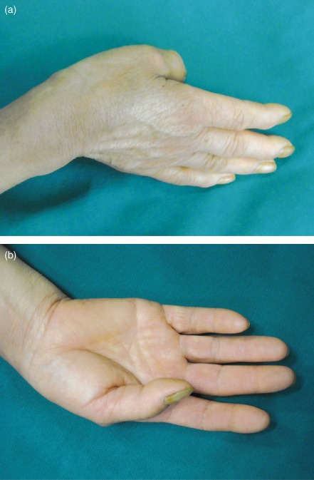
A 65‐year‐old woman presented with pain in the radial side of her wrist on extension and limitation of motion for 4 months. She had originally been diagnosed at her local hospital as having De Quervain's tenosynovitis and been given local injections of 1 mL triamcinolonacetonide 40 mg/mL plus 1 mL lidocaine 2% on six occasions times during the first three months of symptoms. One month prior to presenting to our hospital she had noted loss of extension of the right wrist and fingers after sweeping floors, and these symptoms were not relieved by rest. When she presented to our hospital she complained that she could not actively extend her wrist without considerable pain. She was in good health and had no history of inflammatory arthritis, diabetes, collagen vascular disease, or systemic steroid use. Physical examination revealed ulnar deviation of the wrist, deformity of the thumb (flexion at the metacarpophalangeal joint), a depression on the dorsal radial wrist and that the extensor tendons of the wrist could not be palpated during extension (Fig. 1). Wrist ultrasonography and MRI showed confusing disruption of the dorsal radial wrist extensor tendon structures, suggestive of multiple extensor tendon rupture.
Figure 1.

A 65‐year‐old woman presented with pain in the radial side of her wrist on extension and limitation of motion for 4 months. Photographs of the patient's right hand at the time of presentation to our institution (a) ulnar deviation of the wrist (b) deformity of the thumb (flexion at the metacarpophalangeal joint).
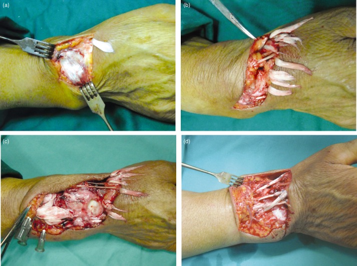
Surgery was performed under brachial plexus block. The extensor retinaculum and tendinous sheaths of the extensors were gray, with evidence of necrosis (Fig. 2a). Ten extensor tendons were completely ruptured at the dorsal radial wrist. The 2 cm long distal ends of all the tendons were dry and grey (Fig. 2b,c). Reconstruction of the extensor‐tendons was performed with autogenous palmaris longus tendon using the standard techniques of Kessler sutures (the tendons of the abductor pollicis longus, extensor pollicis brevis and longus, extensor carpi radialis longus, extensor indicis and long finger were repaired) (Fig. 2d). The patient's hand, including the thumb, was immobilized in a short‐arm plaster with the wrist in 30° of extension. After 5 weeks the plaster was removed and mobilization of the wrist started with passive physiotherapy, and subsequently active movements against resistance. At 6 months follow‐up, the patient had regained a full range of motion of her right hand.
Figure 2.

Operation was performed on the 65‐year‐old woman patient. (a) The extensor retinaculum and tendinous sheath of extensor are gray, with evidence of necrosis. (b, c) Ten extensor tendons on the dorsal radial wrist have completely ruptured. The tendons are dry and grey at their 2 cm distal ends; (d) Extensor‐tendons reconstruction using autogenous palmaris longus tendon (tendons of abductor pollicis longus, extensor pollicis brevis and longus, extensor carpi radialis longus, and extensor indicis and long finger).
Discussion
De Quervain's tenosynovitis, a stenosing tenosynovitis of the first dorsal compartment of the wrist, can cause wrist pain and dysfunction of the wrist and hand. It can be treated by splinting, local corticosteroid injection and surgery1. One or two local injections of 1 mL triamcinolonacetonide 10 mg/mL is an effective method of treating De Quervain's tenosynovitis, with a success‐rate of 78%2. A potential complication of this treatment modality is attritional tendon rupture.
Various mechanisms have been proposed to account for spontaneous tendon ruptures. These include inflammatory conditions such as rheumatoid arthritis3, osteoarthritis of the radio‐ulnar joint4, 5, mixed connective tissue disease6, attrition of tendons by sharp fragments of bone or small osteophytes7, anatomical alteration of Lister's tubercle8, chronic tenosynovitis caused by repetitive activity9, or chronic use of fluoroquinolone antibiotics10, 11 and corticosteroids12, 13, 14. Previously reported tendon ruptures have generally occurred in the patellar or Achilles region11, 15. The diagnosis can easily be made using ultrasound or MRI. To our knowledge, spontaneous rupture of multiple extensor tendons is rare. In advanced Kienböck's disease, a dorsally displaced fragment of the lunate can cause multiple extensor tendon ruptures due to chronic attrition16. In our case, the patient had been diagnosed as having De Quervain's tenosynovitis and had received six local injections of 1 mL triamcinolonacetonide 40 mg/mL plus 1 mL lidocaine 2% during the preceding three months. Our patient demonstrates that steroid abuse can cause multiple extensor tendon ruptures. Local steroids should be administered by an orthopaedic surgeon or trained healthcare practitioner and care taken to prevent injection directly into the substance of the tendon.
Many in vitro and animal studies have been carried out to investigate the relationship between corticosteroid injection and tendon ruptures and it has been suggested that steroids can change the crimp morphology of tendons, which could alter their resistance to rupture and the normal biomechanics of the involved extremity12. Injection of anabolic steroids has been shown to cause collagen necrosis and subsequent tendon weakness. Although corticosteroids make tendons less flexible and elastic, their strength is reportedly unaffected by these medications17, 18. Thus, a distinction should be made between loss of elasticity and actual tendon rupture. One week after a single dose of corticosteroids both intact and injured rat rotator cuff tendons are significantly weakened19. Fragmentation of collagen bundles and inflammatory cell infiltration has been observed after tendon injection with methylprednisolone and betamethasone; the tendons were reportedly abnormally soft and pale in color20. Triamcinolone and dexamethasone have been shown to significantly decrease cell viability, suppress cell proliferation, and reduce collagen synthesis in cultured human tenocytes. Suppressed human tenocyte cellular activity and reduced collagen production may lead to disturbed tendon structure and predispose the tendon to subsequent spontaneous rupture21, 22. Doctor repeatedly injecting steroids into wrist tendons should be aware of the possibility of inducing extensor tendon rupture.
Several methods of management of extensor tendon ruptures have been described. Conservative management may be employed if the patient is not significantly incapacitated or unfit for surgery. However, where there are multiple extensor tendon ruptures, surgery is the best option. This may involve direct end to end repair of tendons or autogenous tendon transfer, such as was performed in this case.
Disclosure: No benefits in any form have been, or will be, received from a commercial party related directly or indirectly to the subject of this manuscript.
References
- 1. Moore JS. De Quervain's tenosynovitis. Stenosing tenosynovitis of the first dorsal compartment. J Occup Environ Med, 1997, 39: 990–1002. [DOI] [PubMed] [Google Scholar]
- 2. Peters‐Veluthamaningal C, Winters JC, Groenier KH, et al Randomised controlled trial of local corticosteroid injections for de Quervain's tenosynovitis in general practice. BMC Musculoskelet Disord, 2009, 10: 131. [DOI] [PMC free article] [PubMed] [Google Scholar]
- 3. Rudge SR. Spontaneous rupture of all three extensor tendons to the thumb in rheumatoid arthritis. Hand, 1980, 12: 154–157. [DOI] [PubMed] [Google Scholar]
- 4. Ohshio I, Ogino T, Minami A, et al Extensor tendon rupture due to osteoarthritis of the distal radio‐ulnar joint. J Hand Surg Br, 1991, 16: 450–453. [DOI] [PubMed] [Google Scholar]
- 5. Carr AJ, Burge PD. Rupture of extensor tendons due to osteoarthritis of the distal radio‐ulnar joint. J Hand Surg Br, 1992, 17: 694–696. [DOI] [PubMed] [Google Scholar]
- 6. Kobayashi A, Futami T, Tadano I, et al Spontaneous rupture of extensor tendons at the wrist in a patient with mixed connective tissue disease. Mod Rheumatol, 2002, 12: 256–258. [DOI] [PubMed] [Google Scholar]
- 7. Kürklü M, Bilgic S, Yurttas Y, et al Spontaneous rupture of the extensor pollicis longus tendon due to a small osteophyte. Acta Reumatol Port, 2009, 34: 555–556. [PubMed] [Google Scholar]
- 8. Perugia D, Ciurluini M, Ferretti A. Spontaneous rupture of the extensor pollicis longus tendon in a young goalkeeper: a case report. Scand J Med Sci Sports, 2009, 19: 257–259. [DOI] [PubMed] [Google Scholar]
- 9. Dawson WJ. Sports‐induced spontaneous rupture of the extensor pollicis longus tendon. J Hand Surg Am, 1992, 17: 457–458. [DOI] [PubMed] [Google Scholar]
- 10. Casparian JM, Luchi M, Moffat RE, et al Quinolones and tendon ruptures. South Med J, 2000, 93: 488–491. [PubMed] [Google Scholar]
- 11. Parmar C, Meda KP. Achilles tendon rupture associated with combination therapy of levofloxacin and steroid in four patients and a review of the literature. Foot Ankle Int, 2007, 28: 1287–1289. [DOI] [PubMed] [Google Scholar]
- 12. Laseter JT, Russell JA. Anabolic steroid‐induced tendon pathology: a review of the literature. Med Sci Sports Exerc, 1991, 23: 1–3. [PubMed] [Google Scholar]
- 13. Mills SP, Charalambous CP, Hayton MJ. Bilateral rupture of the extensor pollicis longus tendon in a professional goalkeeper following steroid injections for extensor tenosynovitis. Hand Surg, 2009, 14: 135–137. [DOI] [PubMed] [Google Scholar]
- 14. Kramhøft M, Solgaard S. Spontaneous rupture of the extensor pollicis longus tendon after anabolic steroids. J Hand Surg Br, 1986, 11: 87. [DOI] [PubMed] [Google Scholar]
- 15. Chen SK, Lu CC, Chou PH, et al Patellar tendon ruptures in weight lifters after local steroid injections. Arch Orthop Trauma Surg, 2009, 129: 369–372. [DOI] [PubMed] [Google Scholar]
- 16. Park JW, Kim SK, Park JH, et al Multiple extensor tendon ruptures with advanced Kienböck's disease. J Hand Surg Am, 2007, 32: 233–235. [DOI] [PubMed] [Google Scholar]
- 17. Inhofe PD, Grana WA, Egle D, et al The effects of anabolic steroids on rat tendon. An ultrastructural, biomechanical, and biochemical analysis. Am J Sports Med, 1995, 23: 227–232. [DOI] [PubMed] [Google Scholar]
- 18. Miles JW, Grana WA, Egle D, et al The effect of anabolic steroids on the biomechanical and histological properties of rat tendon. J Bone Joint Surg Am, 1992, 74: 411–422. [PubMed] [Google Scholar]
- 19. Mikolyzk DK, Wei AS, Tonino P, et al Effect of corticosteroids on the biomechanical strength of rat rotator cuff tendon. J Bone Joint Surg Am, 2009, 91: 1172–1180. [DOI] [PMC free article] [PubMed] [Google Scholar]
- 20. Akpinar S, Hersekli MA, Demirors H, et al Effects of methylprednisolone and betamethasone injections on the rotator cuff: an experimental study in rats. Adv Ther, 2002, 19: 194–201. [DOI] [PubMed] [Google Scholar]
- 21. Wong MW, Tang YN, Fu SC, et al Triamcinolone suppresses human tenocyte cellular activity and collagen synthesis. Clin Orthop Relat Res, 2004, 421: 277–281. [DOI] [PubMed] [Google Scholar]
- 22. Wong MW, Tang YY, Lee SK, et al Effect of dexamethasone on cultured human tenocytes and its reversibility by platelet‐derived growth factor. J Bone Joint Surg Am, 2003, 85: 1914–1920. [DOI] [PubMed] [Google Scholar]


