Abstract
This study explores ultra-widefield angiography with oral fluorescein for the diagnosis of retinal conditions in pediatric patients.
Ultra-widefield (UWF) imaging is one of the new technologies available to ophthalmologists. Current UWF imaging modalities can provide several options for posterior segment documentation and evaluation, including color and red-free photography, fluorescein angiography, and fundus autofluorescence.1 The use of UWF fundus fluorescein angiography (FFA), in particular, has contributed significantly to the detection of peripheral fundus disease in both adult and pediatric patients with retinal conditions.2 Similar to standard field retinal angiography, FFA involves intravenous injection of fluorescein, after which fundus images are obtained. An alternative to intravenous injection of fluorescein is oral fluorescein angiography. It is used in cases in which intravenous injection is difficult to perform or is refused by patients who have a fear of needles. Oral FFA has been successfully used with standard and widefield camera systems.3 Herein, we report our experience with UWF retinal angiography using oral fluorescein in pediatric patients.
Methods
This case series was conducted from August 2016 to August 2017 at the Moorfields Eye Hospital Dubai and Moorfields Eye Hospital Centre in Abu Dhabi in the United Arab Emirates (UAE), and it adhered to the tenets of the Declaration of Helsinki.4 The Ethics Committee of the Moorfields Eye Hospitals UAE approved the study. Parents were educated about the use of oral fluorescein instead of intravenous flourescein and signed informed consent before the procedure. The indication for oral FFA in children was the inability of the health care professional to obtain venous access or a fear of needles on the part of the child. Two ampules of 10% fluorescein dye were mixed with 30 mL of apple juice, and this mixture was ingested by the patient. The patient then ingested 30 mL of apple juice to clear the taste of the dye. The patient’s pupils were pharmacologically dilated before imaging was performed. An imaging system (200Tx; Optos) was used to obtain noncontact high-resolution UWF retinal angiographic images. The feasibility of the procedure was determined by the appearance of dye in the retinal circulation visible on angiography, the ability to obtain images, and the presence or absence of adverse effects.
Results
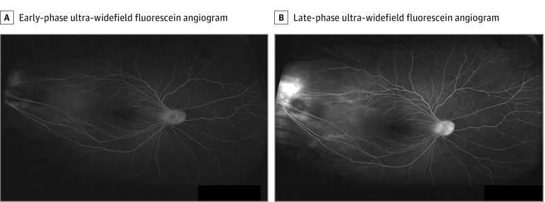
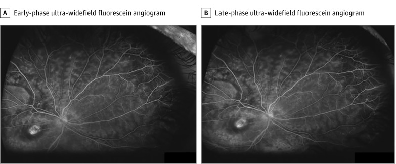
Eighteen patients aged 4 and 16 years received oral fluorescein before UWF imaging was performed. There were 8 girls (44%) and 10 boys (56%), of whom 14 (78%) were Arab and 4 (22%) were white. Clinical diagnoses included 12 cases of posterior uveitis or panuveitis and an additional 6 cases of Coats disease, familial exudative vitreoretinopathy, thalassemia, or tubulointerstitial nephritis and uveitis syndrome. The first images were visible as early as 5 minutes after the patient ingested oral fluorescein and as late as 12 minutes after ingestion. The recirculation phase was visible no later than 15 minutes after ingestion. Seventeen of 18 patients (94%) had images that showed the arterial phase, and 18 (100%) had images that showed the venous and recirculation phases (Figure 1 and Figure 2). All patients tolerated the procedure well and cooperated during the procedure. There were no adverse effects during or after the procedure.
Figure 1. Ultra-Widefield Fluorescein Angiograms of a Pediatric Patient With Familial Exudative Vitreoretinopathy.
A, Ultra-widefield fluorescein angiogram obtained during the early phase of angiography in a pediatric patient with familial exudative vitreoretinopathy that shows filling of dragged retinal vasculature and a lesion in the temporal retinal periphery. B, Angiogram obtained during a late phase of angiography showing peripheral leakage of fluorescein.
Figure 2. Ultra-Widefield Fluorescein Angiograms of a Pediatric Patient With Coats Disease.
A, Ultra-widefield fluorescein angiogram obtained during the early phase of angiography in a pediatric patient with Coats disease that shows filling of retinal vessels, unequal fluorescence, peripheral teleangiectatic retinal vessels, and staining of macular lesion. B, Angiogram obtained during a late phase of angiography that shows a similar picture.
Discussion
Oral FFA was first used to obtain images with a conventional fundus camera,3 before the development of the confocal scanning laser ophthalmoscope, which provided superior image quality.5 Sugimoto et al6 has reported the use of oral FFA with UWF imaging in a cohort of 34 adult patients. More recently, Fung et al7 reported the use of oral FFA to obtain UWF images in 3 premature infants with retinopathy of prematurity. Both reports found oral administration of fluorescein to be safe and a good alternative when intravenous administration is difficult, impossible, or refused. The need for an alternative to intravenous administration is a common scenario in pediatric ophthalmology. We used the same concentration of dye (20 mg/kg of body weight) as reported elsewhere.6 The timing of and success in obtaining images of the early arterial phase as well as the venous and recirculation phases were comparable with published studies.2,6 Imaging artifacts were similar to those found when using UWF technology in patients from all age categories, and these artifacts were easily eliminated. The peripheral retinal anatomy and vascular network were detailed, and necessary clinical information was obtained in all cases.
In summary, these findings suggest that oral fluorescein is an effective alternative to intravenous fluorescein when performing UWF FFA in children. We realize, however, that these few cases cannot definitively determine the effectiveness of oral fluorescein for UWF FFA.
References
- 1.Shoughy SS, Arevalo JF, Kozak I. Update on wide- and ultra-widefield retinal imaging. Indian J Ophthalmol. 2015;63(7):575-581. [DOI] [PMC free article] [PubMed] [Google Scholar]
- 2.Tsui I, Franco-Cardenas V, Hubschman JP, Schwartz SD. Pediatric retinal conditions imaged by ultra wide field fluorescein angiography. Ophthalmic Surg Lasers Imaging Retina. 2013;44(1):59-67. [DOI] [PubMed] [Google Scholar]
- 3.Kelley JS, Kincaid M. Retinal fluorography using oral fluorescein. Arch Ophthalmol. 1979;97(12):2331-2332. [DOI] [PubMed] [Google Scholar]
- 4.World Medical Association World Medical Association Declaration of Helsinki: ethical principles for medical research involving human subjects. JAMA. 2013;310(20):2191-2194. doi: 10.1001/jama.2013.281053 [DOI] [PubMed] [Google Scholar]
- 5.Garcia CR, Rivero ME, Bartsch DU, et al. . Oral fluorescein angiography with the confocal scanning laser ophthalmoscope. Ophthalmology. 1999;106(6):1114-1118. [DOI] [PubMed] [Google Scholar]
- 6.Sugimoto M, Matsubara H, Miyata R, Matsui Y, Ichio A, Kondo M. Ultra-widefield fluorescein angiography by oral administration of fluorescein. Acta Ophthalmol. 2014;92(5):e417-e418. [DOI] [PubMed] [Google Scholar]
- 7.Fung TH, Muqit MM, Mordant DJ, Smith LM, Patel CK. Noncontact high-resolution ultra-wide-field oral fluorescein angiography in premature infants with retinopathy of prematurity. JAMA Ophthalmol. 2014;132(1):108-110. [DOI] [PubMed] [Google Scholar]


