Abstract
Assays were done to assess the ability of 5 methicillin-resistant Staphylococcus pseudintermedius (MRSP) isolates from difference sources to adhere to canine and human corneocytes. Cell wall-associated (CWA) protein gene profiles were examined to look for associations with adherence. Five MRSP strains were studied: 3 with the same CWA protein gene profile (14 genes) and belonging to sequence type (ST) 45 were isolated from a dog, a human, and the environment. The other 2 were an environmental isolate belonging to ST433 that had the lowest number of CWA protein genes (12) and a canine clinical isolate belonging to ST733 that had the greatest number of CWA protein genes (18). The 3 isolates of MRSP ST45, a major clone in Thailand, had the greatest ability to adhere to canine and human corneocytes. Nevertheless, MRSP adherence ability could not be predicted from the profile of genes encoding CWA proteins.
Résumé
Des analyses ont été effectuées afin de déterminer la capacité de cinq isolats de Staphylococcus pseudintermedius résistants à la méthicilline (SPRM) provenant de différentes sources à adhérer à des cornéocytes canins et humains. Les profils de gènes des protéines associées à la paroi cellulaire (APC) furent examinés afin de vérifier des associations avec l’adhérence. Cinq souches de SPRM furent étudiées : trois avec le même profil de gène de protéines APC (14 gènes) et appartenant au type de séquence (ST) 45 isolées d’un chien, un humain et l’environnement. Les deux autres souches étaient un isolat provenant de l’environnement et appartenant à ST433 et qui avait le plus petit nombre de gènes de protéines APC (12) et un isolat clinique canin appartenant au ST733 et qui avait le plus grand nombre de gènes de protéines APC (18). Les trois isolats de ST45, un clone majeur en Thaïlande, avait la plus grande capacité d’adhérer aux cornéocytes canins et humains. Toutefois, la capacité d’adhérence des SPRM ne pouvait être prédite par le profil de gènes codant pour les protéines APC.
(Traduit par Docteur Serge Messier)
Methicillin-resistant Staphylococcus pseudintermedius (MRSP) strains are commonly found on canine skin after treatment with antibiotics and are emerging pathogens in human patients (1). These organisms colonize humans who have contact with dogs, and they can contaminate the environment in veterinary hospitals and households (2). The initial step in colonization and infection is adhesion to skin cells (corneocytes), which may be influenced by bacterial cell wall-associated (CWA) proteins encoded by S. pseudintermedius surface protein genes (sps) (3). The ability of S. pseudintermedius to adhere to both canine and human corneocytes has been used as evidence for zoonotic potential (4). The adhesion ability of individual strains may relate to their source of isolation. This ability was previously evaluated for isolates from dogs and humans, but environmental isolates had not yet been investigated (4,5). Thus, to estimate their pathogenic potential, their ability to adhere to human and canine corneocytes needed to be assessed. Moreover, the association between adherence and genes encoding CWA proteins is still not clear. This study aimed to determine the adherence ability of 5 MRSP isolates and whether there were correlations between the presence of genes for CWA proteins and adherence.
Five MRSP isolates were obtained from the culture collection at Chulalongkorn University, Bangkok, Thailand (Table I). They included 2 isolates each from dogs and the environment and 1 isolate from a human. The sequence type (ST) of these isolates in multilocus sequence typing and their profile of genes encoding CWA proteins had previously been established (1,6).
Table I.
Methicillin–resistant Staphylococcus pseudintermedius isolates used in this study.
| Strain | Source | Site | ST | SCCmec | S. pseudintermedius surface protein genesa | |||||||||||||||||
|---|---|---|---|---|---|---|---|---|---|---|---|---|---|---|---|---|---|---|---|---|---|---|
|
| ||||||||||||||||||||||
| spsA | spsB | spsC | spsD | spsE | spsF | spsG | spsH | spsI | spsJ | spsK | spsL | spsM | spsN | spsO | spsP | spsQ | spsR | |||||
| AP65 | Dog | Perineum | 45 | ΨSCCmec57395 | + | + | + | − | + | − | + | + | + | + | + | + | + | + | + | − | − | + |
| VB88 | Human | Nasal cavity | 45 | ΨSCCmec57395 | + | + | + | − | + | − | + | + | + | + | + | + | + | + | + | − | − | + |
| SW1F4O1 | Environment | Dermatology unit floor | 45 | ΨSCCmec57395 | + | + | + | − | + | − | + | + | + | + | + | + | + | + | + | − | − | + |
| SW1F4O2 | Environment | Dermatology unit floor | 433 | Nontypeable | + | + | + | + | + | + | + | + | + | + | + | + | + | + | + | + | + | + |
| MIC460 | Dog | Pyoderma | 733 | V | + | + | + | − | + | − | + | + | − | + | + | + | + | + | − | − | − | + |
Detected as described previously (6).
ST — sequence type; SCCmec — staphylococcal cassette chromosome mec.
Corneocytes were collected from dogs and humans according to the Animal Care and Use Protocol (no. 1731009) of Chulalongkorn University and with approval of the Research Ethics Review Committee for Research Involving Human Research Participants, Health Science Group, Chulalongkorn University (no. 225.1/59). The corneocytes were collected from the abdominal area of 5 dogs and from the medial biceps area of 5 human volunteers who had little contact with dogs, as previously described (5,7). All dogs and humans were clinically healthy and did not have a history of skin disease. Debris and normal microbiota were removed from the sampling sites by application of 5 successive adhesive tape strips (Scotch Tape; 3M, St. Paul, Minnesota, USA), and then medical adhesive disks 22 mm in diameter (D-Squame; CuDerm Corporation, Dallas, Texas, USA) were placed on the skin to collect corneocytes. The confluence of corneocytes in the disk was examined by light microscopy, and disks with confluent corneocytes were selected for the adherence assays.
The adherence assay was done in duplicate with some minor modifications from previous protocols (5). Briefly, the isolates were cultured overnight in brain–heart infusion (BHI) broth (Oxoid; Thermo Scientific, Basingstoke, England) at 37°C. An aliquot of the MRSP suspension (100 μL) was transferred into 30 mL of BHI broth with shaking, and 25 mL of the culture at mid-exponential phase was centrifuged at 1500 × g and 4°C for 5 min. The bacterial pellet was centrifuged at 800 × g and 4°C for 10 min and washed 3 times with sterile phosphate-buffered saline (PBS). The bacterial suspension was adjusted to an optical density at 600 nm of 0.15, which represented approximately 3 × 10⁷ colony-forming units per milliliter. Two corneocyte disks were incubated with 500 μL of bacterial suspension for 45 min, and 1 disk from each individual was incubated with PBS as a negative control. After incubation the disks were washed with sterile PBS for 20 s, stained with 0.5% crystal violet for 20 s, and then washed with water for 20 s. Ten fields of confluent corneocytes per disk were examined under a microscope (× 1000) to determine bacterial adhesion counts (BACs). Microscopic pictures were captured with Motic Images Plus software, Version 2.0 (Motic China Group Company, Xiamen, China).
The Mann–Whitney U-test was used to compare the median BAC of individual strains for different types of corneocytes. The Kruskal–Wallis test was used to find the difference between strains, with SPSS software, Version 22.0 (IBM Corporation, Armonk, New York, USA).
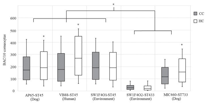
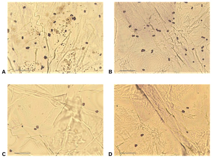
The 3 MRSP strains of ST45, the main clone in Thailand, had significantly higher BACs than the 2 strains of ST733 and ST433 (P < 0.001) (Figure 1). The ST45 isolates from the environment and from a human showed the greatest adherence to canine and human corneocytes (Figure 2). No positive association was found between adherence and the sps gene profiles: MRSP SW1F4O2, belonging to ST433, had the highest number of sps genes (18/18) and the least adherence to both canine and human corneocytes (Figures 1 and 2). Interestingly, the 2 isolates from dogs, belonging to ST45 and ST733, had significantly higher BACs with human corneocytes than with canine corneocytes (P = 0.007 and P < 0.001, respectively). The isolate from a human, belonging to ST45, had a significantly greater BAC with human corneocytes than with canine cells (P < 0.001), suggesting some host specificity. The 2 environmental strains showed no difference in attachment to canine or human cells (Figure 1).
Figure 1.
Box plots representing bacterial adhesion counts (BACs) of 5 methicillin-resistant Staphylococcus pseudintermedius (MRSP) isolates on canine corneocytes (CC) and human corneocytes (HC). The top and bottom of the box represent lower quartile and upper quartile. The line in the box is the median and the whisker is the range of the data. The asterisk indicates a statistically significant difference at P < 0.05.
Figure 2.
Adhesion of MRSP to canine and human corneocytes. A — MRSP SW1F4O1-ST45 adhering to canine corneocytes. B — MRSP VB88-ST45 adhering to human corneocytes. C and D — MRSP SW1F4O2-ST433 adhering to canine and human corneocytes, respectively. Bar — 10 μm.
The adherence assay is a reproducible and rapid method for quantitative measurement of staphylococcal adherence to corneocytes (8,9). The low number of representative isolates might be a limitation of this study. Nevertheless, it is difficult to perform this assay with a high number of isolates because of the large number of host corneocytes needed. Accordingly, many studies using the adherence assay have had limited numbers of isolates (4,9–11).
In this study, MRSP derived from different sources adhered to canine and human corneocytes. The environmental isolate SW1F4O1, belonging to ST45, showed an adhesion ability resembling that of isolates from a dog and a human in this clonal lineage. The MRSP SW1F4O2, belonging to ST433, also adhered to both types of corneocytes, but it had the weakest adherence. The results also showed that strains from the major Thai clone, ST45, had the greatest potential to adhere to canine and human corneocytes, reflecting the situation with the major European type, ST71. This suggests that strains of the predominant STs are better adapted to attaching to canine and human skin cells than are strains belonging to other STs (5). The 3 strains that showed greater adhesion to human than to canine corneocytes might have a greater ability to adapt to human corneocytes in vivo than other strains (5). The strain variation may also influence the adhesion ability. It has been suggested that clinical isolates might adhere to corneocytes more than isolates from healthy dogs (11). In this study, we did not observe this variation since a single clinical isolate was used. Comparison of the adhesion ability of isolates belonging to the dominant clones from clinical and healthy dogs would be useful. In this study no association was found between sps profile and BAC. So far, only 3 out of 18 sps genes — spsD, spsL, and spsO — have been proven to adhere to corneocytes (12). Nevertheless, MRSP SW1F4O2, belonging to ST433 and harboring spsD, spsL, and spsO, had the lowest ability to adhere to canine and human corneocytes in this study. Therefore, the link between sps profile and adhesion ability still needs to be clarified. The adherence of S. pseudintermedius is possibly influenced by the colonization status of the corneocyte donors, and strains with greater adhesion capacity might reside in persistent carriers (10). Moreover, bacterial colonization is a multifactorial process; therefore, products encoded by other gene types may be required for host binding, as described for S. aureus (9). A combination of in vivo experiments, whole genome comparisons, and analysis of surface protein gene expression using canine and human MRSP should provide more understanding of basic bacterial host adaptation (9,13).
In this study, MRSP ST45 isolates from a dog, a human, and the environment adhered to both canine and human corneocytes. The clone type of MRSP may influence the initial stage of colonization and infection but without a direct relationship to the surface protein gene profile.
Acknowledgments
This work was supported by a scholarship from the Graduate School, Chulalongkorn University. Bangkok, Thailand, to commemorate the 72nd anniversary of his Majesty King Bhumibala Aduladeja, the 100th Anniversary of the Chulalongkorn University Fund for Doctoral Scholarship, and the 90th Anniversary of the Chulalongkorn University Fund (Ratchadaphiseksomphot Endowment Fund). We thank Professor David J. Hampson of City University of Hong Kong, Kowloon, for his kind editorial assistance during preparation of this manuscript.
References
- 1.Chanchaithong P, Perreten V, Schwendener S, et al. Strain typing and antimicrobial susceptibility of methicillin-resistant coagulase-positive staphylococcal species in dogs and people associated with dogs in Thailand. J Appl Microbiol. 2014;117:572–586. doi: 10.1111/jam.12545. [DOI] [PubMed] [Google Scholar]
- 2.van Duijkeren E, Catry B, Greko C, et al. Review on methicillin-resistant Staphylococcus pseudintermedius. J Antimicrob Chemother. 2011;66:2705–2714. doi: 10.1093/jac/dkr367. [DOI] [PubMed] [Google Scholar]
- 3.Bannoehr J, Ben Zakour NL, Reglinski M, et al. Genomic and surface proteomic analysis of the canine pathogen Staphylococcus pseudintermedius reveals proteins that mediate adherence to the extracellular matrix. Infect Immun. 2011;79:3074–3086. doi: 10.1128/IAI.00137-11. [DOI] [PMC free article] [PubMed] [Google Scholar]
- 4.Woolley KL, Kelly RF, Fazakerley J, Williams NJ, Nuttall TJ, McEwan NA. Reduced in vitro adherence of Staphylococcus species to feline corneocytes compared to canine and human corneocytes. Vet Dermatol. 2008;19:1–6. doi: 10.1111/j.1365-3164.2007.00649.x. [DOI] [PubMed] [Google Scholar]
- 5.Latronico F, Moodley A, Nielsen SS, Guardabassi L. Enhanced adherence of methicillin resistant Staphylococcus pseudintermedius sequence type 71 to canine and human corneocytes. Vet Res. 2014;45:70. doi: 10.1186/1297-9716-45-70. [DOI] [PMC free article] [PubMed] [Google Scholar]
- 6.Phumthanakorn N, Chanchaithong P, Prapasarakul N. Development of a set of multiplex PCRs for detection of genes encoding cell wall-associated proteins in Staphylococcus pseudintermedius isolates from dogs, humans and the environment. J Microbiol Methods. 2017;142:90–95. doi: 10.1016/j.mimet.2017.09.003. [DOI] [PubMed] [Google Scholar]
- 7.Saijonmaa-Koulumies LE, Lloyd D. Adherence of Staphylococcus intermedius to canine corneocytes in vitro. Vet Dermatol. 2002;13:169–176. doi: 10.1046/j.1365-3164.2002.00294.x. [DOI] [PubMed] [Google Scholar]
- 8.Lu Y-F, McEwan NA. Staphylococcal and micrococcal adherence to canine and feline corneocytes: Quantification using a simple adhesion assay. Vet Dermatol. 2007;18:29–35. doi: 10.1111/j.1365-3164.2007.00567.x. [DOI] [PubMed] [Google Scholar]
- 9.Moodley A, Espinosa-Gongora C, Nielsen SS, McCarthy AJ, Lindsay JA, Guardabassi L. Comparative host specificity of human- and pig-associated Staphylococcus aureus clonal lineages. PLoS One. 2012;7:e49344. doi: 10.1371/journal.pone.0049344. [DOI] [PMC free article] [PubMed] [Google Scholar]
- 10.Paul NC, Latronico F, Moodley A, Nielsen SS, Damborg P, Guardabassi L. In vitro adherence of Staphylococcus pseudintermedius to canine corneocytes is influenced by colonization status of corneocyte donors. Vet Res. 2013;44:52. doi: 10.1186/1297-9716-44-52. [DOI] [PMC free article] [PubMed] [Google Scholar]
- 11.McEwan NA, Kalna G, Mellor D. A comparison of adherence by four strains of Staphylococcus intermedius and Staphylococcus hominis to canine corneocytes collected from normal dogs and dogs suffering from atopic dermatitis. Res Vet Sci. 2005;78:193–198. doi: 10.1016/j.rvsc.2004.09.002. [DOI] [PubMed] [Google Scholar]
- 12.Bannoehr J, Brown JK, Shaw DJ, Fitzgerald RJ, van den Broek AH, Thoday KL. Staphylococccus pseudintermedius surface proteins SpsD and SpsO mediate adherence to ex vivo canine corneocytes. Vet Dermatol. 2012;23:119–124. doi: 10.1111/j.1365-3164.2011.01021.x. [DOI] [PubMed] [Google Scholar]
- 13.Uhlemann AC, Porcella SF, Trivedi S, et al. Identification of a highly transmissible animal-independent Staphylococcus aureus ST398 clone with distinct genomic and cell adhesion properties. MBio. 2012;3:e00027–12. doi: 10.1128/mBio.00027-12. [DOI] [PMC free article] [PubMed] [Google Scholar]


