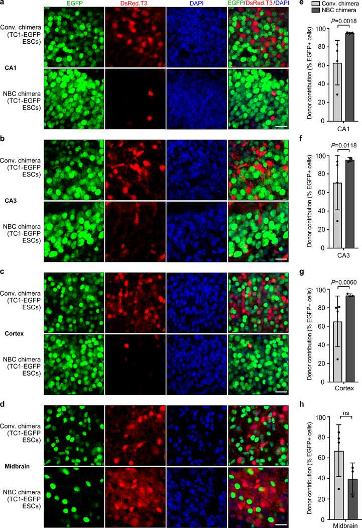
Figure 2. NBC chimeras show consistently high donor contribution in forebrain regions as compared to conventional chimeras.
a-d, Representative images showing extent of TC1-EGFP donor ESC contribution to the indicated brain regions in conventional (conv.) and NBC chimeras at P0, depicting donor-derived cells in green and host-derived cells in red. Nuclei were DAPI stained (blue). CA, cornu ammonis subfields of the hippocampus. Scale bars, 10 μm. e-h, Quantification of donor ESC contribution in conventional (n=4) and NBC chimeras (n=3). Data are mean ± SD. Variance was significantly different between conventional and NBC chimeras for CA1, CA3, and cortex (F test for equality of variances), reflecting the wide variation in donor contribution among individual conventional chimeras, in contrast to the consistently high donor contribution in NBC chimeras. No difference in variance was observed for midbrain (P = 0.5534; ns, not significant; F test for equality of variances), consistent with random donor contribution to this non-ablated brain region in both types of chimeras. See also Extended Data Figures 2 and 3.

