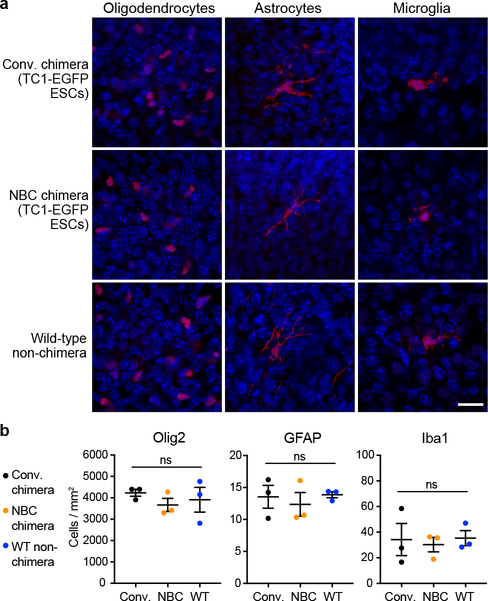
Extended Data Figure 5. NBC mice have normal proportions of non-neuronal cells and do not show signs of neuroinflammation.
a, Representative immunofluorescence images of oligodendrocytes (Olig2), astrocytes (GFAP), and microglia (Iba1) in cortical brain sections of conventional (conv.) and NBC chimeras, with a wild-type, non-chimeric mouse for comparison. The non-neuronal cells are shown in red; DAPI-stained nuclei are in blue. Scale bar, 10 μm. These experiments were repeated on 9 mice (n=3, NBC chimeras; n=3, conventional chimeras; n=3, non-chimeric wild-type mice). b, Quantification Olig2-, GFAP-, or Iba1-positive cells in cortical brain sections of conventional chimeras, NBC chimeras, and wild-type non-chimeric mice (n=3 each). Data represent mean and SEM; ns, not significant, P>0.05 (one-way ANOVA, Tukey’s post hoc correction for multiple comparisons).

