. 2019 Jun 21;8:e47300. doi: 10.7554/eLife.47300

Figure 2. Wnt7a regions that are required for Reck/Gpr124-stimulated signaling.

(A) Backbone model of Wnt7a (N-terminal domain, blue; C-terminal domain, cyan) bound to Fz8 CRD (green) based on the Wnt-CRD crystal structure of Janda et al. (2012). The N-terminal domain of Wnt7a consists of ~270 amino acids (some of which were not resolved in the crystal structure), and the C-terminal domain consists of ~80 amino acids. (B) Beta-catenin signaling assay using STF cells transfected with Reck and Gpr124 (left) or pRK5 vector control (right), together with the indicated Wnts. Inset: summary of STF signaling. Statistical comparisons in (B), (D), and (G) are to WT Wnt7a. (C) Amino acid sequence of the N-terminal domain of mouse Wnt7a, with alanine scanning mutants indicated. (D) Beta-catenin signaling assay using STF cells transfected with Reck, Gpr124, and Fz8, together with WT or mutant Wnt7a. (E) Left, backbone model of Wnt7a bound to Fz8 CRD as in (A), with amino acids that are critical for Wnt7a signaling shown in red. Right, the boxed region is displayed at higher magnification. (F) Single alanine substitution mutants of Wnt7a, indicated in red. (G) Beta-catenin signaling assay as in (D) with WT or the indicated Wnt7a mutants. (H) Left, backbone model of Wnt7a bound to Fz8 CRD as in (A) except rotated 135 degrees, with amino acids that are critical for Wnt7a signaling shown in red. Right, the boxed region is displayed at higher magnification.

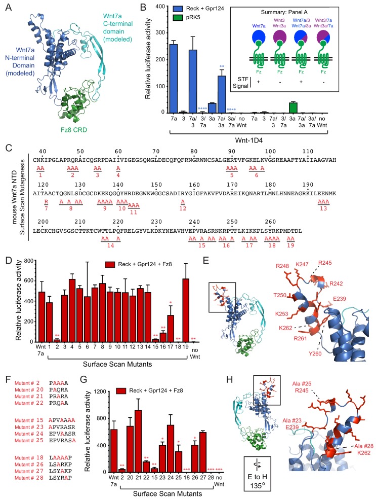
Figure 2—source data 1. Relative luciferase activity for STF experiments in Figure 2 panels B, D, and G.
DOI: 10.7554/eLife.47300.008

Figure 2.

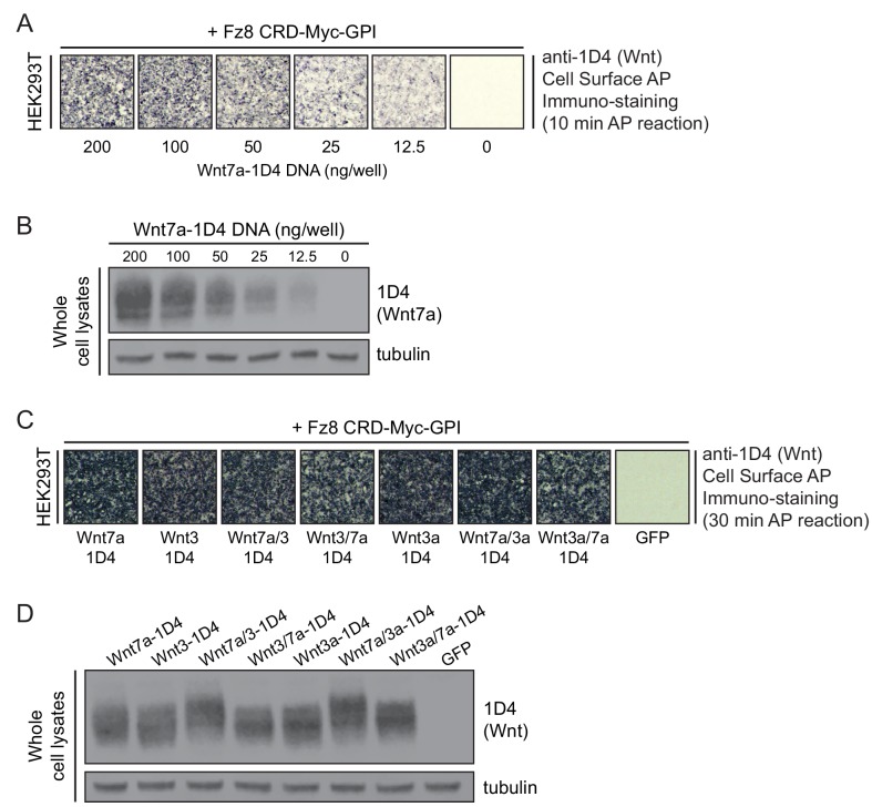
Figure 2—figure supplement 1. Production of intact and chimeric Wnt proteins for Reck- and Gpr124-mediated signaling.

Figure 2—figure supplement 1.

(A) Detecting WT Wnt7a-1D4 proteins on the surface of HEK293T cells co-transfected with Fz8CRD-Myc-GPI and the indicated concentrations of Wnt7a-1D4 plasmid DNA. Live cells were immuno-stained with anti-1D4, fixed, and then incubated with anti-mouse antibody conjugated to AP. (B) Immunoblot of post-nuclear supernatants from HEK293T cells transfected with the indicated concentrations of Wnt7a-1D4 plasmid DNA, probed with mAb 1D4 and anti-tubulin. (C) Detecting Wnt proteins on the surface of HEK293T cells transfected with Fz8CRD-Myc-GPI, together with Wnt7a-1D4, Wnt3-1D4, Wnt3a-1D4, or their chimeric derivatives. Cell-surface AP immuno-staining was performed as described in (A). (D) Immunoblot of post-nuclear supernatants from HEK293T cells transfected with Wnt7a-1D4, Wnt3-1D4, Wnt3a-1D4, or their chimeric derivatives, probed with mAb 1D4 and anti-tubulin.
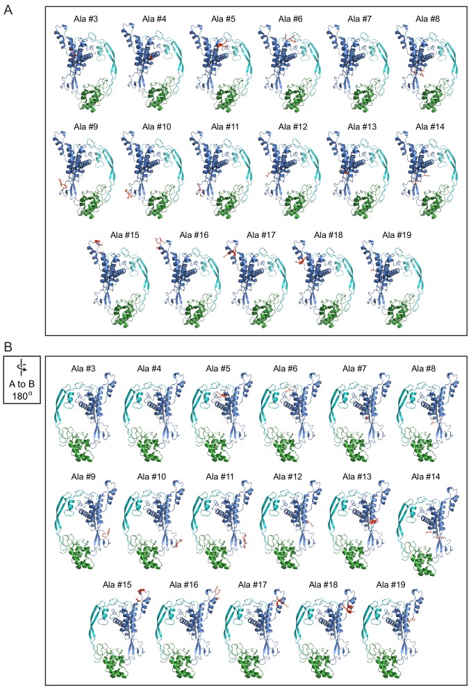
Figure 2—figure supplement 2. Locations of alanine mutations on the Wnt7a-Fz8CRD model.

Figure 2—figure supplement 2.

Front (A) and back (B; rotated 180 degrees) views of the Wnt7a-Fz8CRD model with alanine mutations shown in red.
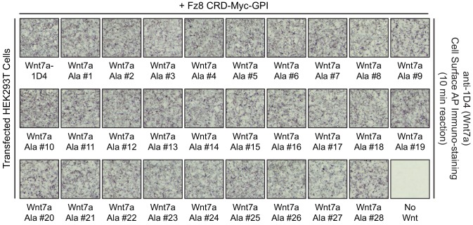
Figure 2—figure supplement 3. Cell-surface CRD binding by Wnt7a alanine mutants.

Figure 2—figure supplement 3.

Detecting WT and mutant Wnt7a-1D4 proteins on the surface of HEK293T cells co-transfected with Fz8CRD-Myc-GPI and the indicated WT or mutant Wnt7a-1D4 plasmids. Live cells were immuno-stained with anti-1D4, fixed, and then incubated with anti-mouse antibody conjugated to AP.